

- •Contents
- •Preface
- •Contributors
- •1 Vessels
- •1.1 Aorta, Vena Cava, and Peripheral Vessels
- •Aorta, Arteries
- •Anomalies and Variant Positions
- •Dilatation
- •Stenosis
- •Wall Thickening
- •Intraluminal Mass
- •Perivascular Mass
- •Vena Cava, Veins
- •Anomalies
- •Dilatation
- •Intraluminal Mass
- •Compression, Infiltration
- •1.2 Portal Vein and Its Tributaries
- •Enlarged Lumen Diameter
- •Portal Hypertension
- •Intraluminal Mass
- •Thrombosis
- •Tumor
- •2 Liver
- •Enlarged Liver
- •Small Liver
- •Homogeneous Hypoechoic Texture
- •Homogeneous Hyperechoic Texture
- •Regionally Inhomogeneous Texture
- •Diffuse Inhomogeneous Texture
- •Anechoic Masses
- •Hypoechoic Masses
- •Isoechoic Masses
- •Hyperechoic Masses
- •Echogenic Masses
- •Irregular Masses
- •Differential Diagnosis of Focal Lesions
- •Diagnostic Methods
- •Suspected Diagnosis
- •3 Biliary Tree and Gallbladder
- •3.1 Biliary Tree
- •Thickening of the Bile Duct Wall
- •Localized and Diffuse
- •Bile Duct Rarefaction
- •Localized and Diffuse
- •Bile Duct Dilatation and Intraductal Pressure
- •Intrahepatic
- •Hilar and Prepancreatic
- •Intrapancreatic
- •Papillary
- •Abnormal Intraluminal Bile Duct Findings
- •Foreign Body
- •The Seven Most Important Questions
- •3.2 Gallbladder
- •Changes in Size
- •Large Gallbladder
- •Small/Missing Gallbladder
- •Wall Changes
- •General Hypoechogenicity
- •General Hyperechogenicity
- •General Tumor
- •Focal Tumor
- •Intraluminal Changes
- •Hyperechoic
- •Hypoechoic
- •Nonvisualized Gallbladder
- •Missing Gallbladder
- •Obscured Gallbladder
- •4 Pancreas
- •Diffuse Pancreatic Change
- •Large Pancreas
- •Small Pancreas
- •Hypoechoic Texture
- •Hyperechoic Texture
- •Focal Changes
- •Anechoic Lesion
- •Hypoechoic Lesion
- •Isoechoic Lesion
- •Hyperechoic Lesion
- •Irregular (Complex Structured) Lesion
- •Dilatation of the Pancreatic Duct
- •Marginal/Mild Dilatation
- •Marked Dilatation
- •5 Spleen
- •Nonfocal Changes of the Spleen
- •Diffuse Parenchymal Changes
- •Large Spleen
- •Small Spleen
- •Focal Changes of the Spleen
- •Anechoic Mass
- •Hypoechoic Mass
- •Hyperechoic Mass
- •Splenic Calcification
- •6 Lymph Nodes
- •Peripheral Lymph Nodes
- •Head/Neck
- •Extremities (Axilla, Groin)
- •Abdominal Lymph Nodes
- •Porta Hepatis
- •Splenic Hilum
- •Mesentery (Celiac, Upper and Lower Mesenteric Station)
- •Stomach
- •Focal Wall Changes
- •Extended Wall Changes
- •Dilated Lumen
- •Narrowed Lumen
- •Small/Large Intestine
- •Focal Wall Changes
- •Extended Wall Changes
- •Dilated Lumen
- •Narrowed Lumen
- •8 Peritoneal Cavity
- •Anechoic Structure
- •Hypoechoic Structure
- •Hyperechoic Structure
- •Anechoic Structure
- •Hypoechoic Structure
- •Hyperechoic Structure
- •Wall Structures
- •Smooth Margin
- •Irregular Margin
- •Intragastric Processes
- •Intraintestinal Processes
- •9 Kidneys
- •Anomalies, Malformations
- •Aplasia, Hypoplasia
- •Cystic Malformation
- •Anomalies of Number, Position, or Rotation
- •Fusion Anomaly
- •Anomalies of the Renal Calices
- •Vascular Anomaly
- •Diffuse Changes
- •Large Kidneys
- •Small Kidneys
- •Hypoechoic Structure
- •Hyperechoic Structure
- •Irregular Structure
- •Circumscribed Changes
- •Anechoic Structure
- •Hypoechoic or Isoechoic Structure
- •Complex Structure
- •Hyperechoic Structure
- •10 Adrenal Glands
- •Enlargement
- •Anechoic Structure
- •Hypoechoic Structure
- •Complex Echo Structure
- •Hyperechoic Structure
- •11 Urinary Tract
- •Malformations
- •Duplication Anomalies
- •Dilatations and Stenoses
- •Dilated Renal Pelvis and Ureter
- •Anechoic
- •Hypoechoic
- •Hypoechoic
- •Hyperechoic
- •Large Bladder
- •Small Bladder
- •Altered Bladder Shape
- •Intracavitary Mass
- •Hypoechoic
- •Hyperechoic
- •Echogenic
- •Wall Changes
- •Diffuse Wall Thickening
- •Circumscribed Wall Thickening
- •Concavities and Convexities
- •12.1 The Prostate
- •Enlarged Prostate
- •Regular
- •Irregular
- •Small Prostate
- •Regular
- •Echogenic
- •Circumscribed Lesion
- •Anechoic
- •Hypoechoic
- •Echogenic
- •12.2 Seminal Vesicles
- •Diffuse Change
- •Hypoechoic
- •Circumscribed Change
- •Anechoic
- •Echogenic
- •Irregular
- •12.3 Testis, Epididymis
- •Diffuse Change
- •Enlargement
- •Decreased Size
- •Circumscribed Lesion
- •Anechoic or Hypoechoic
- •Irregular/Echogenic
- •Epididymal Lesion
- •Anechoic
- •Hypoechoic
- •Intrascrotal Mass
- •Anechoic or Hypoechoic
- •Echogenic
- •13 Female Genital Tract
- •Masses
- •Abnormalities of Size or Shape
- •Uterus
- •Abnormalities of Size or Shape
- •Myometrial Changes
- •Intracavitary Changes
- •Endometrial Changes
- •Fallopian Tubes
- •Hypoechoic Mass
- •Anechoic Cystic Mass
- •Solid Echogenic or Nonhomogeneous Mass
- •14 Thyroid Gland
- •Diffuse Changes
- •Enlarged Thyroid Gland
- •Small Thyroid Gland
- •Hypoechoic Structure
- •Hyperechoic Structure
- •Circumscribed Changes
- •Anechoic
- •Hypoechoic
- •Isoechoic
- •Hyperechoic
- •Irregular
- •Differential Diagnosis of Hyperthyroidism
- •Types of Autonomy
- •15 Pleura and Chest Wall
- •Chest Wall
- •Masses
- •Parietal Pleura
- •Nodular Masses
- •Diffuse Pleural Thickening
- •Pleural Effusion
- •Anechoic Effusion
- •Echogenic Effusion
- •Complex Effusion
- •16 Lung
- •Masses
- •Anechoic Masses
- •Hypoechoic Masses
- •Complex Masses
- •Index
11
Urinary Tract
Hyperechoic
Tract |
|
Malformations |
|
|
|
|
|
Dilated Renal Pelvis and Ureter |
|
|
|
|
|
Renal Pelvic Mass, Ureteral Mass |
Urinary |
|
|
|
Changes in Bladder Size or Shape |
|
|
|
|
|
|
Intracavitary Mass |
|
|
|
|
|
Hypoechoic |
|
|
Hyperechoic |
|
|
Echogenic |
|
|
Wall Changes |
|
|
Urinary Catheter
Blood Clots
Polypoid Bladder Tumor
Benign Prostatic Hyperplasia
Lipoma, Fibroma, Myoma, Hemangioma
Ureterocele
Artifacts
Urinary Catheter
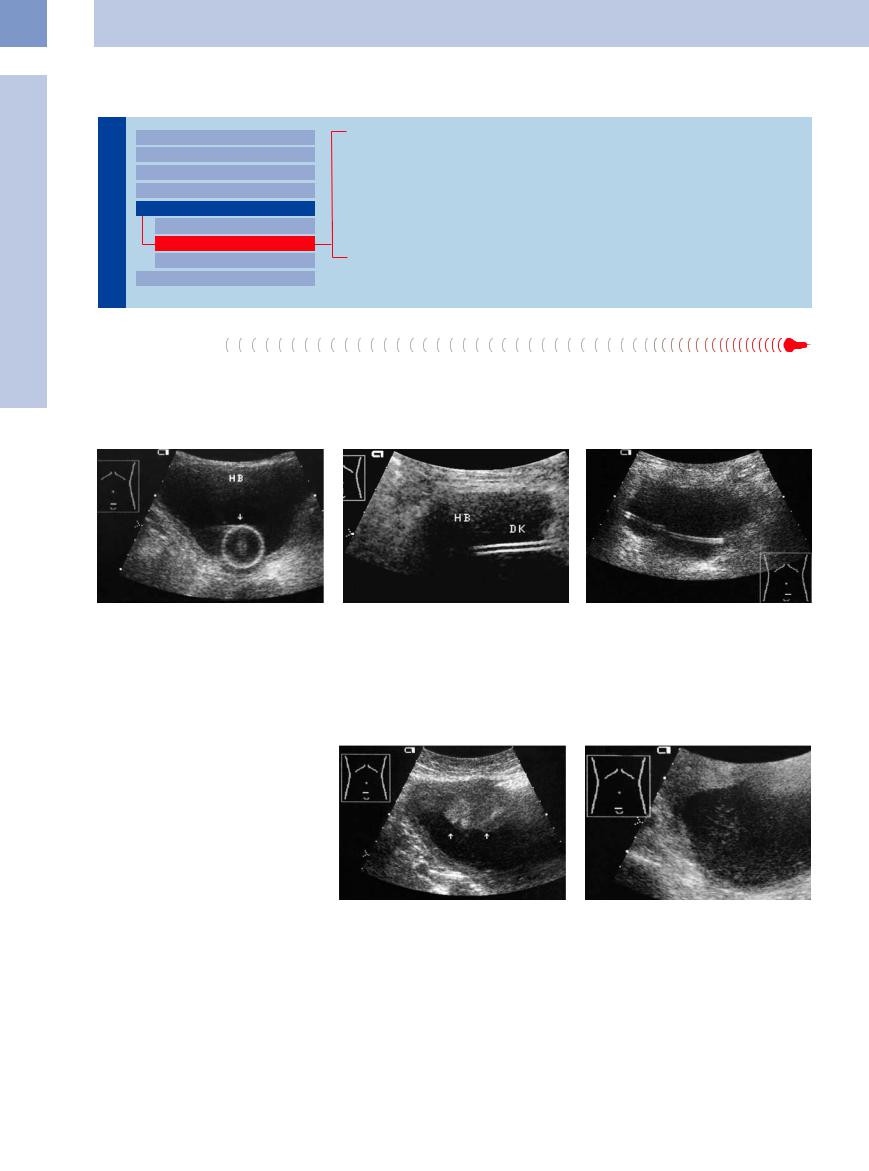
The most common hyperechoic mass seen in |
fluid band. The balloon appears as a target |
Pigtail catheters and catheter fragments ex- |
the bladder is a urinary balloon catheter. The |
figure with a hyperechoic wall and a bright |
hibit only a double wall with a central anechoic |
catheter itself appears sonographically as |
central echo representing the catheter tip |
lumen (Fig.11.79). |
bright parallel walls with a central anechoic |
(Fig.11.77, Fig.11.78). |
|
Fig. 11.77 Urinary catheter balloon (arrow): echogenic balloon wall surrounding a central high-level echo (catheter tip). HB = bladder.
Fig. 11.78 Urinary catheter (DK) in a lower abdominal longitudinal scan: echogenic parallel walls with a central anechoic, fluid-filled lumen. HB = bladder.
Fig. 11.79 Hyperechoic drain, placed to stent a ureter obstructed by urothelial carcinoma.
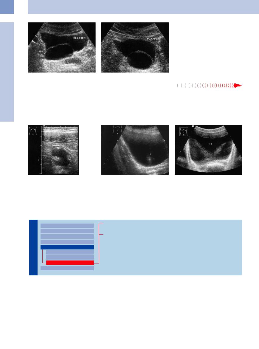
Blood Clots 


Clots with a hyperechoic or irregularly hyperechoic structure do occur but otherwise show the same sonographic features as the more hypoechoic-appearing forms (Fig.11.80). Their diagnosis is described above.
Fig. 11.80 Hyperechoic blood clots.
a Aggregate clot on the bladder roof (arrows).
b After body movement, fragments of the clot appear as bright swirling echoes.
406
Polypoid Bladder Tumor


Like blood clots, benign and malignant bladder tumors may appear hyperechoic owing to the blooming effect—an acoustic enhancement that occurs when sound travels through fluid spaces (Fig.11.81). The true echogenicity of the mass can be appreciated by lowering the time gain compensation (TGC) setting. In other respects the tumors have the same features as the hypoechoic tumors mentioned above.
Some bladder tumors may show a high-am- plitude entry echo (Fig.11.75). Squamous cell carcinomas occasionally contain superficial calcifications.10
Fig. 11.81 Benign bladder papilloma (cursors), histologically confirmed. Typical location on the bladder floor near the ureteral orifices: hyperechoic lobulated mass. Here the echogenicity of the mass is enhanced due to a blooming effect.
Benign Prostatic Hyperplasia


BPH is a mass extrinsic to the bladder. BPH due to a median-lobe adenoma can mimic an exophytic tumor of the bladder floor. BPH may be very hypoechoic as well as hyperechoic, depending on its histology (see Chapter 12).
Unlike bladder carcinoma, BPH shows a visible connection with the prostate on dynamic ultrasound examination. It has a homogeneous echo structure, smooth margins, and a conical or globular shape (Fig.11.82; see also Chapter 12).
Fig. 11.82 Adenoma of the prostatic median lobe (benign |
b Tumor mass protruding into the bladder bottom; thick- |
hyperplasia) appears as a relatively echogenic mass pro- |
ening of the bladder wall: BPH. |
truding into the bladder (HB). |
|
a Polypoid tumor mass (T) in the bladder. |
|
Lipoma, Fibroma, Myoma, Hemangioma 


These somewhat rare benign tumors have smooth margins and high, homogeneous echogenicity. They cannot be reliably classified as benign or malignant by ultrasound.
Ureterocele 


A ureterocele, on the other hand, can be accurately diagnosed sonographically as an intraluminal mass. Its ultrasound appearance is unmistakable: a balloon-like structure with a thin echogenic wall and anechoic lumen, protruding into the bladder from the ureteral ridge. Stone formation is common in ureteroceles, however, and can produce high-level internal echoes with acoustic shadows. Only large ureteroceles are difficult to recognize as arising from the ureteral ridge, appearing as a thin, elliptical membrane within the bladder lumen. A ureterocele may be associated with duplex kidney (Fig.11.83, Fig.11.84).
Fig. 11.83 Ureterocele (Z): anechoic urine and echogenic contents (stone, horizontal arrow, partial acoustic shadow S). The ureteral wall appears as a hyperechoic ring that has herniated into the bladder (HB). The ureter
(U) is partially obstructed.
11
Intracavitary Mass
407
11
Urinary Tract
Fig. 11.84 Large right-sided ureterocele: hyperechoic elliptical membrane in the bladder lumen (images courtesy of Dr. K. Ringewald, Hofgeismar, Germany).
a Lower abdominal transverse scan.
b Lower abdominal longitudinal scan.
Artifacts 


Hyperechoic bladder-wall indentations and |
Ureteral jet. A ureteral jet is a reflection caused |
Side-lobe artifact. A side-lobe artifact is caused |
motion-related or image artifacts can mimic |
by urine flowing into the bladder from the |
by paravesical hyperechoic structures that are |
true masses in the bladder. |
ureteral orifice. It appears as a hyperechoic |
projected into the fluid medium owing to the |
|
cone that exhibits color-flow signals in color |
physics of sound transmission from a convex |
Wall indentations. These often result from in- |
Doppler because of its motion (Fig.11.85b). |
transducer (Fig.11.86). |
complete bladder distension (Fig.11.85a). |
|
|
Fig. 11.85 Hyperechoic wall indentation in a partially filled bladder, with a ureteral jet.
a Slightly filled bladder (HB) with wall indentations mimicking an hourglass bladder.
b In color Doppler, a jet of urine entering the bladder produces a flare of color pixels (see Fig. 11.89a).
Fig. 11.86 Hyperechoic side-lobe artifacts caused by highly reflective structures located lateral to the bladder. HB = bladder.
Echogenic
Tract |
|
Malformations |
|
|
|
|
|
Dilated Renal Pelvis and Ureter |
|
|
|
|
|
Renal Pelvic Mass, Ureteral Mass |
Urinary |
|
|
|
Changes in Bladder Size or Shape |
|
|
|
|
|
|
Intracavitary Mass |
|
|
|
|
|
Hypoechoic |
|
|
Hyperechoic |
|
|
Echogenic |
|
|
Wall Changes |
|
|
Foreign Bodies
Bladder Calculi
408
Foreign Bodies
Foreign bodies such as pins, wires, or small tubes may be inserted into the urethra inadvertently during masturbation. Most foreign bodies found in the bladder are iatrogenic, however. At ultrasound they produce very high-level intraluminal echoes with acoustic shadowing (Fig.11.87, Fig.11.92b).
Bladder Calculi
Stones in the bladder are common, but considerably less so than in the kidney or ureter. Uroliths that have passed through the ureter are occasionally still imaged in the bladder before entering the urethra. Otherwise, bladder calculi are found in association with incom-
plete bladder emptying, bladder diverticula, or a ureterocele. Their ultrasound appearance is like that of other stones: a high-level echo that casts an acoustic shadow and moves when the patient is repositioned.
Fig. 11.87 Echogenic catheter fragment in a not entirely echo-free bladder caused by bleeding after catheter avulsion.
Stones resting on the bladder floor may be missed if they are located in blind spots, but their mobility and acoustic shadows can confirm the diagnosis (Figs. 11.88, 11.89, 11.90).
11
Intracavitary Mass
Fig. 11.88 Recent occurrence of bladder calculi and suppurated cystitis in a known urethral stenosis.
a Two echogenic lesions, fixed, with incomplete shadow. Floating sedimentation.
b Twinkling artifact within the echoes of the calculi.
c 8 months before this scan, only suppurative sedimentation to be seen.
Fig. 11.89 Small bladder stone (arrow) with an acoustic |
b Longitudinal scan. |
shadow (S). |
|
a Transverse scan. HB = bladder; U = ureteral jet. |
|
Fig. 11.90 Hyperechoic mass (left arrow, right ureteral orifice) with an acoustic shadow (S), representing a ureterocele with a stone. A second ureterocele with a thin echogenic membrane appears near the left orifice (right arrow). HB = bladder.
409