

- •Contents
- •Preface
- •Contributors
- •1 Vessels
- •1.1 Aorta, Vena Cava, and Peripheral Vessels
- •Aorta, Arteries
- •Anomalies and Variant Positions
- •Dilatation
- •Stenosis
- •Wall Thickening
- •Intraluminal Mass
- •Perivascular Mass
- •Vena Cava, Veins
- •Anomalies
- •Dilatation
- •Intraluminal Mass
- •Compression, Infiltration
- •1.2 Portal Vein and Its Tributaries
- •Enlarged Lumen Diameter
- •Portal Hypertension
- •Intraluminal Mass
- •Thrombosis
- •Tumor
- •2 Liver
- •Enlarged Liver
- •Small Liver
- •Homogeneous Hypoechoic Texture
- •Homogeneous Hyperechoic Texture
- •Regionally Inhomogeneous Texture
- •Diffuse Inhomogeneous Texture
- •Anechoic Masses
- •Hypoechoic Masses
- •Isoechoic Masses
- •Hyperechoic Masses
- •Echogenic Masses
- •Irregular Masses
- •Differential Diagnosis of Focal Lesions
- •Diagnostic Methods
- •Suspected Diagnosis
- •3 Biliary Tree and Gallbladder
- •3.1 Biliary Tree
- •Thickening of the Bile Duct Wall
- •Localized and Diffuse
- •Bile Duct Rarefaction
- •Localized and Diffuse
- •Bile Duct Dilatation and Intraductal Pressure
- •Intrahepatic
- •Hilar and Prepancreatic
- •Intrapancreatic
- •Papillary
- •Abnormal Intraluminal Bile Duct Findings
- •Foreign Body
- •The Seven Most Important Questions
- •3.2 Gallbladder
- •Changes in Size
- •Large Gallbladder
- •Small/Missing Gallbladder
- •Wall Changes
- •General Hypoechogenicity
- •General Hyperechogenicity
- •General Tumor
- •Focal Tumor
- •Intraluminal Changes
- •Hyperechoic
- •Hypoechoic
- •Nonvisualized Gallbladder
- •Missing Gallbladder
- •Obscured Gallbladder
- •4 Pancreas
- •Diffuse Pancreatic Change
- •Large Pancreas
- •Small Pancreas
- •Hypoechoic Texture
- •Hyperechoic Texture
- •Focal Changes
- •Anechoic Lesion
- •Hypoechoic Lesion
- •Isoechoic Lesion
- •Hyperechoic Lesion
- •Irregular (Complex Structured) Lesion
- •Dilatation of the Pancreatic Duct
- •Marginal/Mild Dilatation
- •Marked Dilatation
- •5 Spleen
- •Nonfocal Changes of the Spleen
- •Diffuse Parenchymal Changes
- •Large Spleen
- •Small Spleen
- •Focal Changes of the Spleen
- •Anechoic Mass
- •Hypoechoic Mass
- •Hyperechoic Mass
- •Splenic Calcification
- •6 Lymph Nodes
- •Peripheral Lymph Nodes
- •Head/Neck
- •Extremities (Axilla, Groin)
- •Abdominal Lymph Nodes
- •Porta Hepatis
- •Splenic Hilum
- •Mesentery (Celiac, Upper and Lower Mesenteric Station)
- •Stomach
- •Focal Wall Changes
- •Extended Wall Changes
- •Dilated Lumen
- •Narrowed Lumen
- •Small/Large Intestine
- •Focal Wall Changes
- •Extended Wall Changes
- •Dilated Lumen
- •Narrowed Lumen
- •8 Peritoneal Cavity
- •Anechoic Structure
- •Hypoechoic Structure
- •Hyperechoic Structure
- •Anechoic Structure
- •Hypoechoic Structure
- •Hyperechoic Structure
- •Wall Structures
- •Smooth Margin
- •Irregular Margin
- •Intragastric Processes
- •Intraintestinal Processes
- •9 Kidneys
- •Anomalies, Malformations
- •Aplasia, Hypoplasia
- •Cystic Malformation
- •Anomalies of Number, Position, or Rotation
- •Fusion Anomaly
- •Anomalies of the Renal Calices
- •Vascular Anomaly
- •Diffuse Changes
- •Large Kidneys
- •Small Kidneys
- •Hypoechoic Structure
- •Hyperechoic Structure
- •Irregular Structure
- •Circumscribed Changes
- •Anechoic Structure
- •Hypoechoic or Isoechoic Structure
- •Complex Structure
- •Hyperechoic Structure
- •10 Adrenal Glands
- •Enlargement
- •Anechoic Structure
- •Hypoechoic Structure
- •Complex Echo Structure
- •Hyperechoic Structure
- •11 Urinary Tract
- •Malformations
- •Duplication Anomalies
- •Dilatations and Stenoses
- •Dilated Renal Pelvis and Ureter
- •Anechoic
- •Hypoechoic
- •Hypoechoic
- •Hyperechoic
- •Large Bladder
- •Small Bladder
- •Altered Bladder Shape
- •Intracavitary Mass
- •Hypoechoic
- •Hyperechoic
- •Echogenic
- •Wall Changes
- •Diffuse Wall Thickening
- •Circumscribed Wall Thickening
- •Concavities and Convexities
- •12.1 The Prostate
- •Enlarged Prostate
- •Regular
- •Irregular
- •Small Prostate
- •Regular
- •Echogenic
- •Circumscribed Lesion
- •Anechoic
- •Hypoechoic
- •Echogenic
- •12.2 Seminal Vesicles
- •Diffuse Change
- •Hypoechoic
- •Circumscribed Change
- •Anechoic
- •Echogenic
- •Irregular
- •12.3 Testis, Epididymis
- •Diffuse Change
- •Enlargement
- •Decreased Size
- •Circumscribed Lesion
- •Anechoic or Hypoechoic
- •Irregular/Echogenic
- •Epididymal Lesion
- •Anechoic
- •Hypoechoic
- •Intrascrotal Mass
- •Anechoic or Hypoechoic
- •Echogenic
- •13 Female Genital Tract
- •Masses
- •Abnormalities of Size or Shape
- •Uterus
- •Abnormalities of Size or Shape
- •Myometrial Changes
- •Intracavitary Changes
- •Endometrial Changes
- •Fallopian Tubes
- •Hypoechoic Mass
- •Anechoic Cystic Mass
- •Solid Echogenic or Nonhomogeneous Mass
- •14 Thyroid Gland
- •Diffuse Changes
- •Enlarged Thyroid Gland
- •Small Thyroid Gland
- •Hypoechoic Structure
- •Hyperechoic Structure
- •Circumscribed Changes
- •Anechoic
- •Hypoechoic
- •Isoechoic
- •Hyperechoic
- •Irregular
- •Differential Diagnosis of Hyperthyroidism
- •Types of Autonomy
- •15 Pleura and Chest Wall
- •Chest Wall
- •Masses
- •Parietal Pleura
- •Nodular Masses
- •Diffuse Pleural Thickening
- •Pleural Effusion
- •Anechoic Effusion
- •Echogenic Effusion
- •Complex Effusion
- •16 Lung
- •Masses
- •Anechoic Masses
- •Hypoechoic Masses
- •Complex Masses
- •Index
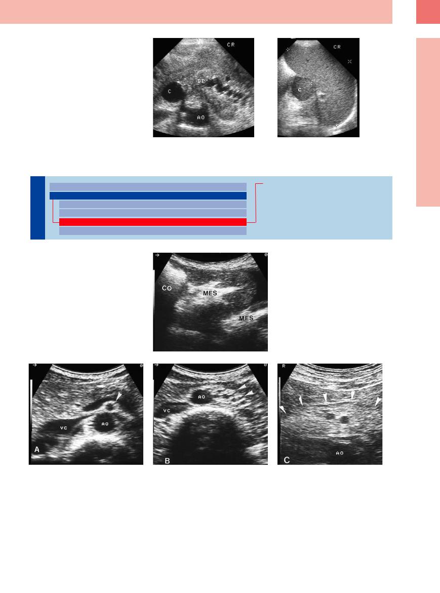
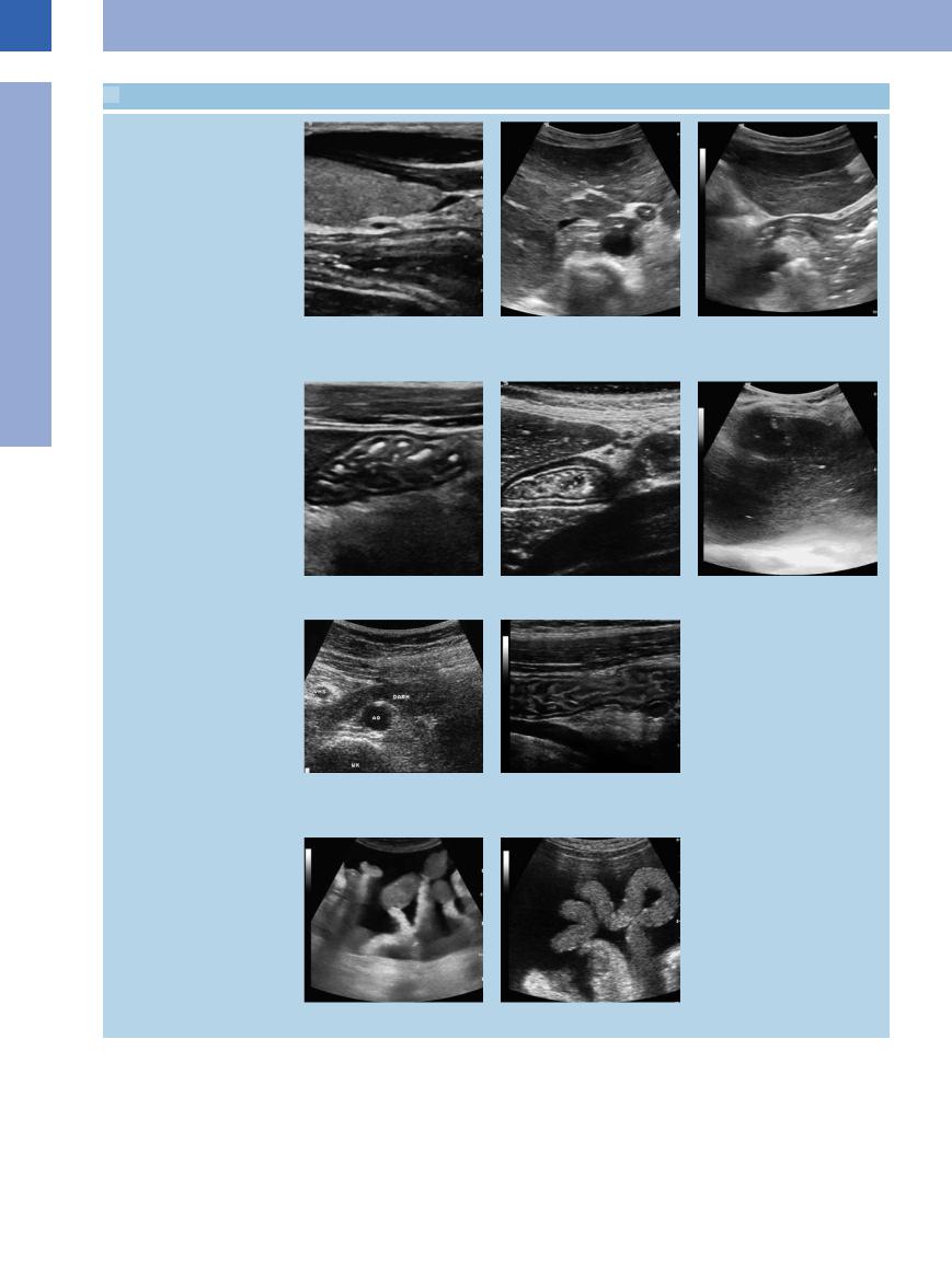
Fig. 6.32
a Signs of chronic pancreatitis with enlarged pancreatic duct (DP) and anechoic pseudocyst (C) at the head of the pancreas. AO = aorta.
b Echogenic pseudocyst (C) at the splenic hilum in the same patient.
Mesentery (Celiac, Upper and Lower Mesenteric Station)
Nodes |
|
|
Peripheral Lymph Nodes |
|
Inflammatory Lymph Nodes |
|
|
||||
|
|
Abdominal Lymph Nodes |
|
Metastases |
|
|
|
|
|
||
Lymph |
|
|
Porta Hepatis |
|
Malignant Lymphoma |
|
|
Splenic Hilum |
|
Other Structures |
|
|
|
|
|
||
|
|
|
Mesentery (Celiac, Upper and Lower Mesenteric Station) |
|
|
|
|
|
|
|
|
|
|
|
Retroperitoneum (Para-Aortic, Paracaval, Aortointercaval, and Iliac Station) |
|
|
Although sometimes the |
mesenteric lymph |
Fig. 6.33 Usually the mesentery (MES) of the individual |
|||
node stations are |
difficult |
to |
visualize |
hypoechoic loop of the small intestines will be visualized |
|
(Fig. 6.33, Fig. 6.34), |
they |
play |
an |
important |
as being homogeneously hyperechoic. CO = colon. |
role in clinical practice.
6
Abdominal Lymph Nodes
Fig. 6.34 Transverse section of the mesentery. AO = aorta; VC = vena cava.
a The mesentery, inferior to the splenic vein at the pancreas, displays a hyperechoic texture.
b Once the mesenteric vessels have branched off the aorta, they rest within this hyperechoic mesentery.
c Mesenteric lipomatosis is characterized by homogeneous echogenic thickening of the mesentery.
251
6
Lymph Nodes
Inflammatory Lymph Nodes 


Most of these lymph nodes are situated in the |
“dirty” pattern. In some cases an enlarged |
mesenteric fat along the unpaired branches of |
lymph node may be several centimeters in di- |
the aorta (celiac axis, superior and inferior |
ameter. One important differential diagnosis of |
mesenteric artery). Since most of these lymph |
acute appendicitis, particularly during child- |
nodes are small, the mesentery displays a |
hood, is mesenteric lymphadenitis, which can |
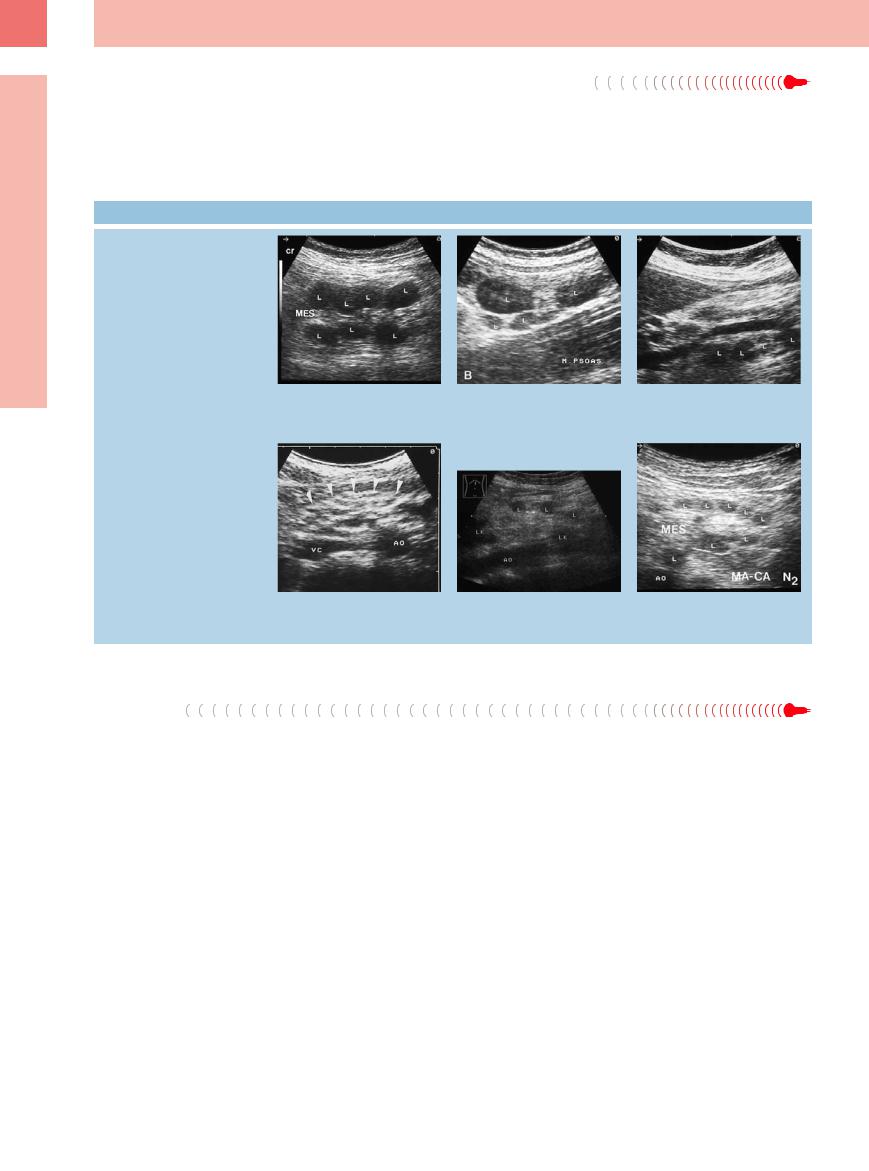
 6.7 Enlargement of the Mesenteric Lymph Nodes
Size, shape, and structure are not criteria for benignancy or malignancy
be assessed as such once ultrasound has demonstrated or excluded other local inflammation (nonspecific gastroenteritis, Crohn disease, ulcerative colitis, yersiniosis, ileocolitis, appendicitis) ( 6.7a–d).
a Marked reactive lymphadenitis (L) along the mesenteric vessels in mesenteric lymphadenitis. MES = mesentery. Longitudinal view.
d Mesenteric invasion of small lymph nodes with spotted structural inhomogeneity (arrows) in gastroenteritis. AO = aorta; VC = vena cava.
b Enlarged individual lymph nodes (L).
e Mesenteric lymph nodes (L, LK) in uterine cancer.8
c In the epigastric longitudinal view, numerous lymph nodes (L) along the mesenteric vein; definite diagnosis: reactive lymphadenitis in tuberculosis.
f Enlarged lymph nodes (L) in gastric cancer.
Metastases
Enlarged mesenteric lymph nodes are a frequent finding in ultrasound studies of abdominal cancer (carcinoma of the stomach, pan-
creas, esophagus, colon, etc.). The definitive diagnosis of malignancy is a prerogative of the pathologist (postoperatively) ( 6.7e,f).
Malignant Lymphoma


Possible involvement of the mesenteric lymph nodes is seen in all the various lymphomas. The sonographic morphology is rather diverse and may lead to problems in the differential diagnosis ( 6.8). In case of primary involvement of
the abdominal lymph nodes, the diagnosis is confirmed by ultrasound-guided needle biopsy; however, sometimes the issue can only be resolved by laparotomy.
252
 6.8 Malignant Lymphomas in Mesenteric Lymph Nodes
Nodular, confluent, extended
a–c Di erent nodular patterns of invasion |
b Numerous confluent lymph nodes in |
in malignant lymphoma. |
CLL. |
a Small solitary lymph node (L) in CLL. AMS |
|
= superior mesenteric artery; MES = mes- |
|
entery. |
|
Atypical patterns
d–f Atypical pattern of mesenteric involve- |
e Cross-sectional view with annular hypo- |
ment in malignant lymphoma: extremely |
echoic tumor formation around the mes- |
di cult diagnosis in case of sole mesen- |
enteric artery in follicular center |
teric involvement. |
lymphoma. AO = aorta; L = liver; MES = |
d Cross-sectional view: hypoechoic mes- |
mesentery. |
enteric tumor formation in high-grade |
|
lymphoma. D = intestine; mes = mesen- |
|
tery; TU = tumor. |
|
Small confluent nodes, bulky formation
g–i Patterns of mesenteric invasion in ma- |
h Extended sheets of lymphomas in lym- |
lignant lymphoma and ascites. |
phoblastic lymphoma (LB-NHL). D = intes- |
g Micronodular confluent lymph nodes (L) |
tine; MES = mesentery. |
in high-grade T-cell lymphoma. D = intes- |
|
tine. |
|
c Extended tumor formation encasing the superior mesenteric artery (AMS) in mantle cell lymphoma.
f Longitudinal epigastric view with extended, partly nodular, ill-defined tumor formation in CLL. AO = aorta; L = liver; MES = mesentery.
i Homogeneous hyperechoic tumor formation invading all of the mesenteric root in Hodgkin disease. A = ascites; AO = aorta; b = bowel; VMS = superior mesenteric vein; WS = vertebral column.
Other Structures
The differential diagnosis of an isolated mesenteric mass covers a whole gamut of possibilities, including:
●Intestinal loop, mesenteric cyst (Fig. 6.35), necrosis in pancreatitis, abscess
●Mesenteric varicosities in portal hypertension, thrombosis of the mesenteric vessels, mesenteric artery aneurysms, hematoma
●Inflammatory pseudotumor (Fig. 6.36), lipoma, primary mesenchymal tumor
Fig. 6.35 Transverse epigastric view with anechoic mesenteric mass corresponding with a cyst (C). AO = aorta; L = liver; MA = stomach; MES = mesentery.
6
Abdominal Lymph Nodes
253
6
Lymph Nodes
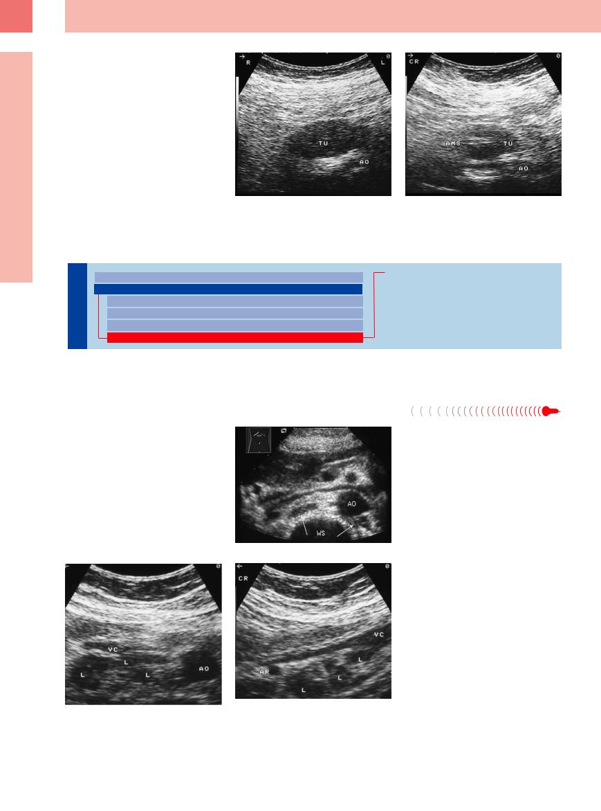
Fig. 6.36 Hypoechoic mesenteric mass (TU); histology confirmed an inflammatory pseudotumor. AO = aorta. a Transverse epigastric view.
b Longitudinal view. AMS = superior mesenteric artery.
Retroperitoneum (Para-Aortic, Paracaval, Aortointercaval, and Iliac Station)
Nodes |
|
Peripheral Lymph Nodes |
|
Inflammatory Lymph Nodes |
|
|
|
||
|
|
Abdominal Lymph Nodes |
|
Metastases |
|
|
|||
Lymph |
|
Porta Hepatis |
|
Malignant Lymphoma |
|
Splenic Hilum |
|
Other Structures |
|
|
|
|
||
|
|
Mesentery (Celiac, Upper and Lower Mesenteric Station) |
|
|
|
|
|
|
|
|
|
Retroperitoneum (Para-Aortic, Paracaval, Aortointercaval, and Iliac Station) |
|
|
In ultrasound studies gas shadowing frequently makes it quite difficult to visualize the retroperitoneal lymph nodes.
Inflammatory Lymph Nodes 


Para-aortic lymph nodes with reactive enlarge- |
Fig. 6.37 Transverse epigastric view with reactive enlarge- |
ment are rare. In the iliac region they may be |
ment of the para-aortic lymph nodes in lupus erythema- |
demonstrated in the presence of local inflam- |
tosus. AO = aorta; WS = spine. |
mation (appendicitis, diverticulitis, adnexitis) |
|
(Fig. 6.37, Fig. 6.38). |
|
Fig. 6.38 Enlarged inflammatory lymph nodes (L) in tuberculosis. AO = aorta; AR = renal artery; VC = vena cava. a Cross-sectional view.
b Longitudinal view.
254
Metastases
Enlarged para-aortic lymph nodes are malig- |
tinguished from malignant lymphoma. Despite |
nant unless proven otherwise. They are seen, |
the principal uncertainty in the assessment of |
for example, in cancer of the kidney, testis, |
possible malignancy, if there is nonregional |
esophagus, prostate, colon, and rectum. Their |
(parietal) lymphadenopathy in cancer of the |
sonographic pattern of invasion cannot be dis- |
stomach, pancreas, gallbladder, or liver, be- |
 6.9 Retroperitoneal Lymph Node Metastasis
Different tumor formations and patterns of involvement
cause of the location alone (para-aortic, aortointercaval, paracaval) it has to be regarded as metastasis with a frequently unfavorable prognosis ( 6.9).
6.9).
6
Abdominal Lymph Nodes
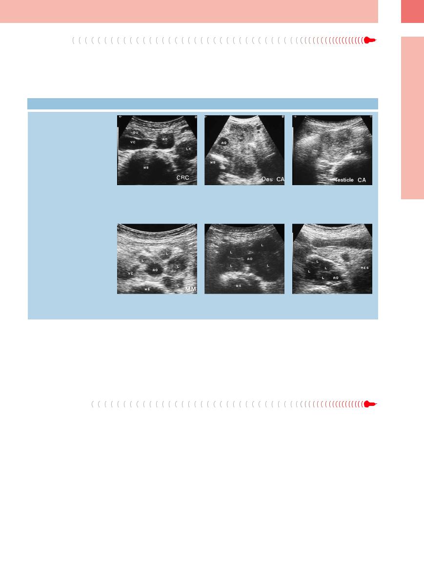
a–d Di erent patterns of invasion of paraaortic lymph nodes in cancer (cross-sec- tional view).
a Individual hypoechoic lymph nodes (LK) in cancer of the colon. AO = aorta; DU = intestine; WS = spine; VC = vena cava.
b Large para-aortic hyperechoic tumor formation encasing the aorta (AO) in esophageal cancer. WS = spine.
c Large para-aortic lymph node to the right of the aorta (AO) in testicular cancer. M = metastasis.
d Numerous para-aortic lymph nodes (L) in malignant melanoma. AO = aorta; VC = vena cava; WS = spine.
e Marked hypoechoic lymph node (L) metastasis in cancer of the bladder. Crosssectional view. AO = aorta; WS = spine.
f Transverse epigastric view with isolated lymph node metastases (L) in ovarian cancer. The mesentery (MES) is not involved. AO = aorta.
Malignant Lymphoma


Quite often as part of a systemic lymphoma the retroperitoneum will be invaded by lymphoma masses. The sonographic patterns of invasion are rather varied. As in all other lymph node stations, here too there is “bulky” disease, as
well as micronodular and macronodular and diffuse involvement. Isolated retroperitoneal manifestation of lymphoma is rare but possible ( 6.9).
6.9).
Other Structures
Particularly in local retroperitoneal tumor formations, the differential diagnosis covers a wide spectrum of possibilities.
●Para-aortic region: crura of diaphragm, horizontal part of the duodenum, horseshoe kidney, hematoma, abscess, aortic aneur-
ysm, retroperitoneal fibrosis, necrosis in pancreatitis, lymphocele, primarily retroperitoneal tumors ( 6.11a–d).
●Iliac region: ovary, undescended testis,
lymphocele, hematoma, abscess (
6.11e–h).
255
6
Lymph Nodes
 6.10 Para-aortic and Iliac Lymph Node Metastasis in Malignant Lymphoma
Different tumor formations and patterns of involvement
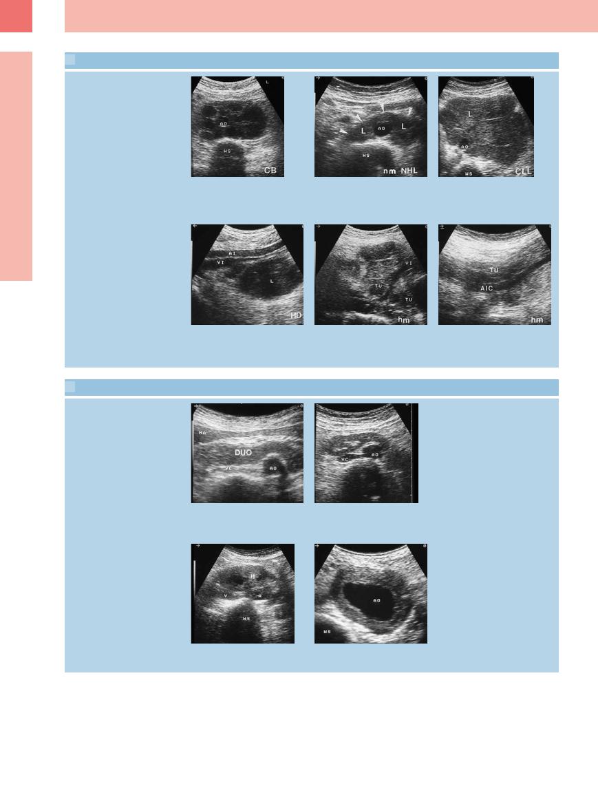
a–c Di erent patterns of invasion in para- |
b Extended tumor formation (arrowheads) |
c “Bulky” tumor formation (L) in CLL. AO |
aortic lymph nodes. |
in low-grade lymphoma (L). AO = aorta; |
= aorta; WS = spine. |
a Confluent lymphomas encasing the aor- |
WS = spine. |
|
ta in centroblastic lymphoma. AO = aorta; |
|
|
WS = spine. |
|
|
d–f Di erent patterns of invasion in iliac |
e Large complexes of well-defined lym- |
lymph nodes. |
phoma masses (TU) in high-grade lym- |
d Large solitary hypoechoic tumor forma- |
phoma. VI = iliac vein. |
tion (L) in Hodgkin disease. AI = iliac artery; |
|
VI = iliac vein. |
|
 6.11 Differential Diagnosis in Enlarged Para-Aortic and Parailiac Lymph Nodes
Para-aortic masses
f Large, faint, ill-defined hypoechoic tumor formation (TU) in high-grade lymphoma. AIC = common iliac artery.
a–d Para-aortic.
a Transverse epigastric view with duodenum (DUO). AO = aorta; MA = stomach; VC = vena cava.
c Transverse view of the lower abdomen with retroperitoneal hematoma (H). A = iliac artery; V = iliac vein; WS = spine.
b Transverse epigastric view with visualization of the parenchymal bridge in a horseshoe kidney. AO = aorta; VC = vena cava.
d Transverse view of the lower abdomen with partially thrombosed aortic aneurysm. AO = aorta; WS = spine.
256
 6.11 Differential Diagnosis in Enlarged Para-Aortic and Parailiac Lymph Nodes (Continued)
Parailiac masses
e–h Parailiac. |
f Transverse view of the lower abdomen |
e Parietal view on the right with the ovary. |
with the inguinal ligament. HB = urinary |
HB = urinary bladder; VI = iliac vein. |
bladder; HD = testicle; SI = sigma. |
6
Abdominal Lymph Nodes
g Transverse view of the right groin with a varicosity (?). AF = femoral artery; VF = femoral vein.
h Transverse view of the right groin with an inguinal hernia. A = iliac artery; V = iliac vein.
Tips, tricks, and pitfalls
A complete sonographical lymph node staging is indicated when
●searching for or staging a malignant lymphoma
●staging other oncological tumors (N-staging)
The following lymph nodes are to be examined and are accessible:
1.Abdominal and retroperitoneal lymph nodes
2.Cervical and nuchal lymph nodes
3.Supraand infraclavicular lymph nodes
4.Axillary lymph nodes
5.Inguinal lymph nodes
6.In special situations further peripheral lymph nodes (e. g., extremities)
Except for the lymph nodes in the abdomen and retroperitoneal space, a high-frequency probe should be used. The more superficially the lymph node is located, the more a probe with a higher frequency might be appropriate (e. g. in the neck region up to 10–13 MHz).
The interenteric lymph nodes are often the ones causing the most difficulties in their assessment. The method of dosed compression of the intestines is helpful in these cases (see Chapter 7).
A fasting patient is not always helpful in this situation, since a full gut is easier to differentiate from lymph nodes, e. g. by its peristalsis.
What size is pathological in lymph nodes? This question cannot be answered, since every new generation of probes detects smaller lymph nodes, especially in superficial regions. The assessment of lymph nodes always includes the clinical assessment (we see what we expect or are looking for) and, as well as size, location, the circulation patterns and the consistency also play an increasing role.
Note: If conspicuous lymph nodes are detected in one region a complete lymph node staging should follow and the spleen should be examined thoroughly (search for infiltration of lymphoma; see Chapter 5, “Invasive Lymphoma,” p. 215). Morphological assessment and visibility by sonography has improved, but it does not replace cytology/histology. Tissue differentiation must be carried out by pathologists/cytologists. If the effort is justifiable (e. g., superficial or peripheral location) the removal of a complete lymph node improves the assessment. Fine-needle aspiration biopsy (usFNA) should be used if access is difficult or longer distances have to be covered, e. g., in abdominal, retroperitoneal or mediastinal lymph nodes. usFNA can set the course and the result can be completed later by a lymph node obtained through surgery.
A surgeon may assist by marking a lymph node that seems particularly suitable because it is conspicuous and easily accessible. A joint preopera-
Fig. 6.39 Endosonography and elastography (EUS) in mediastinal lymph nodes. The green and red coloring indicates soft tissue and argued for a benign lymph node enlargement in this case. FNA supplied the diagnosis of sarcoidosis and confirmed the elastographic result.
tive examination with the surgeon might be helpful.
The elastography (EUS) of lymph nodes, especially in the mediastinal region, gives encouraging results. Even the detection of harder and possibly infiltrated regions within an individual lymph node seems promising (Fig. 6.39).
EUS is preferably used for mediastinal lymph nodes. This is also true for the biopsy of mediastinal lymph nodes, so that the use of mediastinoscopic biopsy for histological examination can be minimized.
257

6
Lymph Nodes
References
[1]Ahuja A, Ying M, Yang WT, Evans R, King W, Metreweli C. The use of sonography in differentiating cervical lymphomatous lymph nodes from cervical metastatic lymph nodes. Clin Radiol 1996;51(3):186–190
[2]Fischer K, Döhner H, Hunstein W. Lymphknotenvergrösserungen [Lymph node hyperplasia]. Internist (Berl) 1994;35(3):301–309
[3]Görg C. Infradiaphragmale Lymphknoten. In: Ultraschalldiagnostic. Lehrbuch und Atlas. 29. Ergänzungslieferung. Landsberg:
ecomed; 2005: 1–74
[4]Gritzmann N, Grasl MC, Helmer M, Steiner E. Invasion of the carotid artery and jugular vein by lymph node metastases: detection with sonography. AJR Am J Roentgenol 1990; 154(2):411–414
[5]Tschammler A, Gunzer U, Reinhart E, et al. Dignitätsbeurteilung vergrößerter Lymphknoten durch qualitative und semiquantitative Auswertung der Lymphknotenperfusion mit der farbkodierten Duplexsonographie. Fortschr Röntgenstr 1991;154:414
[6]Tschammler A, Ott G, Schang T, SeelbachGoebel B, Schwager K, Hahn D. Lymphadenopathy: differentiation of benign from malignant disease—color Doppler US assessment of intranodal angioarchitecture. Radiology 1998;208(1):117–123
[7]Schmidt G, Ed. Checkliste Sonographie. 3. ed Stuttgart, New York: Thieme, 2005
[8]Schmidt G, Görg C. Kursbuch Ultraschall. 5. ed Stuttgart, New York: Thieme, 2008
258
Gastrointestinal |
Tract |
7 |
|
Gastrointestinal Tract 261
|
|
|
Stomach xxx |
266 |
|||||||
|
|
|
|||||||||
|
|
|
|
|
|
Focal Wall Changes |
266 |
||||
|
|
|
|
|
|
||||||
|
|
|
|
|
|
||||||
|
|
|
|
|
|
|
|
|
|
Gastric Polyps |
|
|
|
|
|
|
|
|
|
|
|
|
|
|
|
|
|
|
|
|
|
|
|
Stromal Tumor |
|
|
|
|
|
|
|
|
|
|
|
|
|
|
|
|
|
|
|
|
|
|
|
Carcinoma |
|
|
|
|
|
|
|
|
|
|
|
|
|
|
|
|
|
|
|
|
|
|
|
Lymphoma |
|
|
|
|
|
|
|
|
|
|
|
|
|
|
|
|
|
|
|
|
|
|
|
Gastric Varices |
|
|
|
|
|
|
|
|
|
|
|
|
|
|
|
|
|
|
|
|
|
|
|
GAVE |
|
|
|
|
|
|
|
|
|
|
|
|
|
|
|
|
|
|
|
|
|
|
|
Ulceration |
|
|
|
|
|
|
|
|
|
|
|
|
|
|
|
|
|
|
|
|
|
|
|
Diverticula, Mucosal Folds |
|
|
|
|
|
|
|
|
|
|
|
|
|
|
|
|
|
|
|
|
|
|
|
Hypertrophic Pyloric Stenosis |
|
|
|
|
|
|
Extended Wall Changes |
270 |
|||||
|
|
|
|
|
|
|
|
|
|
Carcinoma/Scirrhus |
|
|
|
|
|
|
|
|
|
|
|
|
|
|
|
|
|
|
|
|
|
|
|
Lymphoma |
|
|
|
|
|
|
|
|
|
|
|
|
|
|
|
|
|
|
|
|
|
|
|
Gastritis |
|
|
|
|
|
|
|
|
|
|
|
|
|
|
|
|
|
|
|
|
|
|
|
Congestion, Edema |
|
|
|
|
|
|
|
|
|
|
|
|
|
|
|
|
|
|
|
|
|
|
|
Peritoneal Carcinomatosis |
|
|
|
|
|
|
|
Dilated Lumen |
272 |
||||
|
|
|
|
|
|
||||||
|
|
|
|
|
|
|
|
|
|
Physiological Dilatation |
|
|
|
|
|
|
|
|
|
|
|
||
|
|
|
|
|
|
|
|
|
|
Inflammation |
|
|
|
|
|
|
|
|
|
|
|
||
|
|
|
|
|
|
|
|
|
|
Gastric Outlet Obstruction |
|
|
|
|
|
|
|
|
|
|
|
||
|
|
|
|
|
|
|
|
|
|
Functional Disorder |
|
|
|
|
|
|
Narrowed Lumen |
273 |
|||||
|
|
|
|
|
|||||||
|
|
|
|
|
|
|
|
|
|
Impression |
|
|
|
|
|
|
|
|
|
|
|
|
|
|
|
|
|
|
|
|
|
|
|
Compression |
|
|
|
|
|
|
|
|
|
|
|
|
|
|
|
|
|
|
|
|
|
|
|
Tumor |
|
|
|
|
|
|
|
|
|
|
|
|
|
|
|
|
|
|
|
|
|
|
|
Postoperative Status |
|
|
|
Small/Large Intestine |
274 |
||||||||
|
|
||||||||||
|
|
|
|
|
Focal Wall Changes |
274 |
|||||
|
|
|
|
|
|||||||
Carcinoma
GIST
Lymphoma
Appendiceal Mucocele
Hematoma
Polyp
Intussusception
Foreign Object/Gallstone/Bezoar
Diverticula
Diverticulitis
Appendix
Appendicitis
Hernia
Extended Wall Changes |
282 |
Enteritis
Celiac Disease (Sprue)
Crohn Disease
Ulcerative Colitis
Amyloidosis
Pseudomembranous Enterocolitis
Ischemia
Hypertrophy
Tumor
Lymphoma
Dilated Lumen |
287 |
||
|
|
Physiological Dilatation |
|
|
|
|
|
|
|
Prepping for the Study |
|
|
|
|
|
|
|
Inflammation |
|
|
|
|
|
|
|
Ileus |
|
|
|
|
|
|
|
Coprostasis |
|
|
|
|
|
|
|
Tumor |
|
|
|
|
|
|
|
Foreign Body |
|
Narrowed Lumen |
290 |
||
|
|
Tumor Stenosis |
|
|
|
|
|
“Starvation Gut”
Ischemia
Atrophy

7Gastrointestinal Tract
M.W.M. Brandt
In humans food is ingested, transported, and stored by the gastrointestinal (GI) tract, which may be subdivided into four major sections: esophagus, stomach, small intestine, and large intestine.
The esophagus propels food taken up in the mouth into the stomach, where it is mixed, ground down, digested, and stored. The ingesta are then passed into the small intestine, where they undergo selective absorption. The residual indigestible parts of our food become thick-
ened in the large intestine and are stored there until evacuation.
In addition, the GI tract plays an important role as defensive barrier, and its Peyer’s patches and lymphocytes make up the most extensive immune system in the human body.
Successful realization of these functions is based on a common wall structure of the GI tube. The innermost mucous membrane secretes and absorbs; the submucous layer in the middle with its blood vessels, lymphatics,
and nerves and most of the enteric lymphatic organ lets the outer tela muscularis slide with respect to the mucous membrane. This muscular coat is characterized by longitudinal and circular muscle fibers arranged in different fashions in the various parts of the GI tract; their peristaltic contraction mixes and propels the intestinal contents as well as controlling this transport by means of special sphincter organs.
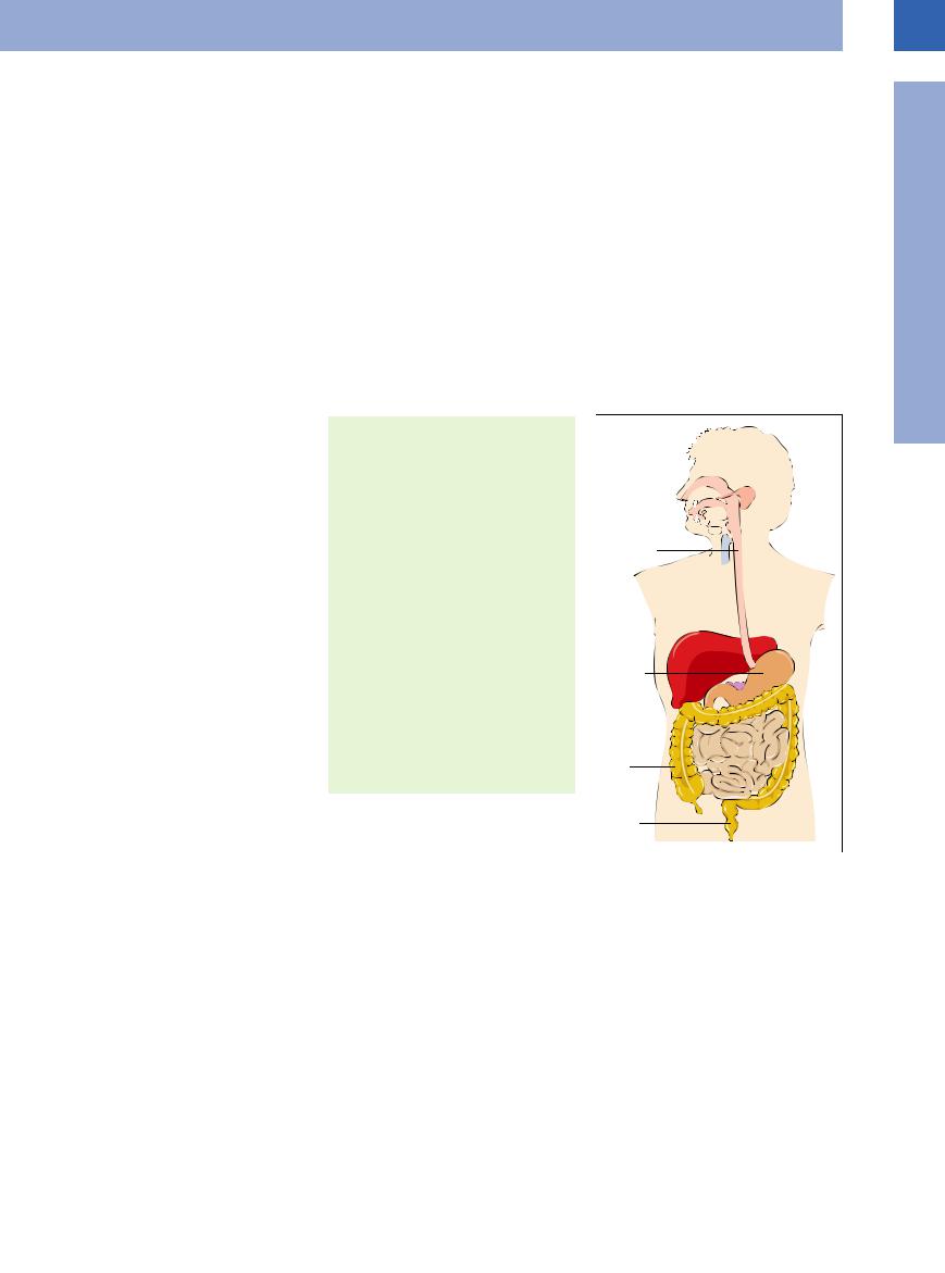
Anatomy and Topography (Fig.
 7.1)
7.1) 


Esophagus. The esophagus is a muscular tube about 40 cm long connecting the pharynx to the stomach and is located in the posterior mediastinum. Its distal segment is anterior and parallel to the aorta. It penetrates the diaphragm through the so-called esophageal hiatus, terminating at the esophagocardiac transition zone.
Stomach. The stomach is one of the intraperitoneal organs and is situated in the left upper quadrant. This hookor comma-shaped hollow organ runs from the cephalad left to the caudad right and has a length of approximately 20 cm. Under fasting conditions it contains about 50 mL of fluid, but will expand physiologically up to a maximum capacity of 2 liters.
The stomach is subdivided into cardia, fundus/fornix, body, and pyloric antrum. The cardiac opening of the stomach is situated anterior to the aorta at the level of the esophageal hiatus; its anterior aspect is covered by the inferior surface of the left hepatic lobe. The fundus/ fornix of the stomach with its round dome fills the space in the left upper quadrant, the superior border of which is formed by the diaphragm, while the left fundic aspect is in contact with the spleen. The distal part of the stomach is made up of the body and the pyloric antrum, which are defined on the anterior aspect by the left hepatic lobe. The muscular opening into the duodenum is the pyloric orifice, which is in direct contact with the head of the pancreas.
Small intestine. The small intestine is subdivided into the duodenum, which is about 30 cm long, and the jejunum plus ileum, which altogether measure approximately 1.5 m. The duodenum is a C-shaped arc (slightly more than a semicircle) that encloses the head of the pancreas, only partially covered by peritoneum. The superior duodenal segment crosses the structures of the porta hepatis, i. e., common bile duct, portal vein, and hepatic artery, while the gallbladder is anterolateral to it. The descending duodenal segment parallels the posterolateral vena cava and borders the right
Esophagus
●Wall thickness 3–4 mm
●Length 40 cm
●Transitory transport of the ingesta
Stomach
●Wall thickness 5–7 mm
●Length 20 cm
●Capacity: fasting 50 mL; postprandial up to 2000 mL
Duodenum
●Wall thickness 3–4 mm
●Length 30 cm
●Transitory transport of the chyme
Jejunum and Ileum
●Wall thickness 3–4 mm
●Total length 150 cm
●Transitory transport of the chyme
Colon
●Wall thickness 2–3 mm
●Length 120 cm
●Scybala/air
kidney. The inferior duodenal segment, situated about two fingerbreadths inferior to the mesenteric root and caudad of the pancreas, crosses anterior to the vena cava and the aorta. Cross-sectional views of the superior mesenteric artery and vein can be demonstrated anterior to the intestine. The retroperitoneal duodenum continues as an intraperitoneal organ at the level of the mesenteric root slightly left of the aorta.
The jejunum and ileum are attached to the posterior abdominal wall by the fan-shaped folds of the peritoneum known as the mesentery proper; most of the coils of the small intestine are found in the left upper (jejunum) and lower (ileum) quadrant. The jejunum and ileum account for most of the intraperitoneal
Fig 7.1 Topographic anatomy of the intestinal tract.
space. The terminal ileum ascends from the pelvis over the right psoas major muscle, the iliac vessels paralleling its medial margin, at the level of McBurney’s point, and ends in the right iliac fossa by opening into the medial side of the cecum at the ileocecal (Bauhin’s) valve.
Colon. The colon is about 120 cm long and runs along the margins of the intraperitoneal cavity. In a clockwise direction (from the subject’s point of view) it is subdivided into cecum, ascending, transverse, descending, and sigmoid colon, and rectum.
The cecum is situated anterior to the psoas major muscle in the right lower quadrant. The ascending colon parallels the right lateral abdominal wall and its superior segment is
7
Gastrointestinal Tract
261

7
Gastrointestinal Tract
anterior to the inferolateral part of the right kidney. The hepatic flexure comprises the terminal part of the ascending colon and the commencement of the transverse colon; usually, the convex part of this flexure hugs the gallbladder and the lower right margin of the liver, but as a variant it may dome up far superior and may even be situated between
Ultrasound Morphology
With enough training and experience the sonographer will be able to identify and demonstrate most regions of the GI tract ( 7.1).
7.1).
Esophagus. The proximal part of the cervical esophagus posterior to the left lobe of the thyroid lends itself to sonographic study ( 7.1a); although for all practical purposes the thoracic segment is beyond the reach of transcutaneous ultrasound, ultrasonography can demonstrate the distal esophagus at the level of the gastric cardia. Endoscopic ultrasonography (EUS) is employed for the sonographic evaluation of the esophagus and the neighboring mediastinum. The esophagocardial junction can be better evaluated by transabdominal ultrasound.
the lower right thoracic wall and the right hepatic lobe (so-called Chilaiditi syndrome). The transverse colon touches the anterior abdominal wall and traverses the upper abdomen from right to left; elongated variants may have a drooping center section reaching all the way into the pelvis. The splenic flexure connects the transverse and descending colon, its convex
Stomach and duodenum. With appropriate techniques all of the stomach and duodenum are accessible to sonographic study.
Jejunum and ileum. In the fasting state the individual loops of the jejunum cannot be differentiated, while their mesenteric attachment and the circular (Kerckring’s) folds characterize the postprandial fluid-filled loops. Later, the postprandial ileum loops in the lower abdomen will also be fluid-filled and display vivid peristalsis.
Colon. Usually, the ileocecum and colon can be visualized reasonably well and, taking into account the anatomic relations, the topography of these parts of the GI tract can be identified
aspect being inferior to the spleen. The proximal part of the descending colon is anterior and lateral to the left kidney. The S-shaped sigmoid colon forms a loop that, depending on its length, frames the roof of the urinary bladder more or less markedly. The rectum descends into the pelvis posterior to the bladder and terminates at the anus.
with certainty. Sometimes an elongated sigmoid colon or drooping transverse colon may interfere and be the reason for misidentification. Almost always the colon will appear as a tube filled with a varying amount of feces and lacking peristalsis.
Gut signature. The morphological criterion to look for in ultrasound studies of the GI tract is the so-called gut signature. At any point in the gut this cross-sectional view will demonstrate the GI tube as an annular structure, while the longitudinal view depicts it as a tubular structure. Assessment of the intestinal wall and differentiation of its characteristic layered structure becomes possible only with high-fre- quency probes (5, 7, or 7.5 MHz) (Table 7.1).
Gut Signature
The normal “gut signature” is known as the “cockade,” a term whose origins date back to Old French and refer to a rosette-shaped badge. In ultrasonography, the gut signature and its pathological variant the “target sign” have come to denote the round crosssectional view of the intestinal wall with its different layers. Although it has also been used to describe the longitudinal view of the GI tube, this usage is reserved exclusively for the GI tract and should not be applied to other rosette-shaped structures, such as hepatic metastasis, which are given their own terms (e. g., “halo” or “bull’s-eye” sign).
Thus, in the cross-sectional view the gut signature/target sign refers to the annular image of the layered intestinal wall and its center, while in the longitudinal view it refers to the parallel arrangement of adjacent intestinal segments with the lumen in between.
Physiological gut signature. A so-called physiological gut signature refers to a lumen that is normal for this segment, a wall structure with characteristic layering, welldefined wall thickness, and normal peristalsis.
Pathological gut signature. A so-called pathological gut signature is characterized by changes in the normal appearance of lumen, wall thickness, and/or peristalsis/pliability (Table 7.2). Pathological wall changes may be circular or eccentric and focal, or they may be continuous or discontinuous along the longitudinal axis. Intussusception will be seen on ultrasound as the “target/doughnut sign” (cross-sectional view) or “pseudokidney sign” (longitudinal view). Adjacent target signs fixed in space are characteristic of a conglomerate.
Table 7.1 Layers of the intestinal wall |
|
Table 7.2 Pathological gut signature: criteria |
|
Sonographic layer |
Histological layer |
Wall |
>4 mm |
Entrance echo—innermost hyperechoic layer |
Mucosal surface |
thickness |
Circumferential thickening |
|
O -center appearance |
||
Innermost hypoechoic layer |
Mucosa |
Wall layering |
Pronounced |
Central hyperechoic layer |
Submucosa |
|
Interrupted |
|
Missing |
||
|
|
|
|
Outermost hypoechoic layer |
Muscularis propria |
Peristalsis |
Rigid wall |
|
|
||
Outermost hyperechoic layer—entrance/exit echo |
Serosal surface |
|
Lack of pliability |
|
Lack of peristalsis |
||
|
|
|
|
|
|
Lumen |
Constricted |
|
|
|
Distended |
|
|
Vascularity |
Missing |
|
|
|
Enhanced |
262

Gut signature criteria. Detection of a gut signature calls for analysis of its ultrasound morphology, lumen, and peristalsis as well as of the surroundings and the intestinal segments upstream and downstream. Only all of these criteria together will permit typing and assessment with a clear-cut diagnosis in many cases. The criteria to be considered are as follows.
●Location/relation. Specifying the position of a gut signature offers the first chance of defining the anatomical relations. It is important to recognize whether the pathology exhibits a fixed location or is mobile.
●Echogenicity. Basically, the echogenicity of the gut signature may be normal, hypoechoic, or hyperechoic; since one is dealing with a layered anatomical structure, this criterion has to be applied to the predominant elements of the wall or should reflect the possible loss of layering.
●Wall thickness. Numerous standard dimensions have been listed for the wall thickness (see above). Measurement of the wall thickness has to account for the degree of filling and the functional state (contraction/dilatation) of the intestines, and also has to specify whether the wall is thickened or thinned out or whether its outline is interrupted or ill defined.
●Wall layering. The wall may display a normal layered structure or may be markedly layered, may demonstrate thickening of just one particular layer, may present with layer defects, or may have lost its layered structure altogether.
●Shape/texture of the gut signature. The cross-sectional view of the gut signature may be round or ovoid, or it may be characterized by asymmetrical changes; one characteristic pathological sign is the socalled target or bull’s-eye sign.
●Extent. In terms of length, short focal or extended lesions have to be differentiated from the diffuse changes in the intestinal wall.
●Internal delineation. Assessment of the luminal surface of the bowel wall will be able to differentiate between normal as well as pathological circular folds and haustra (preserved or lost) and also focal polypoid/tumorous changes, ulcers, and other surface irregularities.
●External delineation. The outside of the GI tube may be smooth or irregular; it may exhibit pod-like extensions or invade other structures.
●Changes in the surroundings. The surroundings of the gut signature may be absolutely normal or there may be free air or fluid; ultrasound may be able to visualize focal reactions in the surrounding tissue, e. g., hyperechoic pannus, abscess and fistula formation, as well as lymphadenopathy.
●Lumen. Assessment of the lumen has to account for the functional state/degree of contraction; it may be normal, distended, or reduced/narrowed.
●Stenosis. A stenosis is a circumscribed functionally narrowed lumen of the bowel with prestenotic distension and poststenotic narrowing of the intestinal tube; usually, a pathological gut signature or target sign will be seen at the stenosis itself.
●Contents. Within the lumen of the gut there may be anechoic fluid, echogenic chyme/ ingesta, hyperechoic feces/scybala, air, and foreign bodies.
●Peristalsis. The assessment of peristalsis differentiates between physiological contraction/distension on the one hand and normal, missing, increased, impaired, or
pathological peristalsis (pendulating peristalsis) on the other.
●Texture/pliability. Normal intestinal loops are compliant; lack of pliability suggests invasive inflammatory or malignant changes.
●Painfulness. A normal gut signature is not painful; if localized pain can be triggered at a focal change in the intestinal wall, this indicates either an inflammatory or a functional disorder (e. g., irritable bowel syndrome).
●Vascularity. The intestinal wall and its surrounding tissues may be hypervascular or avascular, or the vascularity may be normal.
●Changes in intestinal segments upstream. Upstream of a focal lesion one may find characteristic changes in the intestinal lumen (e. g., distension in the presence of ileus), peristalsis, and texture (e. g., thickening of the muscularis propria in case of hypertrophy).
●Changes in intestinal segments downstream. In case of mechanical ileus, a typical sign downstream is the so-called “starvation gut.”
●Changes observed over time. The quality and/or quantity of one or more of the observed criteria may become more or less pronounced.
●Changes in other organs. Additional findings in other organs may help immensely in the correct interpretation of intestinal sonographic observations (e. g., gallbladder, biliary tree, and liver in case of gallstone ileus; assessment of the flow signals in the superior and inferior mesenteric artery in ischemic and inflammatory bowel disease).
Peristalsis
Peristalsis consists of a wave of contraction, followed by immediate relaxation, passing along the longitudinal axis of the intestinal tube; under physiological conditions it may be visualized sonographically at the esophagus, stomach, and small intestine. Next to a segment actively contracting and displaying a thickened intestinal wall with narrowing of the lumen, the segments upstream and downstream will undergo concurrent relaxation of the intestinal wall musculature with thinning of the wall, dilatation, and widening of the lumen.
Esophagus. Rhythmic contractions are deliberately activated during deglutition and can be evaluated typically at the dorsal side of the left thyroid lobe.
Stomach. Gastric peristalsis is best seen around the antrum and is characterized by
rhythmic contractions with a frequency of 2–4 times per minute and a period of 2–20 s. The dilatation phases with discernible passage of chyme subsequent to swallowing are best observed at the esophagogastric junction.
Small intestine. Under fasting conditions, the empty small intestine (the Latin word “jejunus” means empty, dry, barren) is hard to visualize sonographically and thus its peristaltic movement remains hidden. Under physiological conditions the postprandial peristalsis usually appears as a steady and orderly alternation of muscular contraction and dilatation propelling chyme and fluid in a directed fashion.
In inflammatory disorders the lumen of the small intestine will still be filled with some fluid even when fasting; by itself this demonstration of intraluminal fluid (e. g., within
the duodenum in case of pancreatitis or cholecystitis) may be enough to suggest the underlying inflammation. Depending on the severity of the inflammation, the peristalsis may be vigorous, increased or even turbulent (e. g., the so-called “tumbler phenomenon” in celiac disease). Disturbed peristalsis with pathological contractions, mandatory pathological distension of the lumen, and conspicuously uncoordinated movement of the intestinal wall and intraluminal chyme/fluid are characteristic of ileus (see below).
Large intestine. Under physiological conditions, ultrasonography cannot visualize peristaltic activity in the large intestine. However, the characteristically impaired peristalsis in the presence of ileus does become evident on ultrasound.
7
Gastrointestinal Tract
263
7
Gastrointestinal Tract
 7.1 Visualization of the Gastrointestinal Tract
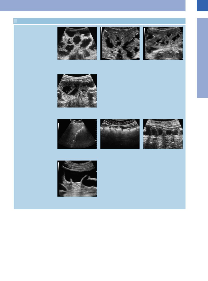
Stomach and duodenum: physiological gut signature
a Demonstration of the cervical normal esophagus posterior to the left thyroid gland.
d Normal gastric corpus folds.
g Normal gut signature of the inferior part of the duodenum where it crosses the aorta (AO). VMS = superior mesenteric vein; WK = spine; Darm = intestine.
b Typical gut signature of the esophagocardiac junction anterior to the aorta at the esophageal hiatus.
e Physiological target sign of the antrum.
h Jejunum, slightly fluid-enhanced, after oral fluid intake; note the clearly delineated circular folds.
c The same segment, longitudinal scan: gut signature of the esophagocardiac junction dorsal to the left liver lobe and transition into the gastic fornix.
f Fluid-filled stomach caused by a pyloric stenosis.
i and j Loops of the small intestine at their mesenteric attachment, floating in ascites of cardiac origin.
264
 7.1 Visualization of the Gastrointestinal Tract (Continued)
The differential diagnosis of fluid-filled intestinal loops has to rely on peristalsis
k Chronic ileus in obstructing tumor of the l and m Celiac disease with so-called “tumbler phenomenon.” hepatic flexure, exhibiting distended in-
testinal loops and abnormally sluggish and ineffective peristaltic movement.
n Mechanical ileus (adhesions) with focal fixation of the intestinal loops, resulting in impaired peristalsis.
Colon
o and p Haustra in colon.
r Hydrocolonic sonography demonstrating the ileocolic valve; the terminal ileum runs into the colon, normal finding with normal haustra.
q Hydrocolonic sonography demonstrating the intestinal wall of the descending colon.
7
Gastrointestinal Tract
265