

- •Contents
- •Preface
- •Contributors
- •1 Vessels
- •1.1 Aorta, Vena Cava, and Peripheral Vessels
- •Aorta, Arteries
- •Anomalies and Variant Positions
- •Dilatation
- •Stenosis
- •Wall Thickening
- •Intraluminal Mass
- •Perivascular Mass
- •Vena Cava, Veins
- •Anomalies
- •Dilatation
- •Intraluminal Mass
- •Compression, Infiltration
- •1.2 Portal Vein and Its Tributaries
- •Enlarged Lumen Diameter
- •Portal Hypertension
- •Intraluminal Mass
- •Thrombosis
- •Tumor
- •2 Liver
- •Enlarged Liver
- •Small Liver
- •Homogeneous Hypoechoic Texture
- •Homogeneous Hyperechoic Texture
- •Regionally Inhomogeneous Texture
- •Diffuse Inhomogeneous Texture
- •Anechoic Masses
- •Hypoechoic Masses
- •Isoechoic Masses
- •Hyperechoic Masses
- •Echogenic Masses
- •Irregular Masses
- •Differential Diagnosis of Focal Lesions
- •Diagnostic Methods
- •Suspected Diagnosis
- •3 Biliary Tree and Gallbladder
- •3.1 Biliary Tree
- •Thickening of the Bile Duct Wall
- •Localized and Diffuse
- •Bile Duct Rarefaction
- •Localized and Diffuse
- •Bile Duct Dilatation and Intraductal Pressure
- •Intrahepatic
- •Hilar and Prepancreatic
- •Intrapancreatic
- •Papillary
- •Abnormal Intraluminal Bile Duct Findings
- •Foreign Body
- •The Seven Most Important Questions
- •3.2 Gallbladder
- •Changes in Size
- •Large Gallbladder
- •Small/Missing Gallbladder
- •Wall Changes
- •General Hypoechogenicity
- •General Hyperechogenicity
- •General Tumor
- •Focal Tumor
- •Intraluminal Changes
- •Hyperechoic
- •Hypoechoic
- •Nonvisualized Gallbladder
- •Missing Gallbladder
- •Obscured Gallbladder
- •4 Pancreas
- •Diffuse Pancreatic Change
- •Large Pancreas
- •Small Pancreas
- •Hypoechoic Texture
- •Hyperechoic Texture
- •Focal Changes
- •Anechoic Lesion
- •Hypoechoic Lesion
- •Isoechoic Lesion
- •Hyperechoic Lesion
- •Irregular (Complex Structured) Lesion
- •Dilatation of the Pancreatic Duct
- •Marginal/Mild Dilatation
- •Marked Dilatation
- •5 Spleen
- •Nonfocal Changes of the Spleen
- •Diffuse Parenchymal Changes
- •Large Spleen
- •Small Spleen
- •Focal Changes of the Spleen
- •Anechoic Mass
- •Hypoechoic Mass
- •Hyperechoic Mass
- •Splenic Calcification
- •6 Lymph Nodes
- •Peripheral Lymph Nodes
- •Head/Neck
- •Extremities (Axilla, Groin)
- •Abdominal Lymph Nodes
- •Porta Hepatis
- •Splenic Hilum
- •Mesentery (Celiac, Upper and Lower Mesenteric Station)
- •Stomach
- •Focal Wall Changes
- •Extended Wall Changes
- •Dilated Lumen
- •Narrowed Lumen
- •Small/Large Intestine
- •Focal Wall Changes
- •Extended Wall Changes
- •Dilated Lumen
- •Narrowed Lumen
- •8 Peritoneal Cavity
- •Anechoic Structure
- •Hypoechoic Structure
- •Hyperechoic Structure
- •Anechoic Structure
- •Hypoechoic Structure
- •Hyperechoic Structure
- •Wall Structures
- •Smooth Margin
- •Irregular Margin
- •Intragastric Processes
- •Intraintestinal Processes
- •9 Kidneys
- •Anomalies, Malformations
- •Aplasia, Hypoplasia
- •Cystic Malformation
- •Anomalies of Number, Position, or Rotation
- •Fusion Anomaly
- •Anomalies of the Renal Calices
- •Vascular Anomaly
- •Diffuse Changes
- •Large Kidneys
- •Small Kidneys
- •Hypoechoic Structure
- •Hyperechoic Structure
- •Irregular Structure
- •Circumscribed Changes
- •Anechoic Structure
- •Hypoechoic or Isoechoic Structure
- •Complex Structure
- •Hyperechoic Structure
- •10 Adrenal Glands
- •Enlargement
- •Anechoic Structure
- •Hypoechoic Structure
- •Complex Echo Structure
- •Hyperechoic Structure
- •11 Urinary Tract
- •Malformations
- •Duplication Anomalies
- •Dilatations and Stenoses
- •Dilated Renal Pelvis and Ureter
- •Anechoic
- •Hypoechoic
- •Hypoechoic
- •Hyperechoic
- •Large Bladder
- •Small Bladder
- •Altered Bladder Shape
- •Intracavitary Mass
- •Hypoechoic
- •Hyperechoic
- •Echogenic
- •Wall Changes
- •Diffuse Wall Thickening
- •Circumscribed Wall Thickening
- •Concavities and Convexities
- •12.1 The Prostate
- •Enlarged Prostate
- •Regular
- •Irregular
- •Small Prostate
- •Regular
- •Echogenic
- •Circumscribed Lesion
- •Anechoic
- •Hypoechoic
- •Echogenic
- •12.2 Seminal Vesicles
- •Diffuse Change
- •Hypoechoic
- •Circumscribed Change
- •Anechoic
- •Echogenic
- •Irregular
- •12.3 Testis, Epididymis
- •Diffuse Change
- •Enlargement
- •Decreased Size
- •Circumscribed Lesion
- •Anechoic or Hypoechoic
- •Irregular/Echogenic
- •Epididymal Lesion
- •Anechoic
- •Hypoechoic
- •Intrascrotal Mass
- •Anechoic or Hypoechoic
- •Echogenic
- •13 Female Genital Tract
- •Masses
- •Abnormalities of Size or Shape
- •Uterus
- •Abnormalities of Size or Shape
- •Myometrial Changes
- •Intracavitary Changes
- •Endometrial Changes
- •Fallopian Tubes
- •Hypoechoic Mass
- •Anechoic Cystic Mass
- •Solid Echogenic or Nonhomogeneous Mass
- •14 Thyroid Gland
- •Diffuse Changes
- •Enlarged Thyroid Gland
- •Small Thyroid Gland
- •Hypoechoic Structure
- •Hyperechoic Structure
- •Circumscribed Changes
- •Anechoic
- •Hypoechoic
- •Isoechoic
- •Hyperechoic
- •Irregular
- •Differential Diagnosis of Hyperthyroidism
- •Types of Autonomy
- •15 Pleura and Chest Wall
- •Chest Wall
- •Masses
- •Parietal Pleura
- •Nodular Masses
- •Diffuse Pleural Thickening
- •Pleural Effusion
- •Anechoic Effusion
- •Echogenic Effusion
- •Complex Effusion
- •16 Lung
- •Masses
- •Anechoic Masses
- •Hypoechoic Masses
- •Complex Masses
- •Index
Isoechoic Masses
|
|
|
Diffuse Changes in Hepatic |
|
Liver |
Parenchyma |
|||
|
|
|
||
|
|
|
|
|
|
|
|
Localized Changes in Hepatic |
|
|
|
|
||
|
|
|
Parenchyma |
|
|
|
|
|
Anechoic Masses |
|
|
|
|
Hypoechoic Masses |
|
|
|
|
Isoechoic Masses |
|
|
|
|
Hyperechoic Masses |
|
|
|
|
Echogenic Masses |
|
|
|
|
Irregular Masses |
|
|
|
Differential Diagnosis of Focal Lesions |
|
|
|
|
||
Focal Nodular Hyperplasia
Adenoma
Hepatocellular Carcinoma
Metastasis
Atypical Hemangioma
Hematoma
“Hepatized” Gallbladder
Bile Ducts/Vessels
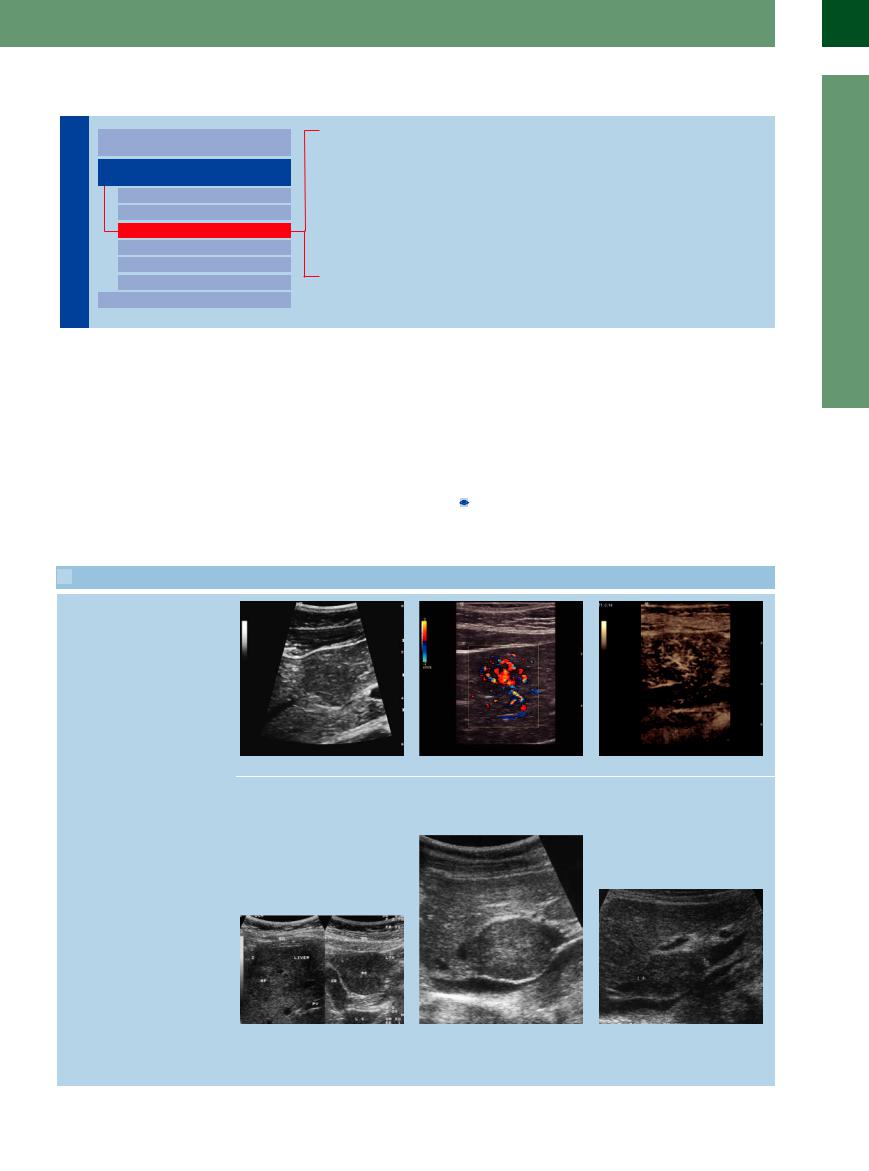
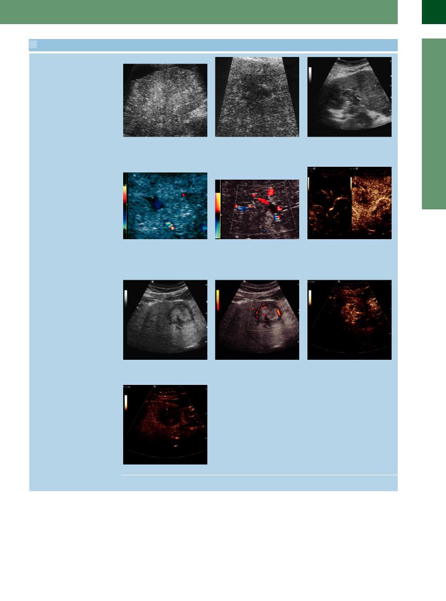
Detection of isoechoic masses in the liver is rather difficult and hinges on slight differences in texture. The most important diagnostic signs are surface and contour changes at the segmental and vascular interfaces.
Focal Nodular Hyperplasia 


In terms of histopathology, FNH is defined as |
only be detected because of the circumscribed |
will unmask them. In CEUS they can be easily |
circumscribed cirrhosis of the liver. Thus, there |
inhomogeneity of their texture ( 2.9 d–i) and |
detected and characterized. |
are FNH variants that hardly differ from the |
their vascular characteristics (if visible at all): |
|
surrounding hepatic parenchyma and may |
in the latter cases, color-flow Doppler scanning |
|
 2.9 Liver Cell Adenoma and Focal Nodular Hyperplasia (FNH)
Liver cell adenoma: adenomas of the liver appear isoechoic or hypoechoic (in fatty liver) to the surrounding parenchyma
2
Localized Changes in Hepatic Parenchyma
FNH is a benign hepatic lesion, its histology corresponding to circumscribed cirrhotic transformation: there is a central vessel where the radial strands of scar tissue terminate; at the rim, increased circular vascularization can be demonstrated
a Liver cell adenoma, histologically proved, with distinct signs of hypervascularity.
d FNH (RF) in segment IV of the left hepatic lobe. BD = abdominal wall; LTH = round ligament of liver; GB = gallbladder; LC = caudate lobe.
b Hypervascularity in CDS (in the central venous vessels, marginal arterial vessels).
e FNH in the caudad lobe.
c Flush-like rapid arterial hyperenhancement, usually initially at the periphery with subsequent very rapid centripetal filling leading to an isoenhancement not distinguishable from the surrounding tissue.
f The same mass as in e, 3 years later after discontinuation of hormone therapy: clearly smaller. Additional presence of a typical circular, spoked-wheel vascular pattern confirms FNH.
101
2
Liver
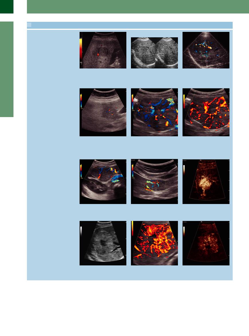
 2.9 Liver Cell Adenoma and Focal Nodular Hyperplasia (FNH) (Continued)
g Isoechoic mass in a fatty liver. Even color- |
h Isoechoic mass of FNH in the right liver |
i The same mass as in h; increased vas- |
flow Doppler imaging was unable to as- |
lobe distinguishable only by the different |
cularization at the margin and barely |
certain its character. Distinctive afferent |
coarse and irregular structure and by the |
visible vascular star-like radiating vessels. |
vascular pedicle and circular vasculariza- |
attenuation of echoes behind the lesion. |
|
tion at the rim. |
|
|
FNH, typical vascularity
Gray-scale imaging and color Doppler techniques strongly support the diagnosis of FNH, more sensitively shown on CEUS
j Central vessels, vascular star.
m–o FNHs are usually vascular malformations and have a strong feeding artery; this may be visible in CDS (m and n) but also in CEUS (o).
k Central vessels, vascular star and radiating vessels.
l Increased vascularization at the margin and barely visible vascular star radiating vessels; as a snapshot for documentation purposes it is possible proof, but during real-time imaging this type of vascularization is definite proof.
p Typical spoke-like image of a FNH with hypoechoic center.
q Color Doppler imaging is helpful to visualize the spoke-wheel vascular pattern which strongly supports the diagnosis of FNH.
r On CEUS, FNH appears as a hyperenhancement in the early arterial phase with rapid fill-in from the center outwards.
102
 2.9 Liver Cell Adenoma and Focal Nodular Hyperplasia (FNH) (Continued)
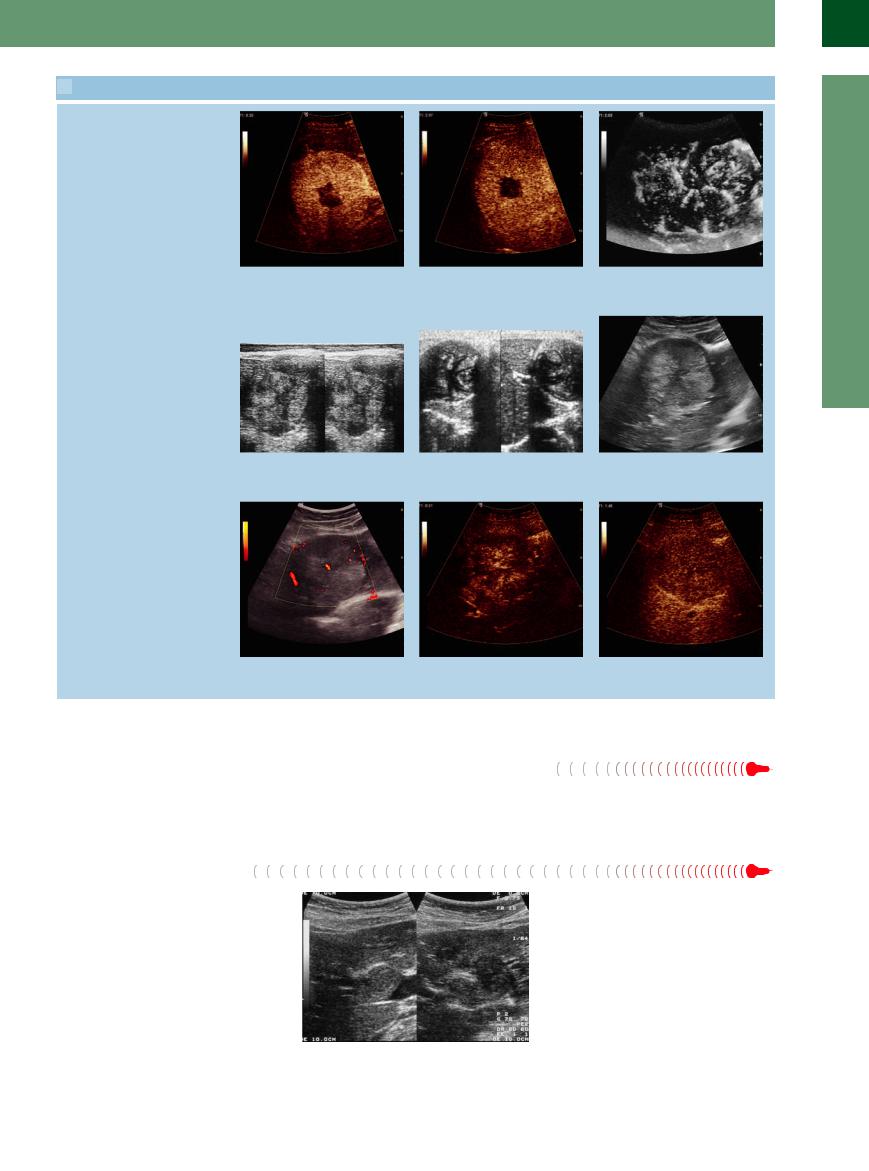
Differential diagnosis for FNH: difficult to decide on gray-scale imaging alone
s Rapid fill-in from a central feeding artery before an enhancement of the surrounding liver parenchyma.
v Spoke-wheel type of vascular pattern in a colonic metastasis.
t During the portal venous and late phases, FNH remains isoenhancing: a centrally located scar is seen.
w Spoke-like representation of a hydatid cyst.
u Vascularization of an FNH in the angio mode.
x A fibrolamellar HCC may also simulate a FNH; further diagnosis by color Doppler and CEUS (y,z left and right).
2
Localized Changes in Hepatic Parenchyma
y Color Doppler shows only a central vessel, but not the typical spoke-like vascularity.
z Left and right: in CEUS, demonstration of chaotic enhancement in the early arterial phase after 21 s (left); in the late parenchymal phase (right; 105 s) hypoenhancement as a sign of malignancy.
Adenoma 


Sometimes it is almost impossible to distinguish between normal parenchyma and adenoma of the liver.
Hepatocellular Carcinoma
One-third of all HCC are isoechoic and may be difficult to detect, particularly when superimposed upon underlying cirrhotic liver disease; this corresponds to the diffuse infiltrative type of hepatocellular carcinoma. In these cases, extra attention should be paid to the typical changes induced by the tumor-invading vessels. In isoechoic HCC, color-flow Doppler scanning will yield bizarre irregular hypervascularization and in CEUS an early irregular enhancement and a rapid wash-out in the late phase (Fig. 2.83, Fig. 2.84,  2.10j–s).
Fig. 2.83 Tumor invasion into the portal vein by an HCC in liver cirrhosis.
103
2
Liver
Fig. 2.84 HCC invading a portal vein with tumor thrombosis, within which tumor vascularization can be demonstrated.
 2.10 Hepatocellular Carcinoma (HCC)
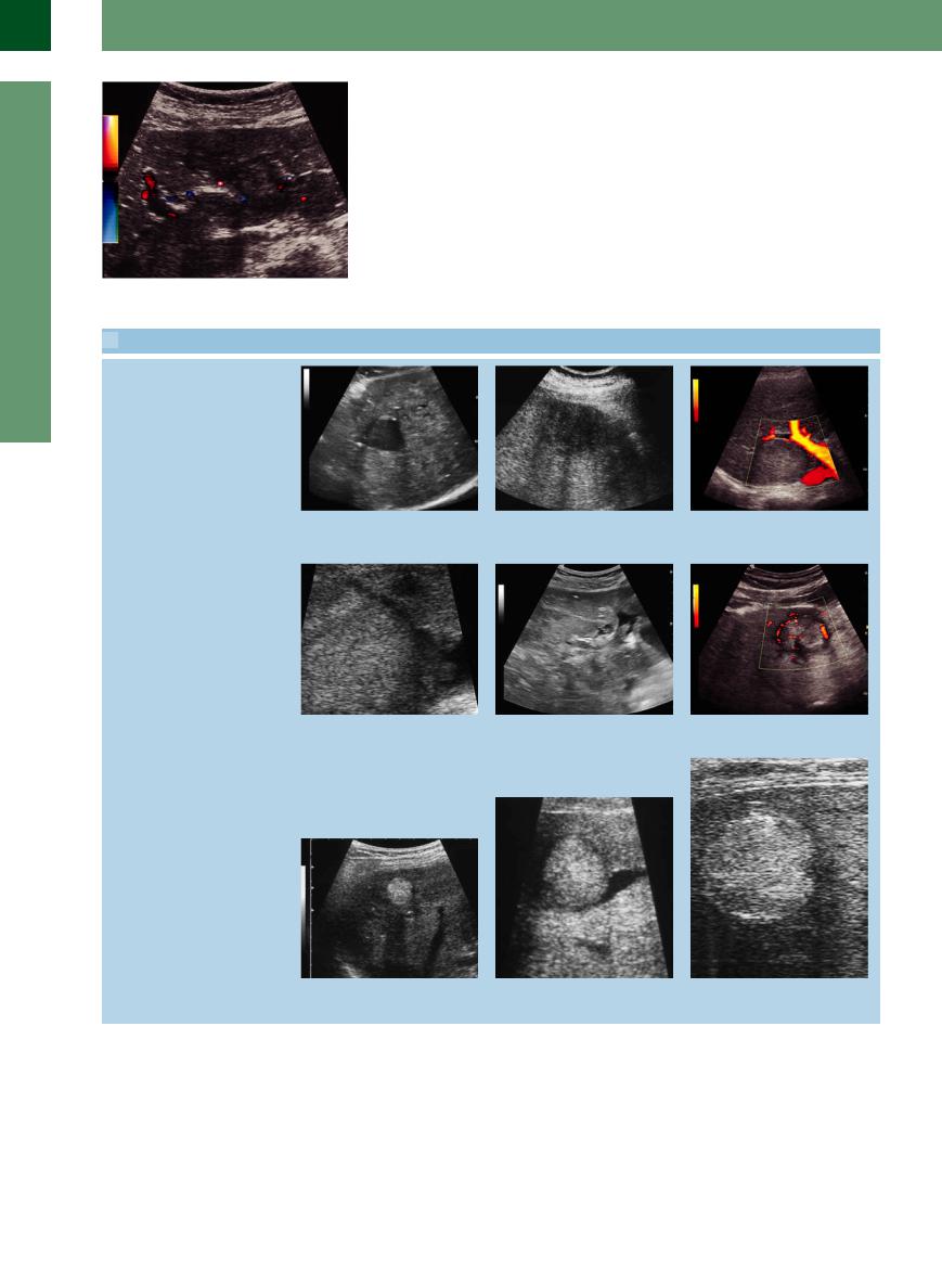
Hypoechoic HCC: one-third of all HCC present as hypoechoic lesions
Isoechoic HCC: about one-third of all HCC are isoechoic to the surrounding liver tissue and may only be detected by their complications or circumscribed irregularities in the texture
Hyperechoic HCC: about one-third of all HCC are hyperechoic to the surrounding liver tissue and may easily be mistaken for hemangiomas, although HCC is characterized by a more pronounced attenuation of the signal
a–c HCC may infiltrate into the parenchyma (often in liver cirrhosis; a and b) or may appear as a circumscribed mass (c).
d Ill-defined invasion of a hepatic vein by HCC.
b Ill-defined infiltration. |
c Circumscribed mass. |
e Invasion of a portal vein by HCC with |
f Isoechoic HCC with halo sign and bizarre |
tumor plug in the lumen. |
irregular hypervascularization. |
g–h Hyperechoic HCC may be mistaken |
h HCC invading a vessel. |
i Hyperechoic HCC with signal attenua- |
for a hemangioma but demonstrates a |
|
tion. |
distinct beam attenuation (g and i). |
|
|
104
 2.10 Hepatocellular Carcinoma (HCC) (Continued)
Diffuse HCC
Vascular complications in HCC: invasion of vessels represents a typical complication of HCC
HCC in CEUS
j HCC may infiltrate the parenchyma diffusely (quite often in previously existing cirrhosis); it is then difficult to recognize and a biopsy is needed.
m HCC invading a hepatic vein.
p and q Small HCC with marked hypervascularization.
s Undifferentiated HCC in the late parenchymal phase demonstrate a rapid washout compared to the surrounding parenchyma (here the same HCC as in p).
k Some suspected HCC with diffuse infiltration can be differentiated by CEUS and directed biopsy.
n HCC invading a portal vein.
q Color Doppler.
l HCC invading a vessel is an unequivocal finding (in this case invasion of a portal vein).
o HCC invading the portal vein: (left) CEUS after 19 s showing the arterial branches; (right) after 26 s. The thrombotic portal vein demonstrates an en- hancement—a typical sign of tumor infiltration.
r The same HCC in CEUS: rapid and irregular enhancement in the arterial phase after 20 s.
2
Localized Changes in Hepatic Parenchyma
105
2
Liver
 2.10 Hepatocellular Carcinoma (HCC) (Continued)
t Large HCC in the right liver lobe posteri- |
u In CEUS, the HCC (t) demonstrates an |
v In the late phase, the same HCC dem- |
or-cranial. |
intensive enhancement in the peripheral |
onstrates typical hypoenhancement. |
|
zone (with a central necrosis). |
|
Metastasis


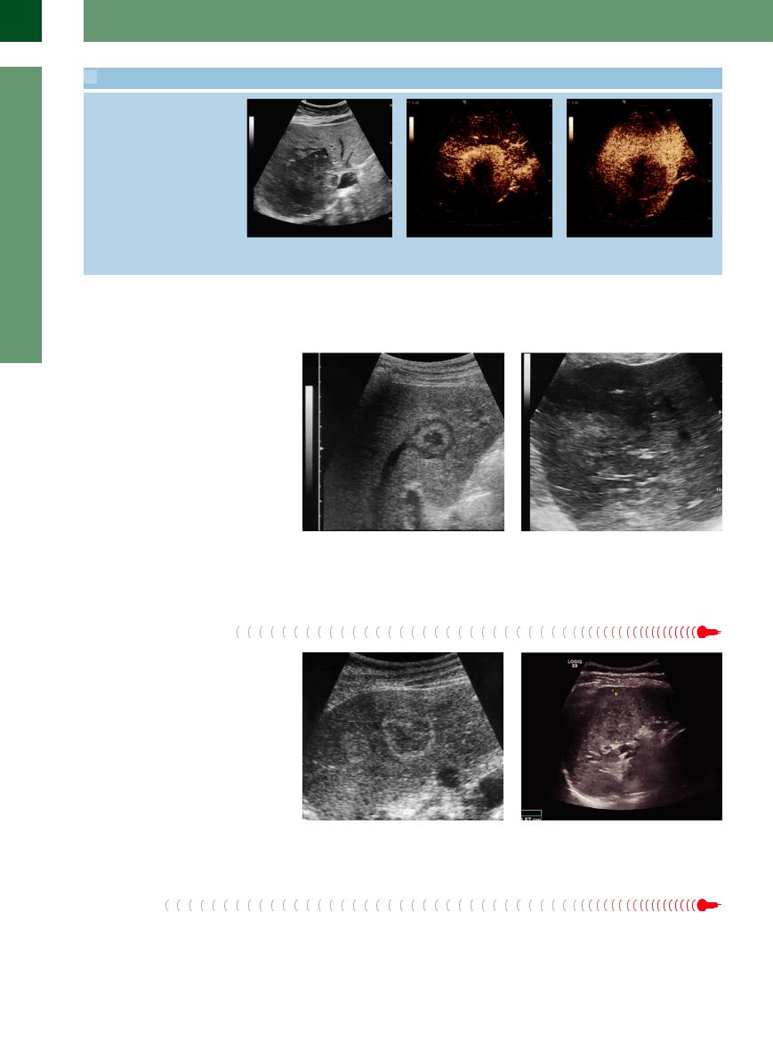
Small hepatic metastases in particular may escape detection because of their possible isoechogenicity. However, when scrutinized more closely their parenchymal texture is somewhat coarser and more echogenic. It is vital to check for any changes in contour at the surfaces and vascular interfaces. Isoechoic metastases are quite typical in diffuse metastasis of gastric and pancreatic cancer (Fig. 2.85, Fig. 2.86), and after successful chemotherapy as remnants of previously confirmed metastases (Fig. 2.56). Their detection occurs routinely by CEUS.
Fig. 2.85 Isoechoic metastasis of pancreatic cancer; the |
Fig. 2.86 Large isoechoic metastasis of cancer of the |
lesion is identified by its vascular invasion, halo sign, and |
colon. |
the central necrosis. |
|
Atypical Hemangioma
Detection and differentiation of hemangiomas depends not only on the texture and echogenicity of the surrounding parenchyma but also on the direction and angle of insonation. This explains why hemangiomas may be detected quite easily at one time but may remain undetected during follow-up studies, and in the more echogenic fatty liver hemangiomas may be masked altogether (Fig. 2.87, Fig. 2.88).
Fig. 2.87 Atypical hemangioma. Two isoechoic masses in |
Fig. 2.88 Atypical isoechoic hemangioma. |
the right hepatic lobe, subphrenic segment, with mar- |
|
ginal echo enhancement. |
|
Hematoma
Organized hemorrhage and hematoma may appear as isoechoic lesions.
106