

1
Vessels
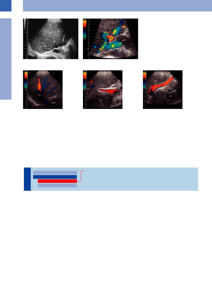
Fig. 1.96 A 24-year-old man with multiple drug dependence: old hepatic veno-occlusive disease (Budd–Chiari syndrome) with esophageal varices. Normal hepatic veins cannot be demonstrated (a); singular varicosities around the junction of the hepatic veins. VC = vena cava; LC = caudate lobe (b).
Fig. 1.97 Patient presenting with primary leiomyoma of |
b Demonstration of the tumor and portal vein (VP) with |
c Marked recanalization of the umbilical vein in post- |
the vena cava. |
normal direction of flow. |
hepatic block. LE = liver; AH = hepatic artery. |
a Intracaval tumor mass (TU); flow reversal in the right |
|
|
hepatic vein. COR = heart. |
|
|
Patients with posthepatic block usually ex- |
ragmatic veins draining in a cephalad direction. |
duced blood flow with a pulsatile profile, |
hibit thickening of the gallbladder wall, and |
Flow reversal in the portal vein may also be |
which in some cases turns into full-fledged |
sometimes it is possible to demonstrate intra- |
seen.17 Quite often in patients with cardiac de- |
pulse-synchronous flow reversal. |
hepatic venovenous collaterals or transdiaph- |
compensation, the portal vein will exhibit re- |
|
■ Intraluminal Mass
Thrombosis
|
|
|
|
Enlarged Lumen Diameter |
|
Portal Vein Thrombosis |
|
|
|
|
|
||||
|
|
|
|
Intraluminal Mass |
|
Splenic Vein Thrombosis |
|
Vessels |
|
||||||
|
|
Thrombosis |
|
Thrombosis of the Superior Mesenteric Vein |
|||
|
|
|
|||||
|
|
Tumor |
|
||||
|
|
|
|
|
|
|
|
Portal Vein Thrombosis


The etiology of portal vein thrombosis is rather varied (Table 1.4). Cirrhosis of the liver is by far the most common cause, although in fresh thrombosis hepatocellular carcinoma must be ruled out.18 Portal vein thrombosis may be subdivided into partial intrahepatic thrombosis, complete or partial thrombosis of the portal trunk, and additional thrombosis of the tributaries (splenic and mesenteric veins).
In acute thrombosis the lumen of the portal vein most often is enlarged and displays a rel-
atively homogeneous anechogenicity ( 1.9 d,g). If color-flow Doppler scanning demonstrates no flow, the diagnosis is confirmed.19 This venous occlusion will open up collaterals, some of which will drain in hepatopetal fashion. Over the course of time the former portal tree will exhibit an atypical vascular architecture; the underlying process is described as cavernous transformation (
 1.9e,f,i). In ultrasonography, marked varices can mimic a tumor (cavernoma). The extent and duration of this
cavernous transformation may be quite varied ( 1.9i,l,m).
Other less frequent collaterals may be seen alongside these cavernous transformations:
●Atypical paraportal collaterals (Fig.1.98)
●Varices around the peribiliary vascular plexus (Fig.1.99)
In addition, there may be hepatofugal collaterals via the right gastric vein or collaterals draining in a caudad direction.20
48
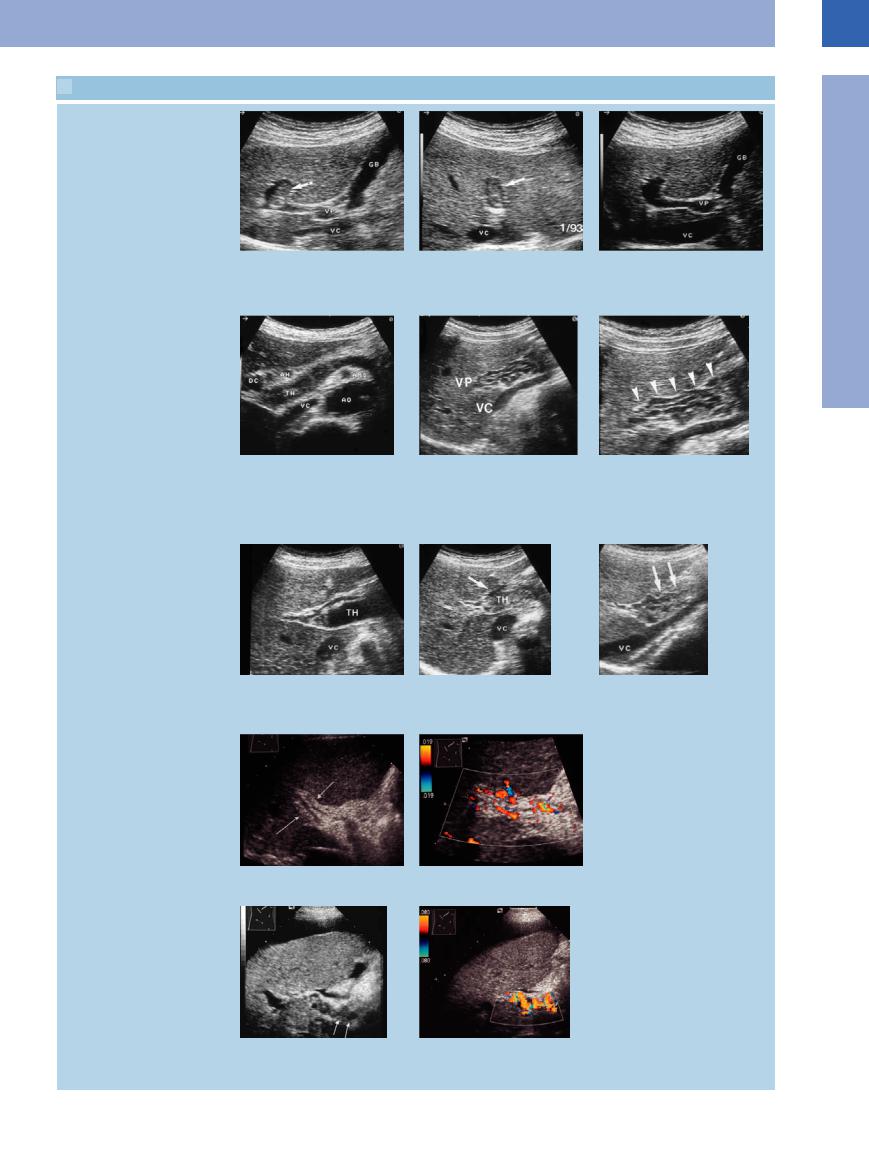
 1.9 Typical Findings in Portal Vein Thrombosis
Portal vein with partial thrombosis
a–c A 71-year-old patient with cancer of the colon. GB = gallbladder; VC = vena cava; VP = portal vein.
a and b Upper abdomen longitudinal and transverse section. Incidental finding of partial thrombosis of the intrahepatic portal vein.
c Spontaneous lysis after 2 months.
Portal vein thrombosis and cavernous transformation of portal vein
d Echogenic image of the portal vein in |
e and f A 24-year-old woman with esoph- f Section magnification. |
thrombosis. DC = bile duct; AH = hepatic |
ageal varices secondary to portal vein |
artery; TH = thrombus; VC = vena cava; AO |
thrombosis. |
= aorta. |
e Varicosities along the portal vein as a |
|
result of cavernous transformation of the |
|
portal vein. VC = vena cava; VP = portal |
|
vein. |
g–i Sonographic course of a fresh portal |
h Increasingly echogenic transformation of i Beginning recanalization after 4 months. |
vein thrombosis (TH). VC = vena cava. |
the thrombosis. |
g Hypoechoic image of the thrombotic |
|
material. |
|
Portal vein thrombosis and partial recanalization
j and k Portal vein thrombosis, 6 years old. |
k CDS: no significant recanalization de- |
j Fibrotic occlusion of the portal vein. |
monstrable. |
l and m Thrombosis of the portal vein with |
m Color-flow duplex scan of the cavernous |
cavernous recanalization. |
recanalization. |
l Cavernous recanalization cephalad of the |
|
portal vein. |
|
1
Intraluminal Mass
49
1
Vessels
Table 1.4 Etiology of portal vein thrombosis22
● Idiopathic
● Liver cirrhosis
● Malignant tumors ● Pancreatic diseases ● Para-infective
● Posttraumatic
● Collagenosis
● Thrombophilia
● Myeloproliferative diseases
Fig. 1.98 A 3-year-old thrombosis of the portal vein with paraportal “recanalization” (collaterals). The portal blood drains toward the gallbladder (GB) and from there into the liver. VP = portal vein.
Fig. 1.99
a Patient with Klatskin tumor, stent palliation, and occlusion of the portal vein (VP).
b Originating from the portal vein there are marked cephalad collaterals around the peribiliary venous plexus. GB = gallbladder.
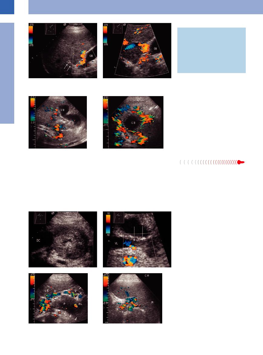
Splenic Vein Thrombosis 


Complete or partial thrombosis of the splenic vein is a common complication of acute pancreatitis and cancer of the pancreas. It may also originate from portal vein thrombosis and grow by apposition.
The ultrasound image depicts a hypoechoic vascular lumen without any flow on color-flow Doppler scanning. Partial thrombosis of the
splenic vein is sometimes observed. One of the sequelae of this vascular obstruction is “segmental” portal hypertension. Collateralization primarily opens the peripancreatic and perigastric veins draining cephalad via esophageal varices as well as veins running in a caudad direction (omental veins draining into the mesenteric veins) (Figs. 1.100, 1.101, 1.102).
A finding of hepatosplenomegaly is not mandatory, and neither is the build-up of ascites. Over time, the splenic vein may become recanalized or undergo cavernous transformation, although quite often the collaterals will persist. In thrombosis of the splenic vein, one rare complication is splenic infarction or even rupture.
Fig. 1.100 Splenic vein thrombosis.
a Cancer (TU) of the pancreatic head. DC = bile duct.
b Demonstration of echogenic material in the splenic vein (VL). AMS = superior mesenteric vein.
Fig. 1.101 Acute pancreatitis.
a The splenic vein cannot be imaged. Marked varices anterior to the pancreas.
b Demonstration of varicosities (V). M = spleen; LE = liver; P = pancreas; AL = splenic artery.
50
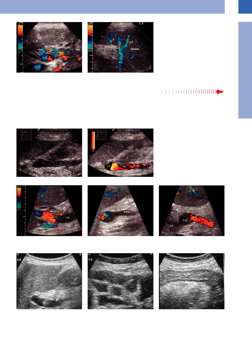
Fig. 1.102 Thrombosis of the splenic vein after mesenteric irradiation of a malignant lymphoma. LE = liver; MA = stomach; CO = venous confluence; VC = vena cava; VL = splenic vein; Milz = spleen.
a The splenic vein cannot be imaged and there is marked peripancreatic collateralization.
b Regular appearance of the splenic vein at the splenic portal.
Thrombosis of the
the Superior Mesenteric Vein


Truly isolated thrombosis of the superior mesenteric vein does not result in portal hypertension. However, it may be associated with splenic and/or portal vein thrombosis. The etiology is primarily inflammatory, in particular acute pancreatitis (Fig.1.103). During the acute
phase, the clinical symptoms with severe abdominal pain are impressive (Fig.1.104). On ultrasound the mesenteric vessels appear dilated and lined with echogenic structures. Color-flow Doppler scanning will not elicit any flow signals. Partial thrombosis is possible.
In rare cases there may be segmental infarction of the intestines. Usually this disorder does not require surgery, and quite often there will be spontaneous recanalization.
Fig. 1.103 Patient with acute pancreatitis.
a The tip of the thrombus within the superior mesenteric vein extends into the confluence of the mesenteric and splenic veins. Color-flow duplex scanning demonstrates echogenic thrombotic material.
b Color-flow duplex scanning depicts partial recanalization. P = tail of pancreas; CO = venous confluence; TH = thrombosis.
Fig. 1.104 A 50-year-old patient with acute abdomen.
a–c Color-flow duplex scanning demonstrates complete thrombosis of the superior mesenteric vein (VMS). DC = common bile duct; TH = thrombosis; VP = portal vein; VC = vena cava; CO = venous confluence; VL = splenic vein.
d–f B-mode imaging also depicts the thrombosis. Massive dilatation of the small intestine. There is thickening of the colonic wall at the level of the sigmoid and a lack of peristalsis (nonsurgical treatment proved successful).
1
Intraluminal Mass
51