

- •Contents
- •Preface
- •Contributors
- •1 Vessels
- •1.1 Aorta, Vena Cava, and Peripheral Vessels
- •Aorta, Arteries
- •Anomalies and Variant Positions
- •Dilatation
- •Stenosis
- •Wall Thickening
- •Intraluminal Mass
- •Perivascular Mass
- •Vena Cava, Veins
- •Anomalies
- •Dilatation
- •Intraluminal Mass
- •Compression, Infiltration
- •1.2 Portal Vein and Its Tributaries
- •Enlarged Lumen Diameter
- •Portal Hypertension
- •Intraluminal Mass
- •Thrombosis
- •Tumor
- •2 Liver
- •Enlarged Liver
- •Small Liver
- •Homogeneous Hypoechoic Texture
- •Homogeneous Hyperechoic Texture
- •Regionally Inhomogeneous Texture
- •Diffuse Inhomogeneous Texture
- •Anechoic Masses
- •Hypoechoic Masses
- •Isoechoic Masses
- •Hyperechoic Masses
- •Echogenic Masses
- •Irregular Masses
- •Differential Diagnosis of Focal Lesions
- •Diagnostic Methods
- •Suspected Diagnosis
- •3 Biliary Tree and Gallbladder
- •3.1 Biliary Tree
- •Thickening of the Bile Duct Wall
- •Localized and Diffuse
- •Bile Duct Rarefaction
- •Localized and Diffuse
- •Bile Duct Dilatation and Intraductal Pressure
- •Intrahepatic
- •Hilar and Prepancreatic
- •Intrapancreatic
- •Papillary
- •Abnormal Intraluminal Bile Duct Findings
- •Foreign Body
- •The Seven Most Important Questions
- •3.2 Gallbladder
- •Changes in Size
- •Large Gallbladder
- •Small/Missing Gallbladder
- •Wall Changes
- •General Hypoechogenicity
- •General Hyperechogenicity
- •General Tumor
- •Focal Tumor
- •Intraluminal Changes
- •Hyperechoic
- •Hypoechoic
- •Nonvisualized Gallbladder
- •Missing Gallbladder
- •Obscured Gallbladder
- •4 Pancreas
- •Diffuse Pancreatic Change
- •Large Pancreas
- •Small Pancreas
- •Hypoechoic Texture
- •Hyperechoic Texture
- •Focal Changes
- •Anechoic Lesion
- •Hypoechoic Lesion
- •Isoechoic Lesion
- •Hyperechoic Lesion
- •Irregular (Complex Structured) Lesion
- •Dilatation of the Pancreatic Duct
- •Marginal/Mild Dilatation
- •Marked Dilatation
- •5 Spleen
- •Nonfocal Changes of the Spleen
- •Diffuse Parenchymal Changes
- •Large Spleen
- •Small Spleen
- •Focal Changes of the Spleen
- •Anechoic Mass
- •Hypoechoic Mass
- •Hyperechoic Mass
- •Splenic Calcification
- •6 Lymph Nodes
- •Peripheral Lymph Nodes
- •Head/Neck
- •Extremities (Axilla, Groin)
- •Abdominal Lymph Nodes
- •Porta Hepatis
- •Splenic Hilum
- •Mesentery (Celiac, Upper and Lower Mesenteric Station)
- •Stomach
- •Focal Wall Changes
- •Extended Wall Changes
- •Dilated Lumen
- •Narrowed Lumen
- •Small/Large Intestine
- •Focal Wall Changes
- •Extended Wall Changes
- •Dilated Lumen
- •Narrowed Lumen
- •8 Peritoneal Cavity
- •Anechoic Structure
- •Hypoechoic Structure
- •Hyperechoic Structure
- •Anechoic Structure
- •Hypoechoic Structure
- •Hyperechoic Structure
- •Wall Structures
- •Smooth Margin
- •Irregular Margin
- •Intragastric Processes
- •Intraintestinal Processes
- •9 Kidneys
- •Anomalies, Malformations
- •Aplasia, Hypoplasia
- •Cystic Malformation
- •Anomalies of Number, Position, or Rotation
- •Fusion Anomaly
- •Anomalies of the Renal Calices
- •Vascular Anomaly
- •Diffuse Changes
- •Large Kidneys
- •Small Kidneys
- •Hypoechoic Structure
- •Hyperechoic Structure
- •Irregular Structure
- •Circumscribed Changes
- •Anechoic Structure
- •Hypoechoic or Isoechoic Structure
- •Complex Structure
- •Hyperechoic Structure
- •10 Adrenal Glands
- •Enlargement
- •Anechoic Structure
- •Hypoechoic Structure
- •Complex Echo Structure
- •Hyperechoic Structure
- •11 Urinary Tract
- •Malformations
- •Duplication Anomalies
- •Dilatations and Stenoses
- •Dilated Renal Pelvis and Ureter
- •Anechoic
- •Hypoechoic
- •Hypoechoic
- •Hyperechoic
- •Large Bladder
- •Small Bladder
- •Altered Bladder Shape
- •Intracavitary Mass
- •Hypoechoic
- •Hyperechoic
- •Echogenic
- •Wall Changes
- •Diffuse Wall Thickening
- •Circumscribed Wall Thickening
- •Concavities and Convexities
- •12.1 The Prostate
- •Enlarged Prostate
- •Regular
- •Irregular
- •Small Prostate
- •Regular
- •Echogenic
- •Circumscribed Lesion
- •Anechoic
- •Hypoechoic
- •Echogenic
- •12.2 Seminal Vesicles
- •Diffuse Change
- •Hypoechoic
- •Circumscribed Change
- •Anechoic
- •Echogenic
- •Irregular
- •12.3 Testis, Epididymis
- •Diffuse Change
- •Enlargement
- •Decreased Size
- •Circumscribed Lesion
- •Anechoic or Hypoechoic
- •Irregular/Echogenic
- •Epididymal Lesion
- •Anechoic
- •Hypoechoic
- •Intrascrotal Mass
- •Anechoic or Hypoechoic
- •Echogenic
- •13 Female Genital Tract
- •Masses
- •Abnormalities of Size or Shape
- •Uterus
- •Abnormalities of Size or Shape
- •Myometrial Changes
- •Intracavitary Changes
- •Endometrial Changes
- •Fallopian Tubes
- •Hypoechoic Mass
- •Anechoic Cystic Mass
- •Solid Echogenic or Nonhomogeneous Mass
- •14 Thyroid Gland
- •Diffuse Changes
- •Enlarged Thyroid Gland
- •Small Thyroid Gland
- •Hypoechoic Structure
- •Hyperechoic Structure
- •Circumscribed Changes
- •Anechoic
- •Hypoechoic
- •Isoechoic
- •Hyperechoic
- •Irregular
- •Differential Diagnosis of Hyperthyroidism
- •Types of Autonomy
- •15 Pleura and Chest Wall
- •Chest Wall
- •Masses
- •Parietal Pleura
- •Nodular Masses
- •Diffuse Pleural Thickening
- •Pleural Effusion
- •Anechoic Effusion
- •Echogenic Effusion
- •Complex Effusion
- •16 Lung
- •Masses
- •Anechoic Masses
- •Hypoechoic Masses
- •Complex Masses
- •Index
Interferon-Induced Thyroiditis 


Hyperand hypothyroidism are prevalent, predominantly hypothyroidism. Antibodies are present in up to 35% of cases. In 50%, spontaneous improvement occurs. Ultrasound often demonstrates increased vascularity in a hypoechoic thyroid.
Hyperechoic Structure
Thyroid Gland

Diffuse Changes
Enlarged Thyroid Gland
Small Thyroid Gland
Hypoechoic Structure
Hyperechoic Structure
Circumscribed Changes
Differential Diagnosis of Hyperthyroidism
Radiation-Induced Hypothyroidism
Radiation-Induced Hypothyroidism


The thyroid gland is located within the radio- |
becomes smaller and undergoes fibrous atro- |
therapy portal when radiation is applied to the |
phy. |
neck region, and it responds with hypothyroid- |
Accordingly, ultrasound demonstrates a |
ism following a long latent period. The thyroid |
small thyroid (see above) that shows a diffuse |
|
or mottled increase in echogenicity (Fig.14.25). |
■ Circumscribed Changes
Anechoic
Gland |
Diffuse Changes |
|
Cysts |
||||
|
|
|
|
||||
|
|
|
|
Circumscribed Changes |
|
Vessels |
|
|
|
|
|
||||
|
|
|
|
||||
|
|
|
|
|
Anechoic |
|
Abscess |
Thyroid |
|
|
|
||||
|
|
|
|
||||
|
|
Hyperechoic |
|
|
|||
|
|
|
|
|
Hypoechoic |
|
|
|
|
|
|
|
Isoechoic |
|
|
|
|
|
|
|
Irregular |
|
|
|
|
|
Differential Diagnosis of Hyperthyroidism |
|
|
||
|
|
|
|
|
|||
Cysts
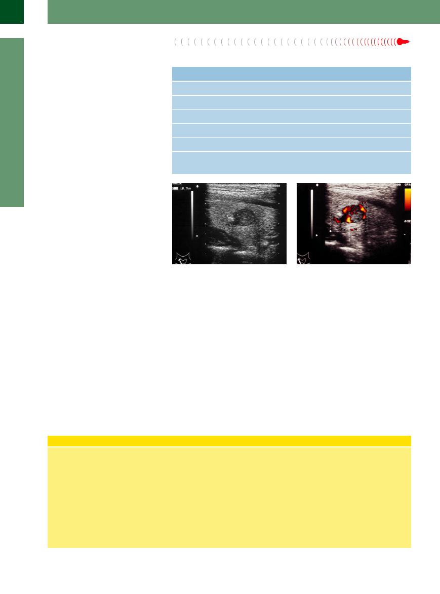
Cystic masses, along with thyroid nodules, are the most common focal changes that are found in the thyroid. Thyroid cysts are not true cysts and therefore do not contain cystic epithelium. Solitary cysts in an otherwise normal thyroid generally result from central hemorrhage in adenomas.
Cyst contents. Depending on their contents, cysts may appear sonographically as anechoic masses or may show a hypoechoic, hyperechoic, or irregular internal echo pattern ( 14.2). Further diagnostic tests consist of color duplex sonography and fine-needle aspiration cytology. In many cases the cyst contents are
unknown until fine-needle aspiration is performed. Cysts may contain various fluids:
●Fresh hemorrhage ( 14.2 d,e)
●Older hemorrhage (chocolate cysts,
Fig.14.49)
●Old hemorrhage (yellow cysts)
●Colloid (colloid cysts,  14.2f)
●Lymph (lymph cysts)
Fresh hemorrhages and colloid cysts generally have a distinctive internal echo pattern and appear hypoechoic or hyperechoic, not anechoic ( 14.2 d,e). With color duplex, cystic tumors show a vascularity that is never seen in cysts or hemorrhages. Other cysts extrinsic to
the thyroid, such as neck cysts, should also be included in the differential diagnosis.
Solitary cysts. Uncomplicated serous cysts appear sonographically as anechoic, rounded or polygonal, sometimes lobulated, scalloped or patchy lesions with smooth margins. Like cysts in other organs, they display several typical cystic features such as absence of internal echoes, smooth borders, and distal acoustic enhancement, while a circular shape is somewhat unusual. Edge shadows are not present because of the absence of a cyst wall. There is an additional sign, however: definite compressibility of the cysts by external transducer pressure.
14
Circumscribed Changes
489
14
Thyroid Gland
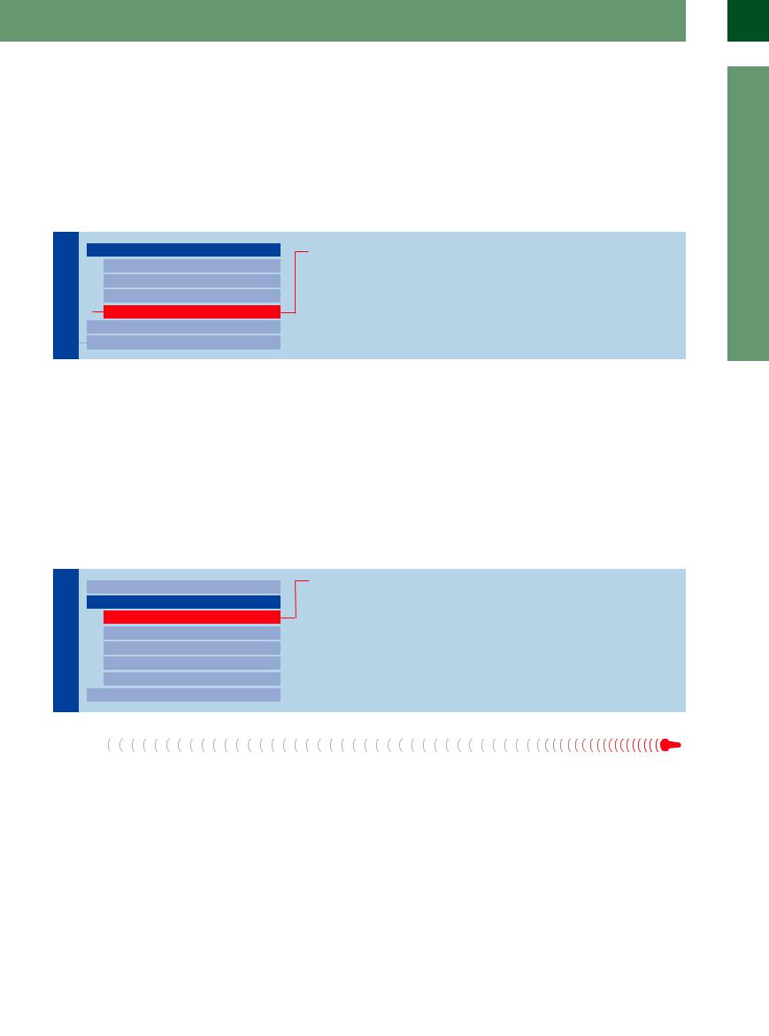
 14.2 Thyroid Cysts
Adenomas/nodules showing cystic regression
a Two cysts (C) in an otherwise un- |
b Cyst (C) in a nodular goiter (TH), the |
changed left thyroid lobe (TH): anechoic |
latter showing some peripheral vascular- |
masses with irregular margins. One shows |
ity. |
septumlike partitions (arrows) and distal |
|
enhancement and creates a bulge in the |
|
thyroid contour. |
|
Hemorrhagic, colloid cysts
d Hemorrhagic cyst with a contour bulge. |
e Hemorrhagic large cyst with moving |
Bright internal floating echoes are typical |
echoes (needle aspiration: “chocolate |
of fresh intracystic hemorrhage. |
cyst”). |
Cysts in nodular goiters
c Anechoic cystic mass in a hyperplastic thyroid nodule surrounded by a vascular rim; no internal blood flow. VJ = jugular vein.
f Two cystic areas (C), one with internal echoes. Fine-needle aspiration yielded a creamy colloidal fluid (confirmed cytologically as colloid). M = musculature, TR = trachea.
g and h Multiple small cysts and fine calcifications in a nodular goiter, interpreted as regressive changes. The more echogenic and more coarsely structured nodules (collagenous connective tissue) are also regressive changes.
i Irregular, regressive cystic mass with indistinct margins and mottled echo structure (C) in a goiter (TH). Fine peripheral vessels rim the nodular area
(N).
Solitary cysts also occur as hemorrhagic areas in diffuse goiters. Peripheral or circular remnants of the old adenomatous structure are also seen in most cases ( 14.2a–c). Color Doppler examination shows a complete absence of vascularity in the cysts (
 14.2b,c,i).
Multiple cysts. Multiple cysts are characteristic of regressive changes in the adenomatous nodules of older nodular goiters. They differ from hemorrhages in true adenomas by their multiplicity and their occurrence in the background structure of the nodular goiter.
Sonographically, they appear in various sizes ranging from microcysts to macrocysts. The latter often present clinically as circumscribed palpable nodules. Fresh bleeding in the cysts leads to the abrupt appearance or enlargement of a goiter.
Hemorrhagic cysts. Fresh bleeding in cysts produces a heterogeneous internal echo pattern that includes high-level echoes (
14.2 d,e).
Lymph cysts. It is not uncommon for small cystic lesions to represent lymph cysts. They do not differ from other cystic lesions, except perhaps by their small size ( 14.2 g).
Colloid cysts. Because they contain colloid, these cysts display an anechoic or more or less finely granular internal echo pattern
( 14.2f).
490

Vessels
Cysts require differentiation from anechoic le- |
ysis (Fig.14.30, Fig.14.31; see also Fig.14.6, |
sions that can be identified as vessels only by |
Fig.14.7). |
color Doppler examination with spectral anal- |
|
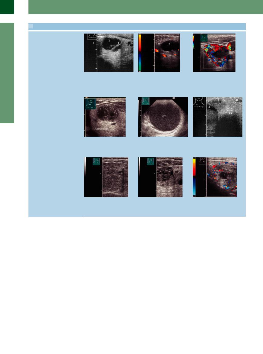
Fig. 14.30 Vessels in the thyroid. AC = common carotid artery; TR = trachea.
a Cystic areas (arrows) in gray-scale image.
b Color Doppler identifies the areas as vessels.
Fig. 14.31 Large, predominantly anechoic cystic mass (cursors) in the left thyroid lobe (TH). The common carotid artery (AC) is posterior. TR = trachea.
a Gray-scale image: the mass may be a large adenoma or a large cyst.
b Color Doppler and spectral analysis identify the mass as a venous vessel (dilated internal jugular vein).
14
Circumscribed Changes
Abscess
Abscesses almost always appear very hypoechoic and occasionally anechoic. The diagnosis is made clinically and/or by percutaneous aspiration (see Fig.14.37).
Hypoechoic
Gland |
|
|
Diffuse Changes |
|
|
|
|
||
|
|
|
Circumscribed Changes |
|
|
|
|||
Thyroid |
|
|
|
Anechoic |
|
|
|
Hyperechoic |
|
|
|
|
|
Hypoechoic |
|
|
|
|
Isoechoic |
|
|
|
|
Irregular |
|
|
|
Differential Diagnosis of Hyperthyroidism |
|
Microfollicular (Papillary) Adenoma Oncocytic Adenoma
Parathyroid Adenoma Abscess
Focal de Quervain Thyroiditis Malignant Lymphoma Carcinoma
Tumor Infiltration or Metastasis
Microfollicular (Papillary)
 Adenoma


Sonographic features. Adenomas may appear as hypoechoic, isoechoic, or hyperechoic masses at ultrasound. The relationships between follicular histology and echogenicity shown in Table 14.2 apply with fair accuracy to the echo structure of adenomas as well as to
adenomatous nodules and regressive changes ( 14.3a–c).
Echogenicity. Hypoechoic nodules generally represent microfollicular adenomas (rarely papillary adenomas), whereas hyperechoic nodules are usually macrofollicular adenomas.
Normofollicular adenomas generally appear isoechoic and are demarcated from the normal thyroid by a hypoechoic rim of displaced vessels, which can be identified with color Doppler. This vascular rim is also a feature of most other adenomas and adenomatous nodules ( 14.3 d–i,k,l). Isoechoic and hypoechoic
491

14
Thyroid Gland
Pathogenesis and Morphology of Thyroid Adenomas and Adenomatous Nodules
Prevalence. Thyroid nodules are found in 35–50% of people living in iodine-deficient areas.
A thyroid nodule is a discrete lesion within the thyroid gland that is palpable and/or ultrasonographically distinct from the surrounding thyroid parenchyma. Nonpalpable nodules detected incidentally with high-resolution technique in ultrasound or other imaging studies are termed “incidentalomas” (in 17–67%, varying regionally11). The exclusion of carcinomas is of clinical significance (depending on several factors up to 5–10%). Nonpalpable nodules have the same risk of malignancy as palpable nodules of the same size.
It is estimated that autonomous nodules are present in 2–6% of the general population12 and in 10–20% of patients with a preexisting goiter.
Pathogenesis. The normal thyroid gland already contains a number of thyrocytes with an autonomous growth tendency that may give rise to adenomas, regardless of the iodine supply. A similar mechanism underlies the development of the predominantly autonomous nodules in a nodular goiter.
Morphology. True adenomas are benign neoplasms. Aside from very rare leiomyomas, they account for virtually all benign neoplasms in the thyroid. Follicular adenomas are the most common type of adenoma. Other variants are papillary adenoma and oncocytoma. Adenomas, unlike the adenomatous nodules in a nodular goiter, do not take part in autoimmune changes. Functionally, microfollicular adenomas consist mainly of autonomously functioning adenomas that appear as hot nodules at scintigraphy, whereas macrofollicular adenomas usually appear as cold nodules.
adenomas are the most common types of adenoma, while hyperechoic adenomas are the least common.
Hypoechoic adenomas. The great majority of hypoechoic adenomas are autonomous adenomas, two-thirds of which are associated with latent hyperthyroidism (suppressed basal TSH with normal fT3/fT4 levels) and only one-third with overt hyperthyroidism.13 The most common microfollicular adenomas are conspicuous by their hypoechoic structure and round shape. The anechoic rim described above is also a general characteristic of adenomas, although it is not seen with every adenoma (see Table 14.2).
Another characteristic of hypoechoic adenomas is their internal vascularity, which signifies autonomy in a high percentage of cases (73%).14 The absence of internal vascularity rules out autonomy with a negative predictive value of 94%.13
A hypoechoic structure is in itself insufficient for evaluating functional autonomy (
14.3a,d,i).
Meanwhile, peripheral and internal vascularity is also found in a high percentage of malignancies, raising problems of differentiation based on ultrasound appearance.
Thyroid adenomas are also subject to regressive changes with intralesional hemorrhage and calcification, resulting in anechoic or hyperechoic internal structures ( 14.3 g–i; see also Fig.14.50). The above points can be summarized as follows:
●The color Doppler examination of thyroid nodules is a useful screening method for autonomous adenomas,12 especially in patients with known hyperthyroidism.
●True adenomas cannot be distinguished from adenomatous nodules by their sonographic features (this cannot even be done histologically in some cases). The sonographic findings can, however, provide clues for differentiation.
Table 14.2 Histomorphology of thyroid nodules correlated with typical echogenicity at ultrasound
Histomorphological finding |
Typical echogenicity |
Predominance of microfollicles |
Hypoechoic |
Predominance of normal-sized follicles |
Normal echo structure |
Predominance of macrofollicles |
Hyperechoic |
Collagenous connective tissue |
Hyperechoic |
Hyaline connective tissue |
Hypoechoic |
Diagnostic Evaluation of Solitary or Multiple Separate Hypoechoic Thyroid Nodules
●Sonography with color duplex scanning
●Generally, nodules > 1 cm should be evaluated, because they have the potential to be clinically significant cancers even if the risk of malignancy is low (< 1%). Nodules < 1 cm should also be explored under the following conditions:
–suspicious ultrasound findings
–whole-body X-irradiation after bonemarrow grafting, or history of head and neck irradiation
–positive family history of thyroid cancer (first-degree relatives)
–rapid growth and hoarseness
–exposure to fallout from Chernobyl under the age of 14 years
●Nodules > 1 cm should be evaluated,15 even when the risk for malignancy is very low (< 1%)
●Nodules 1–1.5 cm: serum TSH (or additional radionuclide thyroid scan)
–TSH in normal range: fine-needle aspiration (FNA) and cytologic evaluation
–TSH subnormal, additional fT3, fT4 if necessary radionuclide thyroid scan whether the nodule is functioning (tracer uptake greater than the surrounding normal thyroid), isofunctioning (“warm”) or nonfunctioning (functioning nodules rarely harbor malignancy)
–TSH elevated: FNA is recommended, because the rate of malignancy in nodules is similar in thyroid glands involved with Hashimoto thyroiditis to normal glands
●With normal TSH but suspected autonomy: suppression scintigraphy.
●With a sonographically hypoechoic nodule that is cold by scintigraphy: always fine-needle aspiration or surgery; also calcitonin assay (medullary C-cell carcinoma).
●FNA is the most accurate and least expensive procedure for evaluating thyroid nodules. Benign nodules should undergo long-term follow-up, with FNA in case of growth.
492

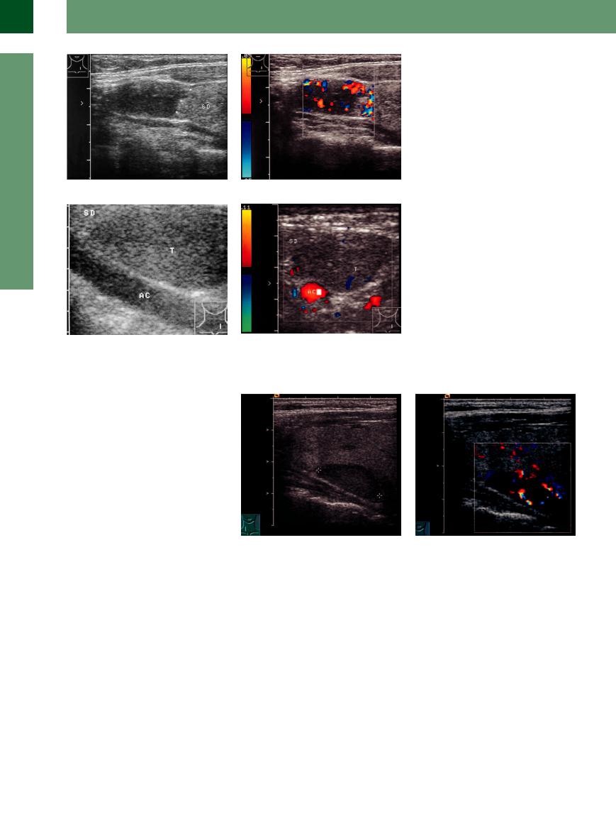
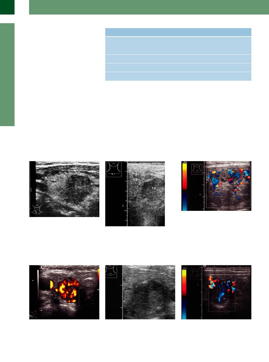
 14.3 Thyroid Adenomas
Types of adenoma
a Hypoechoic (microfollicular) adenoma: |
b Isoechoic adenoma (cursors), delineated |
c Hyperechoic adenoma (A) in a normally |
intensely hypoechoic mass with a subtle |
only by a slightly hypoechoic peripheral |
structured thyroid gland (SD). |
echo-free rim. Patient presented clinically |
vascular rim. |
|
with hyperthyroidism. |
|
|
Adenomas, color duplex
d Hypoechoic mass (T). Di erential diagnosis: adenoma, malignant tumor, hemorrhagic cyst.
g Isoechoic adenoma with hypoechoic areas devoid of internal vascularity: adenoma with fresh intralesional hemorrhage (painful swelling; treated surgically).
Functioning and nonfunctioning nodules in radionuclide scan
j Subclinical hyperthyroidism, radionuclide scan: Functioning nodule left lobe (1), nonfunctioning nodule of the right lobe
(2).
e Further examination with color Doppler shows intense peripheral vascularity as well as internal vessels.
h Hyperechoic adenoma with an old hemorrhagic area and cystic transformation
(C).
k Left thyroid lobe: two large hypoechoic adenomas.
f Several adenomas (A) of varying echogenicity in an otherwise normal thyroid, outlined by peripheral vascularity.
i Thyroid nodule, radionuclide thyroid scan shows a nonfunctioning (“cold”) nodule (see below, j), small calcification with shadow (S), small cystic lesion. This nodule must be examined by FNA and cytology.
l Right lobe, CDS: marked vascularized rim in the caudal nodule; rare internal vascularization, lack of peripheral vascularization in the upper nodule: FNA is required.
Oncocytic Adenoma


Oncocytic adenoma is another, rare type of |
sion of invasive growth in histologically exam- |
adenoma. It is indistinguishable from a well- |
ined surgical material. |
differentiated follicular carcinoma by ultraso- |
Sonographically, oncocytic adenoma shows |
nography or FNA cytology. Benign/malignant |
more or less pronounced internal vascularity |
differentiation relies on the detection or exclu- |
but usually does not have a peripheral vascular |
|
rim (Fig. 14.32, Fig.14.33). |
Differentiation is required from other hypoechoic tumors such as lipoma, malignant lymphoma, carcinoma, and metastases as well as rare abscesses or focal thyroiditis.
14
Circumscribed Changes
493
14
Thyroid Gland
Fig. 14.32 Oncocytoma in a nodular goiter, cytological and surgical diagnosis.
a Very hypoechoic tumor with no detectable halo, relatively well demarcated (arrows) from the rest of the thyroid (TH).
b Color Doppler scan at a low PRF clearly reveals internal vascularity.
Fig. 14.33 Oncocytic adenoma, incidental finding, cytological diagnosis surgically confirmed. AC = common carotid artery.
a Homogeneous tumor (T), slightly hypoechoic to the rest of the thyroid (TH).
b Color Doppler at a relatively high PRF shows almost no vascularity.
Parathyroid Adenoma


Like lipoma, parathyroid adenoma (both primary idiopathic adenoma and secondary parathyroid hyperplasia due to renal failure and hypocalcemia) appears as a hypoechoic mass located next to the thyroid gland. The location of the parathyroid glands has been described above under “Topography.”
Sonographic features. Parathyroid adenoma is markedly less echogenic than the thyroid, has an echo structure ranging from homogeneous to patchy-cystic with increasing size, and presents a round, oval, or polygonal shape. Internal vascularity has been variously described and may be subtle. Peripheral vascularity is believed to be uncommon, but curved vascular rims have been described around the adenomas (Figs. 14.34, 14.35, 14.36).
Fig. 14.34 Superior parathyroid dorsally in the region of |
b CDS: intratumoral vessels. |
the superior to the middle part of the thyroid. Incidentally |
|
found hypercalcemia, elevated parathyroid hormone. |
|
a Longitudinal scan: oval mass dorsally of the thyroid. |
|
494
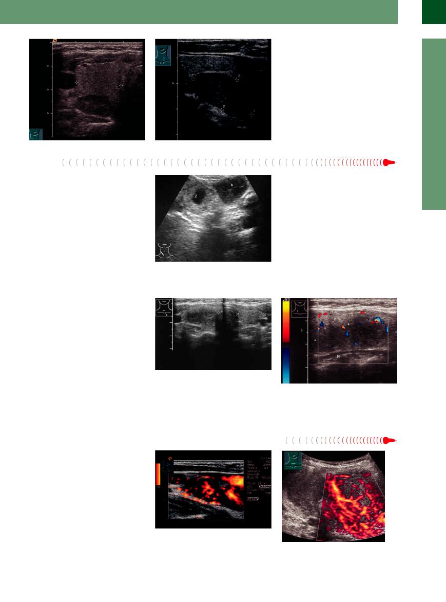
Fig. 14.35 (Same patient as in Fig. 14.34) High transverse scan of the right thyroid lobe: hypoechoic mass dorsally (the normal position of the parathyroid gland is at the dorsal face of the thyroid in 80–85%; ectopic in 15–20%).
f Fig. 14.36 Inferior parathyroid adenoma, longitudinal scan of the left thyroid (cursors): oval mass at the inferior pole (most often medially of the inferior thyroid artery).
Abscess
Rare abscesses of the thyroid gland have a similar hypoechoic appearance to adenomas, cysts, and lymphomas. Their presence signifies an acute purulent thyroiditis.
Thyroid abscess appears sonographically as a mostly anechoic or hypoechoic mass with irregular internal echoes. Color duplex examination shows an absence of vascularity (Fig.14.37).
Fig. 14.37 Thyroid abscess (A) next to a thyroid adenoma with a regressive cyst (C): very hypoechoic mass opposite the cyst with faint internal echoes and blocky contours. Patient presented clinically with marked signs of inflammation and a reddish, tender neck mass.
Focal de Quervain Thyroiditis


De Quervain thyroiditis is usually a diffuse thyroid disease, but infrequent unilateral cases occur. The diagnosis is established by clinical examination and fine-needle aspiration cytology.
Typical ultrasound findings consist of wellcircumscribed or irregular hypoechoic areas that either are hypovascular or, in some reports, show moderate vascularity (Fig.14.38,
Fig.14.39).
Fig. 14.38 Focal de Quervain thyroiditis of the right thyroid lobe, transverse scan through both lobes (cursors). The right lobe contains a central, ill-defined, hypoechoic mass causing an elliptical contour bulge; the left lobe appears normal. Patient had a 3-month history of viral infection, high inflammatory parameters, and a circumscribed bulge with pain radiating to the right side of the neck.
Fig. 14.39 Focal de Quervain thyroiditis. Color Doppler image shows scant peripheral vascularity and no internal vessels. Scintigraphy revealed a cold spot on the right side. Fine-needle aspiration cytology showed no evidence of an abscess or malignancy. Weeks later the left thyroid lobe was also involved. TH = thyroid; M = muscle.
Malignant Lymphoma


Malignant lymphomas of the thyroid gland usually occur in the setting of generalized lymphomatous disease. Tumor clusters in the neck may be distributed about the thyroid gland. They can be difficult to delineate from the normal thyroid structure even when thyroid nodules are present.
Malignant lymphomas are characterized by a very low echogenicity. They show a variable degree of vascularity (Fig.14.40).
Fig. 14.40 Stage IIb Hodgkin lymphoma, nodular sclerosis.
a Highly malignant non-Hodgkin lymphoma on the left side after strumectomy: nodular hypoechoic mass, colorflow Doppler scan: marked vascularization.
b Primary non-Hodgkin lymphoma of the thyroid; the findings are not distinguishable from other tumors/metastases; intense vascularization.
14
Circumscribed Changes
495
14
Thyroid Gland
Carcinoma


Thyroid carcinomas present as a clinically palpable neck mass that produces a cold nodule at scintigraphy. Ultrasound can be an effective adjunct to radionuclide scanning by showing that many cold nodules are not suspicious for cancer (cysts, calcified or hyperechoic nodules). Malignancies always have a hypoechoic or nonhomogeneous hypoechoic structure.
Histology. The following malignant thyroid tumors are listed in the World Health Organization (WHO 2004) classification:
●Carcinoma
–Follicular carcinoma
–Papillary carcinoma
–Medullary thyroid carcinoma
–Poorly differentiated (anaplastic) carcinoma
–Special form: squamous cell carcinoma (Lindsay tumor)
●Nonepithelial tumors
●Primary lymphoma and plasmacytoma
●Secondary tumors of the thyroid
Papillary and follicular carcinoma are more common in the thyroid gland than medullary C-cell carcinoma, while anaplastic and squamous cell carcinoma are rare. Distant metastases are more common with follicular and medullary C-cell carcinoma, while locoregional metastases are more typical of papillary carcinoma. Positions of lymph nodes are the cer- vico-central and parathyroidal region, the cer- vico-lateral and upper mediastinal region.
Medullary C-cell carcinoma grows slowly but often has already metastasized after reaching 10 mm. Undifferentiated anaplastic carcinoma is considered highly malignant because of its propensity for rapid invasive growth and early metastasis. Only about 3% of patients survive longer than 2 years.
Detection. A thyroid malignancy cannot be positively detected by ultrasonography, even when color Doppler is used. FNA cytology is still the most accurate method for cancer diagnosis. This pertains chiefly to papillary and medullary cancers. The diagnosis of follicular carcinoma must generally rely on surgical specimen histology. C-cell carcinoma is best diagnosed by FNA with a calcitonin assay or by direct histologic examination.
Table 14.3 Ultrasound findings for malignant nodules: sensitivity and specifity for detecting thyroid cancer17
Feature |
Sensitivity |
Specificity |
Hypoechoic structure |
+++ |
++ |
Microcalcification |
++ |
+++ |
Ill-defined limits |
++ |
+++ |
Absence of a rim |
++ |
++ |
Internal hypervascularity |
++ |
+++ |
Taller-than-wider diameter (anteriorposterior |
+++ |
+++ |
diameter longer than transverse diameter)18 |
|
|
Fig. 14.41 Thyroid carcinoma (C–cell carcinoma). |
b An intense vascular rim partially surrounds the mass, |
a Hypoechoic mass (cursors) with a hypoechoic rim. It |
which shows internal vascularity. |
contains a small flocculent calcification with an acoustic |
|
shadow. |
|
Sonographic features. Carcinomas are consistently of solid composition and hypoechoic in appearance. Such tumors demonstrate a higher rate of malignancy than mixed-structured or predominantly cystic nodules. Predominantly cystic nodules have a very low probability of malignancy. Nodules with microcalcifications are associated with a threefold increase in the rate of malignancy.16 Acoustic enhancement as well as shadowing may be seen posterior to a carcinomatous lesion. Flocculent calcifications are suggestive of medullary C-cell carcinoma, while microcalcifications are often found in papillary carcinoma as well as in other forms.
CDS demonstrates a marked intranodal vascularity in malignant nodules. Marked internal flow is defined as more flow in the nodule than
in the surrounding thyroid gland and more flow in the central part of the nodule than at the periphery. CEUS has no advantage over CDS. On the whole, the different types of carcinoma cannot be distinguished from one another by their sonographic features (Table 14.3; Figs. 14.41, 14.42, 14.43, 14.63, 14.64).
It can be difficult to detect malignancies in nodular goiters. Patients with multiple thyroid nodules have the same risk of malignancy as those with single nodules. Most cases involve older patients with a prominent goiter.
Sonographic characteristics are superior to nodule size for identifying malignant nodules. These include the presence of microcalcifications, hypoechogenicity and intranodular hypervascularity. In the presence of two or more
Medullary Thyroid Carcinoma
Multiple endocrine neoplasia (MEN) syndrome. Medullary thyroid carcinoma (MTC) is not a carcinoma of the thyroid tissue itself but an endocrine carcinoma located in the thyroid gland. It has a familial occurrence (FMTC) and may be isolated or may occur in the setting of MEN II syndrome: MTC, pheochromocytoma, parathyroid hyperplasia, cutaneous neurofibromatosis). Sporadic cases also occur.
MEN I tumors are also derived from the peripheral endocrine (APUD) system. The diagnosis of this syndrome relies on intesti-
nal polypeptide screening. The age-depend- ent sequence of lesions occurring from early infancy to over age 50 is as follows: insulinomas, parathyroid adenomas, gastrinomas, prolactinomas (ACTH), glucagonomas, and finally VIPomas.
Laboratory values. MTC leads to a rise in basal or stimulated serum calcitonin levels, providing a marker for early diagnosis. Even if the tumor is not detectable, it can be inferred from elevated calcitonin levels. Car-
cinoembryonic antigen (CEA) is also elevated.
Growth characteristics. MTC grows slowly but metastasizes early to locoregional lymph nodes. Standard operative treatment should therefore include locoregional lymph node dissection in addition to total thyroidectomy. This operation is curative in 25% of cases even with positive cervical lymph nodes.15
496
Fig. 14.42 Thyroid carcinoma: squamous cell carcinoma (Lindsay tumor), detected incidentally in a 45-year-old woman undergoing ultrasonography for type 1 diabetes. a Hypoechoic tumor (cursors, microcalcification). S = shadowing.
b Color Doppler shows both peripheral and internal vascularity. AC = common carotid artery.
Fig. 14.43 Papillary thyroid carcinoma, accidental finding, |
b Power Doppler shows partly marginal and spotty inter- |
c CEUS: Hyperenhancement of the tumor with regard to |
29-year-old woman with Chernobyl exposure. |
nal vascularization. |
the rest of the thyroid gland. |
a Longitudinal scan left thyroid: slight distension. Hypo- |
|
|
echoic ill-defined mass, incomplete hypoechoic rim, min- |
|
|
imal microcalcifications. |
|
|
Table 14.4 Differentiation of benign nodules from carcinoma based on common sonographic features |
|
|
|
Benign nodules |
Carcinoma |
Structure |
Hypoechoic, normal echogenicity |
Hypoechoic |
Regressive changes |
Frequent cysts, calcification |
Rarely, cystic elements |
Microcalcifications |
Rare |
Common |
Peripheral rim |
Common, mostly closed |
Variable; rarely closed |
Bounds |
Smooth |
Irregular |
Color Doppler |
Centrally less flow signals than peripheral |
Centrally marked flow signals |
Lymph nodes |
None |
Relatively common |
nodules larger than 1–1.5 cm, those with a suspicious sonographic appearance should preferably be aspirated. A radioiodine scan should be performed in a low serum TSH. FNA should then be considered only for those isofunctioning or nonfunctioning nodules (see
Fig.14.62).17
Differential diagnosis. The most difficult lesion to distinguish sonographically from carcinoma is a hypoechoic adenoma. The former belief that adenoma could be distinguished from carcinoma by its peripheral rim is no longer supported by data in the literature.13 It remains unclear whether color duplex scanning can advance the differential diagnosis. The sonographic features of adenoma are contrasted with those of carcinoma in Table 14.4. Ultrasound is most accurate in diagnosing carcinoma only when multiple signs (absence of a
peripheral halo combined with microcalcifications and intranodal hypervascularity) are simultaneously present in a thyroid nodule (review: see Ref. 16). The presence of microcalcification increases the incidence of malignancy by a factor of three. Microcalcifications are calcified psammoma bodies producing comet-tail artifacts in ultrasound, but usually no shadowing. Sensitivity is low, but the specificity is considerable (see Table 14.3). The combination of several diagnostic signs improves the predictive value, but one criterion alone provides no reliable differentiation between benign and malignant.19 The combination of predominantly solid (< 25% cystic) and hypoechoic nodule with microcalcifications (31.6% malignant) is the most reliable criterion; the least useful criterion is the combination of cystic nodule (> 75% cystic) and absence of calcification (1% malignant).16
Further management.20 When a single nodule is found on thyroid ultrasonography, the presence of at least one of the following malignant findings necessitates FNA regardless of the size of the nodule (see Table 13.3):
●marked hypoechogenicity
●microcalcifications. The definition of a spongiform appearance is the aggregration of multiple microcystic components in more than 50% of the volume of the nodule and macrocalcifications
●a taller-than-wide shape
●a speculated margin
If a nodule has indeterminate findings on ultrasonography and it is larger than 1 cm in diameter, then FNA is recommended because the possibility of malignancy cannot be excluded. If a nodule has indeterminate findings and it is 1 cm or less in size, then a FNA biopsy is not
14
Circumscribed Changes
497
14
Thyroid Gland
necessary and follow-up ultrasonography would suffice.
If a benign-appearing nodule is larger than 1 cm, thenwe recommend perfoming follow-up ultrasonography in 2 years and thereafter at 3–5 year intervals. If a benign-appearing nodule (e. g., a spongiform nodule) is larger than 2 cm, then selective FNA biopsy can be done (the definition of a spongiform appearance is the aggregation of multiple microcystic components in more than 50% of the volume of the nodule).
Table 14.5 shows the recommended diagnostic strategy for a scintigraphically cold nodule that is hypoechoic at ultrasound.
Table 14.5 Diagnosis and treatment of hypoechoic/isoechoic nodule (cold nodule in radioiodine scan)
Nodule |
Diagnosis |
Treatment |
< 10 mm |
No radioiodine scan |
Possibly iodine prophylaxis |
|
12-month ultrasound follow-ups; |
|
|
high-risk groups: FNA |
|
10–20 mm |
FNAB, if benign → |
Possibly iodine prophylaxis, follow-ups |
> 20 mm |
Surgery or FNAB |
|
De novo |
Always evaluate by FNAB |
Surgery if required |
FNAB = fine-needle aspiration biopsy.
Tumor Infiltration or Metastasis


Esophageal and tracheobronchial carcinomas are most likely to infiltrate the thyroid, but hematogenous metastasis is more common with bronchial and renal cell carcinoma, malignant melanoma, and breast cancer.
Contiguous spread. The tumors that most commonly invade the thyroid by direct exten-
sion are esophageal carcinoma and occasionally tracheal carcinoma.
Ultrasonography may show evidence of the primary tumor. Conversely, the trachea may be infiltrated by a primary thyroid carcinoma or very rarely a sarcoma. The growth of a benign nodular goiter into the trachea is extremely difficult to distinguish from the extension of carcinoma (Fig. 14.44, Fig.14.45).
Metastases. Metastases have the same ultrasound appearance as thyroid carcinoma. They may be microscopically small or may grow to a very large size, sometimes acquiring a scalloped shape. Their vascularity at color duplex examination ranges from hypovascular to very hypervascular (Fig.14.46, Fig.14.47).
Fig. 14.44 Carcinomas infiltrating the thyroid gland.
a Tracheal carcinoma invading the thyroid gland (TH). The patient presented with severe stridor. Scan shows a hypoechoic mass with ill-defined margins (T, cursors). VJ = jugular vein.
Fig. 14.46 Metastases from a malignant melanoma. Color Doppler shows marked intralesional vascularity.
b Esophageal carcinoma (TU) invading the lower pole of the thyroid (TH). A = small thyroid adenoma.
Fig. 14.47 Adenomatous nodule or metastasis?
a B-mode image shows a large, suspicious hypoechoic mass following melanoma surgery. Cytology was suspicious for metastatic melanoma, but histology confirmed an adenoma.
Fig. 14.45 This patient presented with a firm nodule on the left side of the neck. Color Doppler demonstrates a vascular rim encircling a large nodule that shows internal vascularity. The resistance index is increased to 0.85. The lesion shows a slightly nonhomogeneous structure with hyperechoic and hypoechoic areas. The morphological features are those of an adenomatous nodule with subtle regressive changes. A similar nodule was found on the right side. Surgery and histology: both were whitish nodules identified as metastases from a trabecular clear-cell carcinoma (renal cell carcinoma 10 years after nephrectomy; see Fig. 1.72b).
b Color Doppler shows prominent internal vascularity.
498