

7. ЛИтература:
1. Хірургічна стоматологія та щелепно-лицева хірургія: підручник; У 2т. – Т.2/В.О.Маланчук, І.П.Логвіненко, Т.О.Маланчук, О.Л.Ціленко – К.: ЛОГОС, 2011. – С. 136-142, 144-147.
2. Стоматологія надзвичайних ситуацій з курсом військової стоматології: [підруч. Для студентів ВМНЗ III-IV рівнів акредитації] / Г.П.Рузін, В.П.Голік, О.В.Рибалов, С.Г.Демяник. – Харків: Торнадо, 2006. – С. 99-162, 206-222.
8. Неотложная диагностика и лечебная тактика в ургентной хирургии: учебное пособие для студ. мед. ВУЗов и врачей-интернов / под ред. В.Д.Шейко – Полтава, 2007. – С. 34-38, 43-44.
9. Ускладнення травматичних пошкоджень щелепно-лицевої ділянки: навч.-метод. посіб. для студ. стомат. факульт. вищих мед. навч. закладів IV рівнів акредитації та інтернів-стоматологів / Рибалов О.В., Ахмеров В.Д. – Полтава: ТОВ «Фірма «Техсервіс»», 2011. – С. 111-115.
Ministry of health of Ukraine
Higher state educational establishment of Ukraine
«Ukrainian Medical Stomatological Academy»
It is «ratified»
at meeting of chair of
surgical stomatology
and maxillofacial surgery
with plastic and reconstructive
surgery of head and neck
Headman of chair
Doctor
medical science Avetikov D.S.![]()
METHODICAL RECOMMENDATIONS
FOR INDividual WORK OF STUDENTS
DURING PREPARATION TO PRACTICAL (SEMINAR) lesson
|
Educational discipline |
Surgical stomatology |
|
Module № |
4 |
|
Rich in content module № |
4 |
|
Theme of the lesson
|
Statistics аnd classification of damages of maxillofacial region. Methods of examination of patients with injures of maxillofacial region. |
|
Course |
IV |
|
Faculty |
Stomatological |
Poltava – 2012
1. ACTUALITY OF THEME. Modern pace of society, technological progress, modernization of production determine the constant growth of injury that requires dentists ability to freely navigate in matters of diagnosing injuries maxillofacial peacetime mastery of first aid in trauma face and further treatment of the victims.
2. СONCRETE AIMS: 1.1. To analyze the etological factors of traumatic injures. 1.2. To analyze statistical datas of the damages of maxillofacial area 1.3. To propose the plan of examination of patients with the damages of maxillofacial area. 1.4.To classify different types of damages of maxillofacial area. 1.5. To analyze the results of laboratoral and instrumental examination. 1.6. To get skills of diagnostics of injures of maxillofacial area.
3. BASIC KNOWLEDGE, ABILITIES, SKILLS, WHICH are NECESSARY FOR STUDY THEMES (intradisciplinary integration)
|
Names of the previous disciplines |
The received skills |
|
1. Topographical anatomy. |
To define the area of damage of maxillofacial localization. |
|
To carry out the temporal stop of bleeding. To lay on the basic types of soft bandages. |
|
To apply the chart of inspection of patient and to describe a hospital chart. |
|
To provide the first aid. |
|
5. Pharmacology. |
To apply the chart of drug treatment. |
TASKs FOR INDividual WORK DURING PREPARATION TO Lesson.
List of basic terms, parameters, characteristic, which a student must master at preparation to lesson:
|
Term |
Definition |
|
1.Isolated trauma. |
This is damage of one anatomical area by single injured factor. |
|
2. Compleх trauma. |
This is damage of two and more anatomical areas by single injured factor. |
|
3. Combined trauma. |
This is damage by several injured factors. |
4.2. Theoretical questions to lesson:1. What is trauma? 2.Statistics of traumatic injures of maxillofacial area. 3. Classification of injures of bones of maxillofacial area. 4. Classification of injures of soft tissues of maxillofacial area. 5. Basic methods of examination of patients at policlinic. 6. Additional methods of victim’s examination.
4.3. Practical works (task) which are executed on lesson:
1. Conduct palpation of maxillofacial region of a patient with a suspected fracture of the upper jaw. 2. Conduct a test of indirect loads in a patient with a suspected fracture of the lower jaw.
5. TABLE OF CONTENTS OF THEME:
The facial skeleton can be roughly divided into three areas: the lower third or mandible, the upper third, which is formed by the frontal bone, and the middle third, an area extending downwards from the frontal bone to the level of the upper teeth or, if the patient is edentulous, the upper alveolus.
Fractures of the middle third area have also been called «upper jaw fractures» or «fractures of the maxillа», but in view of the fact that bones adjacent to the upper jaw are almost invariably involved in such injuries, these terms are not strictly accurate. It is better to use the term «mid-facial». Fractures of the facial skeleton are but one component of a spectrum of «maxillofacial injuries» and they are associated with varying degrees of involvement of the overlying soft tissues and such neighbouring structures as the eyes, nasal airways, paranasal sinuses and tongue. They can vary in severity from a simple crack in the upper alveolus to a major disruption of the entire facial skeleton.
Fracture of the mandible worldwide occurs more frequently than any other fracture of the facial skeleton apart from the nose. Fractures of the zygomatic complex are also common and are often associated with facial lacerations. All doctors working in Accident and Emergency departments should therefore be able to recognize these injuries and be familiar with the basic management. Fractures of the lower jaw or alveolus may present to a dental surgeon in his practice or, albeit rarely, be a complication of a difficult tooth extraction. The study of the management of facial bone fractures has therefore a real practical application which is not merely relevant to those studying for higher qualifications or pursuing a career in oral and maxillofacial surgery.
The mandible has a basic structure similar to a long bone with a strong outer cortex and a cancellous centre. In contrast, the bones of the middle third, while presenting a superficial appearance of strength, are in fact comparatively fragile and they fragment and comminute easily. In view of the fact that they articulate and interdigitate in a most complex fashion, it is difficult to fracture one bone without disrupting its neighbours. This gross comminution is difficult to visualize, for mid-facial injuries are usually closed injuries, but in a severe fracture the skeleton may be comminuted into 60 or 70 separate fragments.
Fractures of the facial skeleton may broadly be divided into two main groups:
Fractures with no gross comminution of the bone and without significant loss of hard or soft tissue.
Fractures with gross comminution of the bone and with extensive loss of both hard and soft tissue.
The majority of fractures fall into the first category. Those in the second group typically result from missile injuries in war situations, industrial injuries involving machinery or major road accidents where there is direct injury from sharp objects moving at relatively high velocity. Although arbitrary, this broad division is useful because the general management of the second group is entirely different from the first, both in the primary and in the reconstructive phases.
Aetiology. The contemporary causes of fracture of the facial bones are, in order of frequency: interpersonal violence, sporting injuries, falls, road traffic accidents, and industrial trauma. For 30 years after the Second World War road traffic accidents were found to be the major cause of these injuries, accounting for between 35 and 60 per cent of fractures of the facial bones (Rowe and Killey, 1968; Vincent-Townend and Shepherd, 1994). Perkins and Layton (1988) reviewed the aetiology of maxillofacial injuries in general and emphasized the changes which had occurred during the previous 20 years. More recently this changing pattern of maxillofacial trauma has been reviewed by van Beek and Merkx (1999), who have compared their own longitudinal studies from The Netherlands with similar data from Hamburg and Great Britain. Economically prosperous countries all show a striking reduction in the broad category of road traffic accidents and the increasing influence of interpersonal violence and sports injuries.
The relative importance of the various factors which affect the incidence of facial bone fractures is influenced by:
Geography.
Social trends.
Alcohol and drug abuse.
4. Road traffic legislation. 5 Seasons.
History and local examination
History of the injury and description of the patient's symptoms
If the patient is unconscious or confused, any relevant facts concerning the accident and the subsequent management of the patient must be obtained from eye-witnesses, ambulance persons, or medical and dental practitioners who may have attended the patient following the injury.
If the patient is conscious and co-operative a history can be obtained, but as patients with facial injuries may experience some difficulty in talking owing to the discomfort and mobility of the fractures the interrogation should be brief at this stage:
Loss of consciousness:
It is essential to ask if loss of consciousness has occurred and, in that event, whether the patient can remember up to the moment of the accident or whether there is a memory gap. Pre-traumatic or anterograde amnesia is failure to remember up to the time of injury and post-traumatic or retrograde amnesia is loss of memory following the accident - both are indicative of cerebral damage.
2. Symptoms:
It is also important to enquire whether the patient has any difficulty in breathing or swallowing and whether he or she has a headache or pain elsewhere in the body.
3. Medication:
If possible all current medication should be recorded. Information as to whether the patient was being treated with insulin, steroids or anticoagulants prior to the accident is particularly important.
4. Consumption of food or drink:
Recent alcohol consumption and a history of recent food intake should be obtained from either the patient or accompanying persons.
A detailed history is obtained when the patient can talk more comfortably.
Local clinical examination of the facial injury
The examination of a patient with a recent severe injury to the facial skeleton will be greatly facilitated if the patient's face is gently washed with warm water and cotton-wool swabs to remove caked blood. The congealed blood in the palate and buccal sulcus can be removed with cotton wool held in non-toothed forceps. Sometimes cotton-wool swabs dipped in hydrogen peroxide will facilitate the removal of any particularly tenacious clots in the mouth and upon the teeth. If a denture is fractured, the fragments should be assembled to make sure that no portion is missing - possibly displaced down the throat. Only after careful cleaning has been carried out, both extra-orally and intra-orally, is it possible to evaluate the full extent of the injury. It is surprising how often the magnitude of the surgical problem diminishes as the overlying blood is removed and accurate visualization becomes possible.
External examination
The operator should take careful note of oedema, ecchymosis and soft-tissue lacerations. Any obvious bony deformities, haemorrhage or cerebrospinal fluid leak should be recorded. Many of the physical signs of a fractured bone result from the extravasation of blood from the damaged bone ends. This results in rapid early swelling from the accumulation of blood within the tissues and subsequent even greater swelling resulting from increased capillary permeability and oedema. Swelling and ecchymosis often indicate the site of individual fractures, particularly of the mandible or zygoma. There may be obvious deformity in the bony contour of the mandible, and if considerable displacement has occurred the patient is unable to close the anterior teeth together and the mouth hangs open. A conscious patient may seek to support the lower jaw with his hand. Many fractures are compound into the mouth and blood-stained saliva is frequently observed dribbling from the corners of the mouth, particularly if the fracture is recent.
The eyelids are gently separated and, if the patient is conscious, visual acuity is tested in each eye. The patient is asked to follow the clinician's finger with his or her eyes and to report if diplopia occurs. A note is made of any alteration in the size of the two pupils, and the light reflex is tested. The extent of any sub-conjunctival ecchymosis is recorded (see below).
Gentle palpation should begin at the back of the head, and the cranium should be explored for wounds and bony injuries. The fingers should then be run lightly over the zygomatic bones and arch, and around the rim of the orbits. Areas of tenderness, step deformities, and unnatural mobility are noted. The nasal complex is next examined in the same manner.
Palpation should continue bilaterally in the condylar region and continue downwards and along the lower border of the mandible. Bone tenderness is almost pathognomonic of a fracture, even an undisplaced crack, but if there is more displacement it may be possible to palpate deformity or elicit bony crepitus.
Areas of loss of skin sensation should be noted. The infraorbital nerve is frequently contused when the zygomatic complex has been fractured producing anaesthesia or paraesthesia of the cheek, lateral aspect of the nose, and half of the upper lip. Fractures of the body of the mandible are often associated with injury to the inferior dental nerve, in which case there will be reduced or absent sensation on one or both sides of the lower lip.
Intra-oral examination
It is impossible to assess intra-oral damage if the parts are obscured by blood. Conscious cooperative patients may be given a lukewarm mouthwash but in most cases the clinician will have to remove the clotted blood by gently cleaning the whole area with moistened swabs. Congealed blood and any fragments of teeth, alveolus or dentures are removed carefully by forceps, assisted by gentle suction if available.
A good light is essential. The buccal and lingual sulci are examined for ecchymosis. Sub-mucosal extravasation of blood is often indicative of underlying fracture, particularly on the lingual side.
Ecchymosis in the buccal sulcus is not necessarily the result of a fracture as there is considerable soft tissue overlying the bone in this area and extensive bruising may follow a blow insufficient to cause a fracture. However, on the lingual side the mucosa of the floor of the mouth overlies the periosteum of the mandible which, if breached following a fracture, will invariably be the cause of any leakage of blood into the lingual submucosa. This then is a most valuable sign of bony injury in the body of the mandible.
Small linear haematomas, particularly in the third molar region, are reliable indicators of adjacent fracture. The mucosa overlying the root of the zygoma should be carefully examined as fractures of the zygomatic complex and Le Fort I fractures frequently produce a haematoma in this area. A haematoma in the palate is a reliable sign of a bony split associated with a fracture of the mid-face.
The occlusion of the teeth is next examined or, if the patient is edentulous, the alveolar ridge. Premature contact of the posterior teeth with defects in the occlusion or alveolus are noted along with any obvious lacerations of the overlying mucosa. It is important to examine all the individual teeth and to note any luxation or subluxation along with missing crowns, bridges or fillings. Individually fractured teeth must be assessed for involvement of the dentine or pulp. Finally, all teeth should be carefully examined with a mirror and probe to detect loose fillings, fine cracks or splits in the tooth substance. If teeth, portions of teeth, dentures, fillings, etc. are not accounted for, a radiograph of the chest must be ordered in case they have been inhaled.
Possible fracture sites in the mandible are gently tested by placing a finger and thumb on each side and using pressure to elicit unnatural mobility. If the patient can co-operate, he or she is asked to carry out a full range of mandibular movements and any pain or limitation of movement recorded. Occasionally, even this detailed examination fails to confirm a mandibular fracture which is thought to be present from the history and presence of haematoma. In such cases the flat of both hands should be placed over the two angles of the mandible and gentle pressure exerted. This manoeuvre will always elicit pain when even a crack fracture is present, but the procedure should be one of last resort as it produces extreme discomfort if a mobile fracture is present.
In the upper jaw the tooth-bearing segment is gently manipulated to elicit unnatural mobility. A finger and thumb are then placed over the frontonasal suture line and any mobility of the facial skeleton tested by pressure from the fingers in the palate. A false impression of mobility of the mid-facial skeleton can be obtained, especially in the unconscious patient, by pressure in the palate alone, for the upper part of the head moves inside the epicranial aponeurosis producing the illusion of movement of the mid-facial skeleton. If the dento-alveolar segment moves independently of the remainder of the mid-facial skeleton, particularly if crepitus is elicited, it is indicative of a Le Fort I type fracture. The upper teeth should be tapped with the handle of a dental mirror. A characteristic «cracked-pot» sound is elicited if there is a fracture above the teeth.
6. MATERIALS FOR SELF-CONTROL:
A. Questions for self-control:
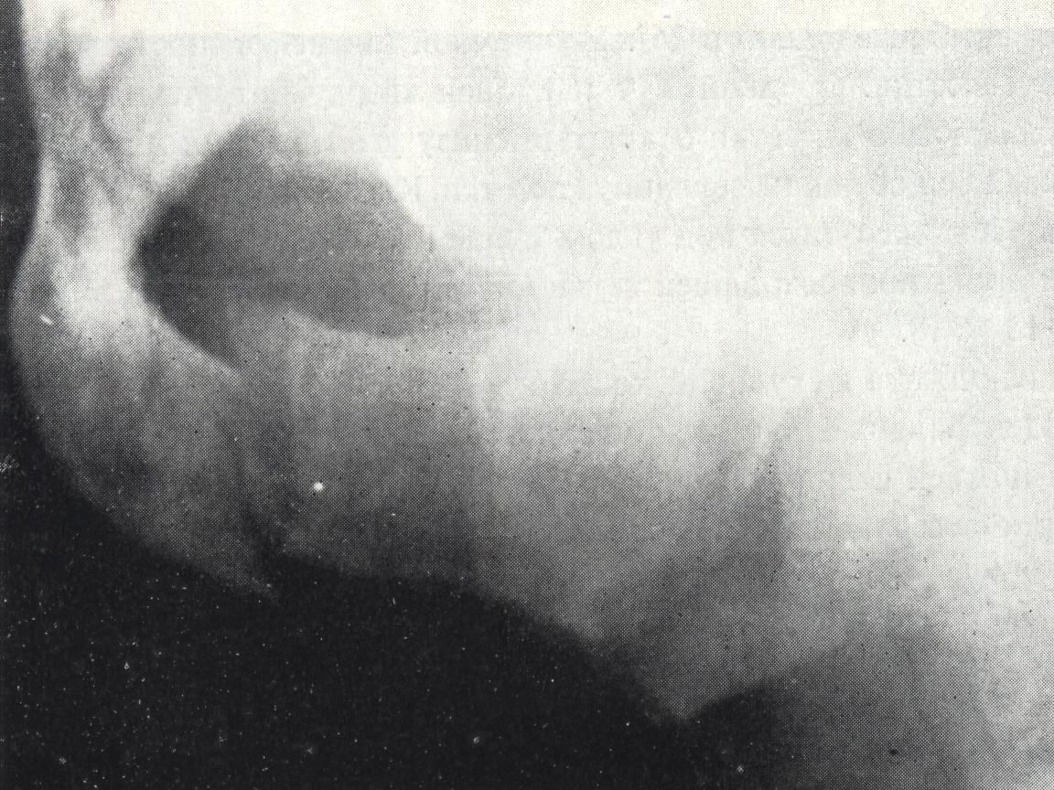
Symptom of glasses at fracture of maxilla

Direct and lateral projection of facial bones.


Axial and semi-axial projection of skull.


Lateral projection of mandible.
B. Tasks for self-control: 1. The patient asked to the doctor with the trauma of the nose, resulting in sports training. After examination doctor diagnosis nasal bone fracture without displacement of fragments. How to classify this injury etiology, given that the patient worked as a sports coach? (Answer: work injury).
2. Worker got hit the board in the region of upper jaw during the home renovation at the weekend. Doctor diagnosis contused wound of the upper lip, partial dislocation of the maxillary incisors. Qualify as an injury received in such circumstances? (Answer: consumer injury).
3. The patient complained of pain and impaired chewing due blow to the angle of the lower jaw to the right, headache, nausea. Set a preliminary diagnosis of fracture of the lower jaw. What additional studies should be carried out for the diagnosis? (Answer: X-ray, consultation neurologist).
C. Materials for test control. Test tasks with the single right answer (a=II):1. What damage is izolated? A. Burn of face and neck. B. Fracture of maхilla and mandible. C. Fracture of mandible. D. Wound of check and inframandible area. E. Fracture of nose and concussion of cerebrum. (Correct answer: С).
2. What damage is combined? A. Wound of check. B.Wound of chine. C. Fracture of nose and concussion of cerebrum. D. Fracture of maхilla and mandible. E. Fracture of mandible and termical burn of face. (Correct answer: Е).
D. Educational tasks of 3th levels (atypical tasks): 1. A patient with an open fracture of the body of the mandible is brought to Maxillofacial department from the place of accident. The general state of the patient is moderate severity, at anamnesis of desease: unconsciousness for a few minutes and vomiting. The patient complains of nausea, weakness, pain in the left side of the abdomen. Blood pressure – 100/60 mm Hg. Art., pulse 100 beats / min., weak filling. Under the block anesthesia maxillo-facial surgeon produced reposition of fragments and fiхated their by Tigershtedt’s tires with toe loops, and then appointed a consultation of therapists. Find the mistakes in the actions of the doctor: (Answer: the victim have the complex trauma with signs of traumatic shock, in this state surgery manipulation performed due bleeding or asphyxia only, should be consultation of surgeon traumatologist and a neurologist).
2. The patient complains of pain in the angle of the lower jaw at chewing, headache, dizziness, nausea. It is established that the patient was beaten 24 hours ago, there was a loss of consciousness for a few minutes. After clinical and radiographic examination, the doctor ruled out the diagnosis of a fracture of the lower jaw, doctor appointed Ketanov and inspected in 3 days. What is wrong in the actions of the doctor. (Answer: at this clinical picture neurologist consultation was necessary).
3. At examination of the victim dentist diagnosis bilateral fracture of the lower jaw in the chin with a displacement of bone fragments. After that doctor perfomed pressing bandage for transport immobilization. During transport, the patient showed signs of respiratory disorders. What type of asphyxia occurred in the patient and what is wrong in the actions of the doctor? (Answer: compressive bandage is contraindicated in fractures of the lower jaw with displacement, as it leads to further displacement of bone fragments. This patient had dislocation asphyxia).
Theme 2. SUBJECT AND GOALS OF MILITARY STOMATOLOGY. ORGANIZATION OF SURGICAL AID FOR MAXILLOFACIAL VICTIMS AT PEACE TIME AND EXTREME CONDITIONS. MILITARY-MEDICAL DOCTRINE. GENERAL PRINCIPLES OF ORGANIZATION, CONTENT AND RENDERING OF MEDICAL AID, STAGES OF MEDICAL EVACUATION AND MEDICAL SCREEN OF WOUNDED IN MAXILLOFACIAL AREA.
ACTUALITY OF THEME: The military stomatology is a section of the military medicine which task is the organization and granting all types of the stomatologic aid to staff of Armed forces of Ukraine in the conditions of peace and a war time. Knowledge of features of gunshot and not gunshot wounds, diseases of tissues and bodies of a maxillofacial site, the organization of medical care, prevention of complications and treatment wounded in the conditions of extreme situations and a war time - a direct duty of the stomatologist. The knowledge of the principles of medical sorting, volume of medical care at stages of medical evacuation of the wounded will allow the stomatologist to organize rationally providing medical care to the maxillofacial wounded.
2. СONCRETE AIMS: 1.1. To analyze provisions of the military-medical doctrine in historical aspect. 1.2. To explain basic provisions of the military-medical doctrine. 1.3. To offer forces and means of a health service for rendering the stomatological surgical aid to wounded in maxillofacial area. 1.4. To classify the basic principles of the organization, content and rendering of medical aid at stages of medical evacuation by the wounded in maxillofacial area. 1.5. To treat basic provisions of a medical deontology and medical ethics at assistance to wounded in maxillofacial area. 1.6. To draw the scheme of medical screen of wounded in maxillofacial area. 1.7. To analyze the principles of medical screen of wounded in maxillofacial area. 1.8. To make the scheme of medical evacuation of wounded in maxillofacial area.
3. BASIC KNOWLEDGE, ABILITIES, SKILLS, WHICH are NECESSARY FOR STUDY THEMES (intradisciplinary integration).
|
Names of the previous disciplines |
The received skills |
|
1. Medicine of catastrophes |
To organize rendering the first medical, pre-medical and first medical aid to wounded. |
|
2. Topographic anatomy and operative surgesy |
To apply knowledge of surgical anatomy of the head and a neck. To represent schematically a surgery technique at assistance to maxillofacial wounded. To show practical skills of imposing of different types of seams at aid to wounded with traumatic damages of maxillofacial localization. |
|
3. Surgical diseases |
To describe the history of illness of wounded with traumatic damages of maxillofacial localization. To show skills of carrying out primary surgical treatment (PST) of wounds of maxillofacial area and a neck. To define a condition in which there is a wounded with traumatic damage of maxillofacial area (traumatic shock, hemorrhagic shock, etc.). To define character of a gunshot wound, to examine and survey wounded, to define sequence and an order of rendering medical care to the wounded, an order and sequence of evacuation of the wounded. |
4.5. TYPES OF INDIVIDUAL WORK OF STUDENTS DURING PREPARATION TO LESSON.
4.1. Basic therms, parametrs, characterics whith are nessesery during preparation to lesson.
|
Therm |
Definition |
|
1. Military-medical doctrine.
|
It is set of scientifically reasonable principles defining system and methods of medical ensuring operations of armies in specific historical conditions, containing means and methods of the armed fight, level of medical science and practice, a condition of forces and means, and also features of battle fields. |
|
2. Medical situation. |
It is set of factors and the conditions characterizing forces and means of a health service, the contents and volume of future work, a sanitary and epidemic condition of provided armies and the area of their operations, and also other factors and conditions which can affect preparation and a course of medical providing armies. |
|
3. Medical sorting. |
This distribution of victims and patients to groups, proceeding from needs for priority and uniform actions (medical, preventive, evacuation) in a concrete situation. Even two victims who have arrived at the same time, require carrying out medical sorting. When carrying out medical sorting hyper diagnostics is allowed. Sorting basis: uniform ideas of diagnostics, medical actions and forecasts of results of treatment. Sorting purpose: to provide timely rendering medical care to the maximum number of victims in optimum volume. The condition of injured children and pregnant women without visible damages is always estimated as heavy, evacuation and assistance is carried out at first the stage. |
|
4. Stages of medical evacuation.
|
Medical aid stations and the medical institutions developed on ways of evacuation with a task of reception, medical sorting of victims and patients, rendering to them medical care, treatment and their preparation for further evacuation; in medicine of catastrophes – expansion of only two stages is provided, as a rule; the first – groups of the first medical care, the second – hospital base in military medicine. |
4.2. Theoretical questions for lesson: 1. Organizational structure of health service of Ukrainian Armed forces; 2. Structure of stages of medical evacuation. 3. Equipment of stages of medical evacuation. 4. Problems of stomatology of extreme situations. 5. Types of medical care. 6. Forces of a health service of Armed forces of Ukraine according to types of medical care. 7. Means of a health service of Armed forces of Ukraine according to types of medical care.
4.3. Practical skills for lesson: 1. To get skills of the organization of rendering medical care maxillofacial wounded at stages of medical evacuation. 2. To get skills of medical sorting of the maxillofacial wounded. 3. To acquire skills of providing medical care maxillofacial wounded at stages of medical evacuation. 4. To carry out a kuration of the maxillofacial wounded.
5. TABLE OF CONTENTS OF THEME:
Principles of military doctrine (time of Second World war)
1. All gunshot wounds are initially bacterial infected.
2. The only reliable method of the prevention of development of an infection is early surgical treatment of wounds which should as soon as possible.
3. Early surgical treatment of wounds is demanded by the greatest part of wounded.
4. The prognosis of treatment and consequence of wound is the best if early surgical treatment of a wound is carried out.
5. The volume of medical care, choice of medical actions and order of evacuation depends not only on especially surgical indications, but, mainly, on a military and medical situation.
Medical situation is the number of wounded who get on this stage, their condition, number of surgeons, existence transport, medical equipment and so forth means.
In the Great Patriotic War profile of medical care by the maxillofacial wounded was approached to a front line. Even at the advanced stages - medical care in the conditions of a regiment appeared taking into account features of this group of wounded (a thirst satisfying, imposing of gauze and standard bandages and so forth).
There is actual a problem of treatment of victims with burns of face and with combined injures.
At the combined radiation injures there are peculiar features of a current both radiation illness, and wound process.
Development of anesthesiology and resuscitation opened new opportunities in fight against traumatic shock and expanded borders of surgical treatment of wounded in maxillofacial area.
At explosion of a ball bomb from different places of the spherical case 300 balls take off (diameter of 5,56 mm, weight of 0,7 g), the bomb has big striking force and causes emergence of multiple wounds. Wounds are very life-threatening and are difficult for treatment.
Bullets of caliber of 5,56 mm at hit they overturn and move to tissues in the cross direction, causing in depth and in the field of an exhaust outlet big destruction of tissues. There are also artillery shells, each of which is filled with small arrows (length of 3-4 cm, thickness of 0,1-0,2 cm) in quantity to 10 thousand.
The above and other new types of weapon are characterized by their damaging shells is less on diameter and easier on weight in comparison with last, but they possess enormous initial speed (from 700 to 1500 m/s). Emergence of wound channels often also has a talk with the freakish directions, and considerable damages of tissues and organs.
In this regard the principles of the organization of help to wounded in modern conditions of war have to change.
Use of the nuclear missile weapon is accompanied by emergence in very short periods of mass sanitary losses in a wartime and in front areas in quantities which are exceeded considerably by that took place in the period of the Great Patriotic War.
The structure and nature of fighting damages will change also in these conditions: on the first place there will be burns, traumas and the combined damages (mainly radiation), the specific weight of seriously wounded (shock, a crush-syndrome, radiation injuries) will increase. Gunshot wounds thus lose the dominating value.
At mass receipt of victims there is a need for evacuation maneuver which has the purpose dispersal of victims between several stages and which is carried out on the basis of careful sorting. Overloads of the next to the front (the defeat center) stages at which there is the first medical assistance, quite often are involved by reduction of indications to intervention (reduction of volume of medical care). In these cases it is necessary to be limited only to actions which provide elimination of direct threat of life or heavy complications (the help according to vital indications).
Important element of the organization of surgical work of each stage of medical evacuation is the "conveyor" principle (O.O.Vishnevsky) according to which all process of medical aid e to each victim is subdivided into some parts, is carried out in the different ways by different doctors (removal of a bandage, surgical treatment, imposing of tires and bandages and so forth).
The conveyor principle promotes increase of capacity operational and dressing.
Modern provisions of the military-medical:
the volume of medical care, order of evacuation and choice of medical actions depend not only on especially medical indications, but also, mainly, on fighting and medical circumstances;
creation of system of medical and evacuation actions is carried out with the maximum reduction of quantity of stages of medical evacuation;
objective assessment of weight of wound and condition of the wounded introduction of criteria of the forecast of treatment;
preservation of uniform approach to treatment of wounds;
priority value of emergency medical service actions, treatment of shock and to blood loss completion at all stages of medical evacuation;
specialization of the surgical help;
accurate organization and sequence in rendering the surgical help at the combined, multiple and combined injures;
increase in a role of the reanimatological and anesteziological help at treatment of wounded at stages of medical evacuation.
The stomatologic help in a wartime will be organized by the Head Military-medical department of the Ministry of Defence of Ukraine through the Chief stomatologist of Armed forces. At military-medical management of each front there is a regular chief stomatologist of the front, he is a deputy chief surgeon of the front. The military-medical service of Ukraine is valid also means which allow qualitatively and to render in due time medical care by the wounded in maxillofacial area in the battlefield and at stages of medical evacuation.
The help to wounded in maxillofacial area which appears in the battlefield in the center of mass sanitary losses, is called as the first medical care. It appears in the form of most and mutual aid or shooters-hospital attendants and sanitary instructors, and also staff of divisions who carry out rescue efforts. It should be noted that the self-help among wounded in maxillofacial area appears very seldom because of complexity of definition of localization, the amount of damage and insolvency of imposing of a bandage on face of victim. For most and mutual aid all military personnel is provided with individual prophylactics and rendering medical care. Them treat:
package the dressing individual;
first-aid antigas kit;
individual first-aid kit ;
tablets for water disinfecting (Pantotsid, Akvasept).
Shooters-hospital attendants and sanitary instructor for search and carrying out of wounded, rendering the first medical care have special equipment:
Sanitary bag;
bag the medical military;
violent and special medical strap.
The pre-medical (medical assistant's) help appears the paramedic of a battalion in close proximity to a wound place, on the medical aid station of a battalion (MASB). It supplements the first medical care. However, possibilities of the paramedic on assistance, including elements of the stomatological help, much wider. Organic equipment of a medical aid station of a battalion, and also medical equipment of staff is applied to rendering the medical assistant's help. To complete organic equipment of MASB belong:
medical assistant's field;
sterile dressing means;
tires;
medical military bag;
bags of hospital attendants;
the first-aid kit military, and also, medical and medical means, devices (the device of artificial ventilation of lungs a portable, oxygen inhaler of KI-4, the mental tire with head bandages to them, we send for wounded to the head, a cape medical on 10 people, etc.). It is necessary to emphasize that the medical aid station of a battalion in defense when it works at a place, carries out functions of a stage of medical evacuation.
In a medical company of crew (MCC) there is the first medical and qualified aid. In structure MCC except doctors of an all-surgical and therapeutic profile is the atomatology. Main task of stomatologist of MCC is rendering the stomatological aid to regiment staff. During operations the stomatologist works as a part of an operative-dressing platoon in operational tent or as a part of sorting and evacuation office in dressing tent for lightly wounded, renders together with doctors of an all-surgical profile the first medical and qualified medical care, including the stomatologic. On MCC there can be patients and wounded for treatment with term of recovery of 3-5 days.
Specialized medical care to persons with damages and wounds of maxillofacial area render in maxillofacial offices of specialized hospitals for wounded in the head, neck and a backbone, in hospitals for treatment lightly wounded, and also in stomatologic offices of other hospitals of base of the front in which wounded with damages of maxillofacial area are on treatment concerning the main, more essential wound.
The significant part is assigned to stomatologic group of the front. The stomatologic group of the front is created for:
rendering the specialized help to maxillofacial wounded and patients who are on treatment in front hospitals;
rendering the stomatologic and dentoprosthetic aid to staff of parts, connections and establishments;
rendering the advisory help to dentists of medical divisions, parts and medical institutions.
The stomatologic group is headed by the doctor-stomatologist. Group structure: management; main divisions; providing divisions.
The main divisions, in turn, have also three divisions: stomatologic office - basic (3 stomatologist); dentoprosthetic office with dentoprosthetic laboratory (3 orthopedists, 5 dental technicians); ten mobile stomatologic offices (in each 2 stomatologist and the dental technician).
Mobile stomatologic offices go to medical institutions of the advanced hospital base for assistance to maxillofacial wounded, oral cavity sanitation by the serviceman, the patient and the wounded. These offices can go to connections, parts where there are no regular stomatologists for planned work on sanitation and oral cavity prosthetics.
All listed forces and means of a health service which carry out rendering the stomatologic help to maxillofacial wounded, at their rational and clever use, allow qualitatively and to carry out objectives in due time.
Medical sorting is a distribution of wounded and patients on groups and signs of requirement for uniform medical and evacuation and preventive actions according to medical indications, volume of medical care and the accepted order of evacuation.
Medical sorting of wounded is carried out by doctors of the sorting and evacuation office (SEO) of a medical company of crew. Main objective of medical sorting in SEO is selection from the general flow of wounded and patients who need medical care in MCCr, and, first of all, in the urgent medical and qualified medical care and sanitary processing, and also, wounded and patients to whom the qualified medical care can be delayed and which in these conditions are subject to further evacuation to the back without their direction in other functional divisions MCC.
If on MPP many wounded arrive, in sorting and evacuation office two crews one of which heads the doctor, and the second – the most skilled paramedic (nurse) can be created.
Wounded in maxillofacial area share on such groups:
І. Proceeding from need for sanitary processing and need for isolation: need partial sanitary processing; are subject to isolation; don't need sanitary processing and isolation.
II. Proceeding from need for medical care, place and sequence of its rendering:
1. Need rendering medical care in the dressing.
2. Don't need medical care or need medical care which can be rendered in a reception and classifying section. For wounded and patients who are subject to the direction in dressing, the turn – first of all, in the second turn is appointed.
III. Wounded and patients who don't need the medical help on MPP, and also what received it, are distributed on the following groups:
evacuations are subject;
are subject to return to the divisions.
In the course of medical sorting which is carried out on the sorting platform, all wounded and patients divide into groups:
In the first group enlist wounded who needs urgent actions of the first medical assistance (wounded with bleeding, asphyxia, shock, etc.). Them send directly to the dressing.
To the second group carry wounded then medical care can be rendered in a reception and classifying section (for example, injured with the closed fracture of the top jaw) at a satisfactory general condition of wounded who needs to give drink by means of a poilnik from the rubber tube put on his nose). After performance of the simple actions stated above this group of wounded is subject to the direction in SMB or SMG.
To the third group carry those wounded who without assistance on MPP go on the following stage of medical evacuation (for example the victim with the closed change of a malar arch and nose bones in the absence of bleeding).
To the fourth group carry lightly wounded which are subject to return to parts after rendering medical care to them (for example, the victim with wound of soft tissues of a face without the expressed hematoma).
To the fifth group carry those wounded and patients who are in an agony, or have traumas incompatible with life (for example, wounds of face and neck with a rupture of an internal carotid and noncompensated blood loss).
To clothes of the wounded or the patient who ate medical sorting, attach sorting brand in which specify, to what functional division it is necessary to send it. The sorting brand is the main reference point for hospital attendants-porters and the medical personnel.
6. MATHERIALS FOR SELF-CHECKING:
А. Questions for self-checking: 1. Types of medical aid. 2. Means of medical service. 3. Principles of medical sorting. 4. Organization of medical aid for maxillofacial wounded at stages of medical evacuation.
B. Tasks for self-checking: 1. The wound of the left cheek at wounded polluted by radioactive materials. There is bandage from a dressing package on a wound the, is carried out pain relief and antibacterial therapy. At what stage of medical evacuation it is necessary to replace a bandage? (Answer: at each stage of evacuation)
2. The general condition of wounded is moderate severity, consciousness is kept, skin pale with a cyanosis shade, lips cyanotic. Breath is complicated, 29 in a minute. There is a suspicion on existence of dislocational asphyxia. At what stage of medical evacuation wounded its tongue will be stitched by a silk ligature? (Answer: at a medical aid station of a battalion).
3. Two wounded come in hospital with wounds of the face, one – from a zone of radiation pollution are delivered. Who from them needs to carry out wound PST first of all? (Answer: wounded with the combined meсhano-radiation defeat).
C. Matherials for test control. Test tasks with one right answer (α = II):
1. What frequency of gunshot wounds of face and jaws by Great Patriotic War experience: A. around 1%; B. around 4%; C. around 50%; D. around 60%; E. around 90%. (Correct answer: В).
2. What is the mean of military-medical doctrine? A. Stages treatment of the military personnel and dispensary supervision; B. evacuation of wounded in rear country hospital; C. evacuation wounded out of limits of the center of defeat; D. system of stages treatment with evacuation to destination; E. rendering the specialized aid to victims in the war zone and return them in a system. (Correct answer: D).
3. Types of medical care in a wartime: A. mutual aid, first medical aid, specialized aid; B. first medical aid, pre-medical aid, first medical aid, qualified medical aid; C. pre-medical help, first medical aid, qualified medical aid, rehabilitation; D. first medical aid, medical aid, surgical aid, specialized aid; E. first medical aid, pre-medical care, first medical aid, qualified medical aid, specialized medical aid. (Correct answer: Е).
D. Educational tasks of 3th levels (atypical tasks):1. The wound of the left cheek at wounded polluted by radioactive materials. There is bandage from a dressing package on a wound the, is carried out pain relief and antibacterial therapy. At what stage of medical evacuation it is necessary to replace a bandage? (Answer: at each stage of evacuation).
2. The general condition of wounded is moderate severity, consciousness is kept, skin pale with a cyanosis shade, lips cyanotic. Breath is complicated, 29 in a minute. There is a suspicion on existence of dislocational asphyxia. At what stage of medical evacuation wounded its tongue will be stitched by a silk ligature? (Answer: at a medical aid station of a battalion).
3. Two wounded come in hospital with wounds of the face, one – from a zone of radiation pollution are delivered. Who from them needs to carry out wound PST first of all? (Answer: wounded with the combined meсhano-radiation defeat).
Theme 3. GENERAL CHARACTERISTIC, CLINICAL COARCE, DIAGNOSTIC OF GUNSHOT WOUNDS AND DAMAGES OF SOFT TISSUES, BONES OF FACE IN PEACE TIME: CLASSIFICATION, CLINICAL FEATERS, DIAGNOSTIC, MEDICAL AID, METHODS OF SURGICAL TREATMENT OF WOUNDS OF SOFT TISSUES OF FACE. INFLUENCE OF MALFUNCTION OF FACE ESTETIC AT PSYHIC OF WOUNDED. SELF WORK – PRACTICAL SKILLS OF FORMING OF SOFT BANDAGES.
1. ACTUALITY OF THEME: Damages of maxillofacial area at pease time and its treatment is very interesting and difficult branch of surgical stomatology. The insufficient awareness of the doctor in questions of surgical processing of such wounds leads to negative consequences in the form of a disfiguration of appearance of the patient, violation of such functions as speech, breath, chewing and so forth. For this reason the stomatologist has to know well features of surgical processing of such damages.
2. SPECIFIC GOALS: 1.1 . To analyze clinical manifestations of traumatic damages of maxillofacial area at peace time. 1.2. To explain etiological and pathogenetical factors of emergence of traumatic damages of maxillofacial area at peace time. 1.3. To offer the plan of inspection of victims with a trauma maxillofacial area. 1.4. To classify traumatic damages of maxillofacial area. 1.5. To treat the principles of diagnostics and treatment of traumatic damages of maxillofacial area at peace time. 1.6. To draw the graphological scheme of lesson. 1.7. To analyze results of laboratory and instrumental investigations. 1.8. To make the scheme of treatment of patient with trauma of maxillofacial area.
3. BASIC KNOWLEDGE, ABILITIES, SKILLS, WHICH are NECESSARY FOR STUDY THEMES (intradisciplinary integration).
|
Names of the previous disciplines |
The received skills |
|
1. Topographic anatomy |
To define anatomic areas of damage. |
|
2. General surgery |
To define type of hemorrhage |
|
3. Internal diseases |
To define the diagnosis of syncope, shock |
|
4. Pharmacology |
To prescribe the scheme of medical treatment |
|
5. X-ray research |
To define necessary method of investigation |
4.5. TYPES OF INDIVIDUAL WORK OF STUDENTS DURING PREPARATION TO LESSON.
4.1. Basic therms, parametrs, characterics whith are nessesery during preparation to lesson.
|
Therm |
Definition |
|
1. Primiry surgical treatment of wound |
This is first intervention of wound which is carried out for the purpose of prevention of development of a wound infection and creating favorable conditions for wound healing. |
|
2. Secondary surgical treatment of wound |
This is secondary intervention after primiry surgical treatment of wound |
4.2. Theoretical questions for lesson: 1. defined notion “abrasion”“contution”, “wound”. 2. The layerwise structure of soft tissues at different thopographic areas. 3. Blood supply of face. 4. Innervation of face. 5. Classification of wounds. 6. Clinic of damages of soft tossues and bones of maxillofacial area at pease time. 7. The features of diagnostic of traumatic damages of soft tissues of maxillofacial area at pease time. 8. Classification of bleeding. 9. Methods of temporal artherial stop bleeding. 10. Final stop bleeding. 11. What is “primary surgical treatment of wound”. 12. Features of primary surgical treatment of wound. 13. Types of primary surgical treatment of wound. 14. Types of sutures. 15. Methods of temporal immobilization at damages of facial bones.
4.3. Practical skills for lesson: 1. To make digital occlusion of general carotid arthery. 2. To make the mental sling bandage.
5. TABLE OF CONTENTS OF THEME:
Combine
Injures
of
soft tissues at peace time


Open
(scratch,
wounds)
Complex
Close
(contusion)
Isolated
Wounds

Etiology

Profundity
of injury
Type
of vulnerary projectile
Penetrating
to oral cavity
Cut


Superficial
Penetrating
Gunshot
Chopped


Deep
Non-penetrating
Non-gunshot
Compound

Lacerated

Bruising

Stab

Degloving
Bite
Injures
of
bones at peace time

Contusion
Fracture

-
open;
-
close;
-
one-side;
-
two-side;
-
direct;
-
non-direct;
-
single;
-
double;
-
multiple;
-
line;
-
comminuted;
-
with displacement;
-
without
displacement;
-
traumatical;
-
pathological.
Bleeding
control


Continuous
Temporary


Digital
occlusion
Ligation
of vessels
application
of tourniquet

Tamponade
Vessel-suture
Clipping
in wound
Compressive
bandage
Principles
of surgical treatment of wound

Early

Single-step

With
using of primary plastic

Exhaustive
Radical
Types
of surgical treatment of wound

Early
primary surgical treatment of wound
Before
signs of inflammation (to 24 hours after injures)
Primary
blind suturing
Deffered
primary surgical treatment of wound
Before
signs of inflammation (from 24 to 72 hours after injures in case of
using of antibiotics)
Primary
deffered
suturing
Late
primary surgical treatment of wound
First
operation in advancet inflammation (after 72 hours after injures)
Primary
deffered
suturing
Secondary
or reccurent surgical treatment of wound
Secondary
operation after primary surgical treatment
Secondary
early or late suturing
Kinds
of sutures
Time
of suturing
Way
of suturing
Kind
of suture material
Primary
blind sutures
Primary
deffered
sutures
Secondary
sutures
Early
Late
Nodular
Continuous
Plate
From
naturals materials
Resorbable
Not-resorbable
From
synthetic materials


PSIHO-EMOTIONAL VIOLATIONS AT DAMAGES OF MAXILLOFACIAL AREA
Psycho-emotional violations which are observed at victims with a trauma of maxillofacial area, are caused by injury of a brain, and emotional reaction to trauma and the related disfiguration of the face.
Mental disorders which arise in connection with an injury of a brain differ considerable polymorphism. They clinical picture depends on localization of damage of brain.
Direct result of injury of a brain are deep frustration of consciousness in the form of a sopor or a coma. The exit from this condition comes not at once. Usually patients are in a condition of devocalization for long time and remind people who finally didn't wake up: they don’t understand at once questions which are raised, long repeat one and a touch the phrase, happen whimsical and whining. Thus patients also complain of a headache, dizziness, noise and weight in the head, nausea; in some cases there is a vomiting. Weakness of storing, fast exhaustion of attention, causeless mood swings are noted. All these phenomena connected with concussion, gradually abate and by the end of the second week usually disappear.
However, in certain cases, after an exit from a coma, there are signs of a delirious condition of consciousness: patients don't remember people around, aren't guided in a situation, don't supervise the behavior. Except disorder of orientation, there are hallucinations, mainly visual, the alarm, fear, develops motive excitement. Contents of hallucinations most often concern the subject closest to the patient: episodes of road incidents, the scenes connected with a profession and so forth. Duration of such condition 2 - 3 days though cases long a delirium after a trauma till 2 weeks.
In some cases sharp traumatic psychosis is characterized by signs of a peculiar twilight condition of consciousness. Into the forefront orientation violation, motive excitement with feeling of fear and unconscious alarm acts. It is promoted by premature transportation. That is why it is important to abstain at a heavy craniocereberal trauma from transportation of patients within 2 - 3 weeks.
That often meet in a wartime - a surdomutizm carry to sharp frustration of the psychological sphere also. This type of pathology is usually connected with an air contusion.
On character of emotional reaction to the received damage of maxillofacial area of victims it is possible to divide into two groups.
In the first group sharpness of reaction isn't proportional to weight of injury of a face that is connected with hypererethism of nervous system.
In the second group mental depression of victims corresponds to extent of functional frustration. Especially heavy frustration cause wounds of the face penetrated into an oral cavity with injury of jaws, tongue, big palate defects, lateral area of the face, a mouth floor and a chin site with a lower lip.
Expressiveness of mental depression depends also on such factors as a profession of the victim, education, the social status and so on.
Psychogenic frustration at patients with injury of a face and jaws proceed differently at different stages of a course of wound process, and are functionally reversible process. It is characteristic that persons who lost sight at wound, don't react at all to a disfiguration, even when absence, for example, a nose or lips is realized and without sight on functional violations.
At the heavy course of wound process with high temperature and intoxication phenomena, the dream owing to fatigue which interrupts only bandaging, food and oral cavity washings, promotes that mental depression of victims considerably decreases and cases of neurotic reaction arise seldom.
On the contrary, when the general provision of the patient satisfactory, consciousness is kept, intoxication is a little expressed, drowsiness is absent, and he is in chamber where others freely talk, eat not from a poilnik, smoke etc., mental depression and neurotic reaction are observed rather often.
As a whole, in development of psychogenic frustration the next moments matter:
• the mental trauma which has arisen at the time of wound at accurate understanding of the received disfiguration of face;
• mental trauma which the same repeats, at contact with people around, especially at the wrong behavior of the they rather to sick;
• psychotrauma which arises each time when the victim sees his face in a mirror;
• repeated mental trauma in connection with repeated expeditious reconstructive and cosmetic interventions;
• mental trauma in connection with loss of expressiveness of a facial expression or defects of speech (teachers, actors, lecturers, workers of brainwork);
• mental trauma in connection with problems in private life.
Usually at the time of release of the face of the victim from a bandage it has an irresistible desire to look at itself in a mirror. Very often it strengthens neurotic reaction which and without that takes place. At heavy damage, especially if the doctor couldn't prepare the patient or underestimated this moment, the impression can be very negative. The victim starts retiring, becoming reserved, refusing communication with relatives.
In pathogenesis of emotional shifts at a disfiguration of face the consciousness of ugliness becomes the reason of heavy mental depression which can lead to a depression, psychosis and even to suicide.
In some cases neurotic reaction at wounded in a face is possible also as a result of functional or organic changes which result from an injury of a brain. Therefore even in the absence of mental depression wounded in a face differently, not as at wound of other sites of a body, perceive both the condition, and result of treatment.
For prevention of development of serious psycho-emotional conditions the victim needs to provide consultation of the psychiatrist or the psychotherapist in due time. It is desirable to place such wounded in chamber with the victims having similar damages, in every possible way to support in it belief in recovery (psychotherapeutic conversations, communication with the patients who have already transferred recovery operations with satisfactory cosmetic effect, demonstrations of photos on which results of successful plastic surgeries, etc. are fixed). In hard cases it is necessary to provide continuous supervision for wounded or even to transfer it to a psychiatric institution.
6. MATHERIALS FOR SELF-CHECKING:
А. Questions for self-checking:
Damages of soft tissues and facial bones at pease time (by screw of helicopter).


B. Tasks for self-checking: 1. A victim came to maxillofacial department with damage of soft tissues of face, that was happened more then day ago in time of working with chain saw. Objective: There is big wound faulty form in left buccal and parotid area, its borders are impregnated by blood, wound connected with oral cavity. What is diagnosis and kind of surgical aid? (Answer: lasereted wound in left buccal and parotid area, connecting with oral cavity, late primary surgical treatment).
2. The wounded was hospitalizated after 2 days after damage. General condition is normal, there is a wound in left infraorbital area 2х1, 5 cm, without inflammation. What the kind of seam is necessary in this case. (Answer: primary late seam).
C. Matherials for test control. Test tasks with one right answer (α = II): 1. Where shod be ligated the carotid artery? A. Lower that artery thyroid superior. B. Between artery thyroid superior and artery lingual. C. Between lingual and facial artery. D. Higher then facial artery. E. In bifurcation of carotid artery. (Correct answer: B).
2. Primary surgical treatment of wound is carrying out: A. Not after than 4-6 hours after trauma. B. Not after than 6-8 hours after trauma. C. Not after than 8-10 hours after trauma. D. Not after than 12 hours after trauma. E. Not after than 24 hours after trauma. (Correct answer: E).
3. Where is put in first seam on face at surgical treatment of wound: A. In parotid-masseteral area. B. In buccal area. C. In mental area. D. Near natural foramens of face. E. Near ear. (Correct answer: D).
D. Educational tasks of 3th levels (atypical tasks):1. Patient was getting to maxillofacial department with complains of a headache, dizziness, pain in the top jaw, bleeding from a nose. Objectively: insignificant hypostasis of soft tissues in an upper lip, is defined a "step" symptom in the field of lower marge of an eye-socket from both parties, and also pathological mobility of the top jaw together with nose bones. The lower jaw is fixed to maxilla by Ayvi’s ligature wire across. Define the preliminary diagnosis and a mistake in the previous actions of the stomatologist. (Answer: fracture of the maxilla on midle type Le Fort; intermaxillary fixing by a ligature wire is contraindicated. ) .
2. The patient was getting to hospital in an unconsciousness by ambulance car in a reception in an irresponsible condition from a place of transport incident. There is the bandage impregnated with blood in the head. Tongue sinks down, breath complicated, pulse of 50 beats per minute, is intense, joint stock company of 100/60 mm. The rigidnost of muscles of a nape, tone of extremities on the right side normal is noted, with left - is sharply lowered. The pupil of the right eye is expanded. Establish the preliminary diagnosis, define an office profile to which it is necessary to send the victim. (Answer: open craniocereberal trauma; neurosurgical department).
3. The patient was getting to reception in 3 hours after a gunshot wound is delivered during hunting. According to accompanying, the victim lost a lot of blood. The condition heavy, consciousness is absent. On the head a bandage impregnated with blood, multiple damages of soft tissues of a face. Breath frequent, superficial, pulse of nitevideny 40 blows in min., the blood pressure is 70/10 mm. pupils are expanded. Make the preliminary diagnosis, define an office profile to which it is necessary to send the victim. (Answer: combined craniocereberal gunshot wound, hemorrhagic shock; neurosurgical department.).
Theme 4. GENERAL CHARACTERISTIC, CLINIC, DIAGNOSTIC OF GUNSHOT WOUNDS AND DAMAGES OF SOFT TISSUES AND BONES OF FACE IN EXTRYM CONDITIONS: CLASSIFICATION, CLINIC, DIAGNOSTIC OF DAMAGES AT STAGES OF MEDICAL EVACUATION. PLASTIC SURGERY AT TREATMENT OF DAMAGES OF FACE. MODERN GUNSHOT WOUND, ITS TREATMENT.
1. Actuality of theme: Knowlegess of general characteristic, clinic, diagnostic and complications of gunshot damages of maxillofacial area and traumatic illness is necessary for stomatologist for rational organization of medical aid for victims at pease, war time and extrime conditions.
2. SPECIFIC GOALS: 1. To analyze clinical manifestations of traumatic damages of maxillofacial area at extreme condition. 2. To explain etiological and pathogenetical factors of emergence of traumatic damages of maxillofacial area at extreme condition. 3. To offer the plan of inspection of victims with a trauma maxillofacial area. 4. To classify traumatic damages of maxillofacial area at wartime. 5. To treat the principles of diagnostics and treatment of traumatic damages of maxillofacial area at extreme condition. 6. To draw the graphological scheme of lesson. 7. To analyze results of laboratory and instrumental investigations. 8. To make the scheme of treatment of patient with trauma of maxillofacial area.
3. BASIC KNOWLEDGE, ABILITIES, SKILLS, WHICH are NECESSARY FOR STUDY THEMES (intradisciplinary integration).
|
Names of the previous disciplines |
The received skills |
|
1. Topographic anatomy |
To define anatomic areas of damage. |
|
2. General surgery |
To define type of hemorrhage |
|
3. Military surgery |
To define the character of gunshot wound, medical aid, stages of evacuation. |
|
4. Pharmacology |
To prescribe the scheme of medical treatment |
|
5. X-ray research |
To define necessary method of investigation |
4.5. TYPES OF INDIVIDUAL WORK OF STUDENTS DURING PREPARATION TO LESSON.
4.1. Basic therms, parametrs, characterics whith are nessesery during preparation to lesson.
|
Therm |
Definition |
|
1. Primary suture |
This is suture, which is used before development of inflammation. |
|
2. Secondary suture |
This is suture, which is used after secondary surgical treatment. |
4.2. Theoretical questions for lesson: 1. General characterizes of gunshot wounds. 2. Features of gunshot damages of maxillofacial area. 3. Classification of gunshot damages of maxillofacial area. 4. Methods of diagnostic of gunshot damages of maxillofacial area. 5. Traumatic illness. 6. Periods of traumatic illness. 7. Features of traumatic illness at maxillofacial wounded. 8. Classification of complication of gunshot damages of maxillofacial area. 9. Clinic of immediate complications at gunshot damages of maxillofacial area. 10. Clinic of early complications at gunshot damages of maxillofacial area. 11. Volume of medical aid at stages of evacuation.
4.3. Practical skills for lesson: 1. To make the bandage “cap”. 2. To make the 8-like bandage.
5. TABLE OF CONTENTS OF THEME:
Classification of damages of maxillofacial area at war-time (by Kabakov)
I. Mechanical damages of upper, middle, low and lateral parts of face
Localization:
А. Damages of soft tissues:
1. isolated with damaged of:
а) tongue; b) salivary glands; c) big nerves; г) big vessels;
2. complex.
B. Damages of bones:
а) mandible; b) maxilla; c) zygomatic bones; d) nasal bones; e) two and more bones.
Character of damage: perforating, blind, tangent wounds;
Wounds witch penetrate in to nasal cavity, maxillary sinus.
Mechanism:
1. Gun-shot wounds: missile, shrapnel, ball-like and arrow-like elements.
2. Non-gunshot wounds: contused, stab, cut, chopped.
II. Combine damages.
III. Burns.
IV. Frostbites.
All damages can be isolates single, isolated multiple, complex single and complex multiple.
Anatomic, topographic and physiologic features of maxillofacial area
Good blood supply, that makes big blood lose, damages of big and magistral vessels;
Good lymphatic system, that makes quick forming of edema and high regenerative and contrainfectional possibilities of tissues;
Good innervation of area, that forms evident pain syndrome and shock, and paresis, paralyses of sensitive and motional nerves;
Mimic muscles, that forms discrepancy of appearance and severity of damages, because of gaping of wound and distortion of face;
Closeness of vital importance organs – brain, organ of vision, hearing, smell, witch forms the syndrome of mutual additional burden and complex traumas;
Trauma of salivary glands makes salivary cysts, fistulas, stenosis and obliteration of ducts;
Cellular areas can cause inflammatory complications;
Teeth and orthopedic constructions can be secondary vulnerary shells and can cause to asphyxia;
Micro flora of oral cavity can cause inflammatory complications;
Non-possibility of self food taking;
Non-possibility of using simple respirators.
Periods of clinical course of gunshot damages of soft tissues of face:
I period - 48 hours after damage – dominancy of traumatic edema in wound without sings of infectional inflammation. The most favorable period for primary surgical treatment and primary plastic operations.
II period – after 3 days after damage before visible granulations. There is inflammatory process in wound, infiltration of nearest tissues, exudation, purulence. Treatment is limitation of inflammatory process.
III period – granulation of wound. Treatment is acceleration of granulation growth. Possible to use early secondary surgical treatment.
IV period – epitelization and formation of scars.
Evacuation-stages system of treatment of victims (by Opel)
Field of hate (first medical aid):
Deliverance of victims under obstruction;
Temporal external hemostasis;
Use of aseptic bandage;
Position of wounded of face down to prevents asphyxia;
Deliverance of upper respiratory airways;
Using of anesthetics;
Using of respirators;
Slaking of thrist;
Using of antidotes to poisonous substance.
Medical office of battalion (paramedical aid):
Control and correction of bandages;
Using of analgesics and heart drugs;
Struggle with asphyxia;
Temporal hemostasis;
Using of antibiotics;
Using of contra-vomiting drugs;
Slaking of thrist;
Preparation to evacuation.
Optimal time for paramedical aid 1,5-2 hours after damage.
Medical office of regiment (first medical aid):
hemostasis;
elimination of all types of asphyxia;
transport immobilization at laceration wounds of soft tissues of face;
correction of bandages;
Using of antibiotics, analgesics and heart drugs;
Anti-shock actions;
Using of tetanus anatoxine at open wounds of maxillofacial area;
Slaking of thrist;
Filling of primary medical card;
Preparation to evacuation.
Optimal time for first medical aid 4-5 hours after damage.
Detached medical battalion (qualified medical aid):
elimination of all types of asphyxia;
final hemostasis;
Anti-shock actions;
Surgical treatment of lacerated wounds of face;
Nutrition of victims;
Preparation to next stage of evacuation.
All wounded at maxillofacial area should be inspected by stomatologist. Stomatologist ascertains the degree of wound, diagnosis and turn of medical aid.
I turn – maxillofacial wounded, who needs qualified medical aid at vital indications (shock, bleeding, asphyxia);
II turn – wounded with moderate disturbances of breath, speech and wounded with combine and complex damages;
Ш turn – wounded with blind wounds;
ІV turn – all other victims with easy damages.
Optimal time for qualified medical aid 5-12 hours after damage.
Special surgical field movable hospital (special medical aid):
Exhaustive medical aid at bleeding, shock, asphyxia;
final hemostasis and its prevention;
radical primary surgical treatment of wound of soft tissues of face and oral cavity;
final treatment of all wounds of soft tissues;
treatment of early complications and prevention of late complications;
early reconstructive operations with local tissues and method of free skin transplantation;
special care and nutrition of victims.
Victims, who has damages of soft tissues with big defects and disfiguration of face, malfunctions, who needs long treatment (more than 2 months) with using of multistage reconstructive operations are guided to special logistic hospitals.
6. MATHERIALS FOR SELF-CHECKING:
А. Questions for self-checking:
Gunshot splinter injury of mental area of mandible.


Before surgical treatment After surgical treatment
(there are lamellar sutures at wound.)
B. Tasks for self-checking: 1. Patient 45 years has salivary fistula at parotid area after gunshot damage. What kind of surgical treatment is useful if the scar is cuted out, at depth of wound make seam. And the wound was close layer-by-layer after it. (Answer: method of Sapozhnikov).
2. A wounded with perforated gunshot wound of soft tissues without damages of bones, big vessels and nerves was inspected by stomatologist at stage of qualified medical aid. What is the necessary treatment? (Answer: early surgical treatment of wound).
3. The wounded with tangent gunshot wound of maxillofacial area with big defect of right cheek was get to medical office of regiment. Blood pressure 95/65, mental confusion, moderate bleeding from wound. What the volume of first medical aid? (Answer: stop bleeding, compressive bandage, anti-shock therapy).
C. Matherials for test control. Test tasks with one right answer (α = II): 1. Fixation of tongue as first medical aid is used in case such asphyxia. A. Aspirational. B. Obturational. C. Dislocational. D. Stenotic. E. Valvate. (Correct answer: C).
2. Primary surgical treatment of gunshot wound is carrying out: A. Before than 4-6 hours after trauma. B. Before than 6-8 hours after trauma. C. Before than 8-12 hours after trauma. D. Before than 12-24 hours after trauma. E. Before than 48 hours after trauma. (Correct answer: D).
3. Medical sorting of wounded is become at such stage of evacuation: A. Field of hate. B. Medical office of battalion. C. Medical office of regiment. D. Detached medical battalion. E. Special surgical hospital. (Correct answer: C).
D. Educational tasks of 3th levels (atypical tasks):1. The patient, 23 years old, was getting to polyclinic to the surgeon-stomatologist with complaints to pain in a chin on the right, bleeding from an oral cavity, the complicated meal. Patient ago foll and hit a chin two days. He didn't lose consciousness. Objective: there is asymmetry of the face at the expense of hypostasis of soft tissues in a chin on the right. Opening of a mouth is a little limited. "Step" symptoms between 42 and 45, a rupture of a mucous membrane, pathological mobility of fragments of the mandible. Define the diagnosis, inspection volume, a type of a temporary immobilization. (Answer: fracture of the mandible in a chin on the right with shift fragments; X-ray research of the mandible; fundiform bandage).
2. The serviceman was getting to maxillofacial department of specialized hospital with a gunshot wound of maxillofacial area which is delivered more than days ago. Objectively: in a chin a wound of the wrong form of 3х5 cm in the depth of which the shattered bone is visible. Bleeding is absent, breath violation takes place. Establish the diagnosis; specify a mistake which was made at the previous stage of evacuation and a type of surgical processing which will be carried out. (Answer: gunshot fracture of the mandible in a chin; dislocation asphyxia; at the previous stage of evacuation the tongue wasn't fixated; the deferred primary surgical processing will be carried out).
3. The serviceman was getting to pecialized hospital in 42 hours after wound in a satisfactory condition. Objectively: there is a wound of the considerable sizes with defect of tissues, penentrate into an oral cavity in the left buccal area with obvious signs of an inflammation. Define a type of surgical treatment and sutures which will be imposed. (Answer: secondary surgical treatment, lamellar sutures).
Theme № 5. TRUMATIC ILLNESS: PATHOGENY, FEATURES AT DAMAGES OF MAXILLOFACIAL AREA.
1. Actuality of theme: Knowlegess of general characteristic, clinic, diagnostic and complications of gunshot damages of maxillofacial area and traumatic illness is necessary for stomatologist for rational organization of medical aid for victims at pease, war time and extrime conditions.
2. СONCRETE AIMS:1. To analyze reasons of traumatic illness. 2. To explain clinical features of traumatic illness at maxillofacial wounded. 3. To propose general characteristic of gunshot wounds of maxillofacial area, its complications. 4. To classify gunshot wounds of maxillofacial area, its complications. 5. To treat the principles of diagnostics of traumatic illness at maxillofacial wounded. 6. To draw the scheme of organization of treatment of wounded. 7. To analyze principles of prophylaxis of traumatic illness at maxillofacial wounded. 8. To make the scheme of treatment of patient with traumatic illness.
3. BASIC KNOWLEDGE, ABILITIES, SKILLS, WHICH are NECESSARY FOR STUDY THEMES (intradisciplinary integration).
|
Names of the previous disciplines |
The received skills |
|
1. Disaster medicine. |
To demonstrate organization of medical aid to maxillofacial wounded |
|
2. Operative surgery and topographic anatomy |
To use knowledges at surgical anatomy of head and neck. To make scheme of surgical treatment to maxillofacial wounded. |
|
3. General surgery |
To define the condition of victim with traumatic illness. To define the character of gunshot wound. To demonstrate practical skills at primary surgical treatment of wound. |
4.5. TYPES OF INDIVIDUAL WORK OF STUDENTS DURING PREPARATION TO LESSON.
4.1. Basic therms, parametrs, characterics whith are nessesery during preparation to lesson.
|
Therm |
Definition |
|
1. Traumatis illness |
It is the special clinical category, which is allowing to consider separate stages and complications (shock, the postshock period, infectious complications, the reconvalescence and rehabilitation periods), and all process as a whole to definition of an outcome of a disease. |
|
2. Traumatic shock |
This serious condition caused by a trauma, being accompanied the expressed violations of functions of vitals, first of all blood circulation and breath. It is characterized by two phases of a current – erectile (tension, active reaction of an organism to a trauma) and torpent (exhaustions of the main functions of life support). Shock isn't a nosological form. It represents a phase of a traumatic illness. |
|
3. Hemorrhagic shock |
This condition is similar on a clinical picture to traumatic shock. Arises owing to massive blood loss in case of a trauma (external bleeding) or at internal bleedings. |
|
4. Hypovolemic shock |
This condition is developing at considerable losses by an organism of plasma of blood and other liquids (the considerable burn surfaces, exhausting vomiting, diarrhea, etc.) when volume of circulated blood is directly decreases. |
|
5. The mutual additional burden syndrome |
This condition, is characterized by such symptoms: large quantity of the centers afferentny pathological (first of all painful) impulsation; large quantity of the centers of bleedings; violation of coordinating function of the central nervous system at heavy shock and a craniocereberal trauma; a large quantity of the centers primary, and then and a secondary necrosis of tissues that leads to intoxication development. |
4.2. Theoretical questions for lesson: traumatic illness, pathogeny, classification, prognosis, treatment, complications.
4.3. Practical skills for lesson: 1. To investigate the patient with trauma of maxillofacial area, make diagnosis, prescribe treatment. 2. Fill in medical documentation. 3. To make primary surgical treatment of wound. 4. To make the temporal immobilization of jaws.
5. TABLE OF CONTENTS OF THEME:
Polytrauma and traumatic illness of the maxillofacial wounded
Polytrauma is monotonous defeats of two and more anatomic areas at which violations of the vital functions of an organism. It is a heavy combined trauma which is accompanied by development of clinic of traumatic shock, that is, it is a shockogenic combined trauma. Polytrauma allocation in separate category is important in connection with certain features of such wounds. Combination of damages of several anatomic areas of a body - the pathological condition connected with emergence of a syndrome of mutual burdening. The essence of a syndrome is that each of damages worsens the general pathological situation and each concrete damage at a polytrauma proceeds more hard, with big risk of development of complications, than at the isolated trauma. Despite of a big variety of clinical options of a shockogenic combined trauma, at a polytrauma the traumatic illness which has the general regularities, its defining main features of pathogenesis and principles of medical tactics develops.
Traumatic illness– complex of symptoms of the injured patients with heavy, multiple damages; it is the pathological process predetermined by a heavy shockogenic mechanical trauma in which consecutive change of key factors of pathogenesis defines natural sequence of the periods of a clinical current. The concept about a traumatic illness was formed on the basis of the doctrine about traumatic shock. At the heart of a traumatic illness protective and reflex reaction of an organism, first of all, central nervous system, endocrine and other systems on action of the injuring agent, nervous impulses and absorptions of products of disintegration of tissues in the damage center, blood loss lies.
The clinical symptomatology and the course of a traumatic illness depends on localization, character and weight of damage.
Periods of traumatic illness:
The shock period - the period of sharp functional and system frustration which are predetermined by a heavy trauma, duration of 12-48 hours.
The early post-shock period - the period of threat of emergence of organ and polyorgan insufficiency - duration is 3-7 days after a trauma.
The period of infectious complications or big risk of their development (the period of a secondary immunodeficiency) - duration from 1 week - till 1 month.
The reconvalescence period - duration is from several weeks about several months.
A.V.Kaplan allocates such periods of a traumatic illness:
I - period of traumatic shock and bleeding;
II - the period of a heat of changes of a homeostasis (the general changes together with pathomorphological changes of the damaged tissues);
III - the reconvalescence period with development of conditions for reparative process of the damaged tissues and bodies.
Pathogenesis of a traumatic illness is predetermined by interaction of the traumatic agent with organism tissues owing to what there are their damages which influence different systems of an organism, thus the main links of pathogenesis include blood loss (the different volume, speed), specific disorders of functions of the damaged bodies, a hypoxia, a toxemia and a pain syndrome. As a result, there is a mono - or polyorgan insufficiency.
At a trauma of reaction of damage are replaced with protection reactions - a stress, or the general adaptation syndrome in reply to a trauma. Distinguish two phases of a metabolism after a trauma: the catabolism - lasts 2-4 days, is characterized by destruction, loss of proteins that not directed reduction of damages of structures and preservation of the main functions of an organism; anabolic the phase - lasts 1-2 weeks and more, for it renewal of the damaged structures and bodies and renewal of their function is inherent.
"Primary" changes result from direct anatomic defeat of body. In process of increase in power of injuring effect decreases weight of primary changes. For example bruise, gap, concussion.
6. MATHERIALS FOR SELF-CHECKING:
А. Questions for self-checking:
1. Fill in colunm:
|
№ |
Task |
Answer | ||
|
1. |
General characteristic of gunshot wounds. |
| ||
|
2. |
What is “traumatic illness”? |
| ||
|
3. |
Periods of traumatic illness. |
| ||
|
4. |
Clinical features of traumatic illness at maxillofacial wounded. |
| ||
|
№ |
Characteristic of phase of shock |
The name of phase of shock | ||
|
1. |
It is arises directly after extraordinary mechanical influence and characterized by motive and speech excitement of the victim, tachycardia, paleness of skin integument, increase systolic blood pressure, sometimes spontaneous urination and defekation. |
| ||
|
2. |
It is characterized by dispny, an oliguriya, increase of rectal-skin gradient of temperature, block of the victim, tachycardia, decrease of systolic blood pressure. |
| ||
|
Periods of traumatic illness. |
Answer | |||
|
First |
| |||
|
Second |
| |||
|
third |
| |||
|
fourth |
| |||
B. Tasks for self-checking: 1. Patient A., 42y was caught under blockage at quarry. The left half of face and left leg were smash by stones for 4 hours. Well-being is normal, blood pressure is 110/70 mm m c. Pulse 80 at minute. The doctor was at place when the patient was getting under blockage. The condition gets worse. Complaints become at strong pain at left half of face and leg. Patient is inconvenient. Skin is pallid. blood pressure is 80/50 mm m c. Silk pulse 130 at minute. Patient was getting to the hospital. What is the reason of worse of patient’s condition? (Answer: patient has traumatic illness (syndrome of mutual additional burden). Because of doctor’s mistake the “tourniquet” shock becomes).
2. Patient B., 51 y, was getting to hospital at 40 min after damage in bed condition. The fracture of maxilla Le Fort II, fracture of right VІ, VІІ, VІІІ ribs traumatic shock are diagnosed. What the succession of measures at first hours of treatment? (Answer: to get patient out from shock: use anti-shock, analgetic and blood drugs. Patient should be consulted by maxillofacial surgeon, neurosurgeon, traumatologist. It is possible to use the apparatus of Zbarazh).
3. Patient B., was getting to hospital after fight. Behavior of patient is inadequate. Vomiting was twice. Skin is pallid, hematomas are at infraorbital areas, there are abrasions. Nasal bleeding was. The middle face is long. Blood pressure is 100/60 mm m c, pulse 104 at min temperature is normal. What is diagnosis? (Answer: traumatic illness, complex trauma: close brain damage, fracture of maxilla Le Fort ІІ).
C. Matherials for test control. Test tasks with one right answer (α = II): 1. Typical features of trauma of maxillofacial area are: А. disfiguration of face; В. Discrepancy of appearance of victim to hardness of trauma; С. Complex trauma of head and brain; D. traumatic shock; Е. All answers are correct. (correct answer: Е).
2. Specific signs of traumatic illness: А. surprise of beginning, etiological agent and morphological substrate (damage of organ or tissue); В. surprise of beginning, absence of latent period, malfunction of mastication; С. Traumatic shock, massive blood loss; D. Hemorrhagic shock, pain syndrome; Е. surprise of beginning, absence of latent period, etiological agent and morphological substrate (damage of organ or tissue), pain syndrome, multiple organ failure. (correct answer: Е).
3. Non-specific signs of traumatic illness: А. Traumatic shock, stress; В. Hemorrhagic shock, aseptic or purulent inflammation; С. fever, pain, general malaise, physical and emotional weakness; D. periods of disease – initiate, height and result; Е. All answers are correct. (correct answer: Е).
D. Educational tasks of 3th levels (atypical tasks):1. The patient was getting in a reception of hospital after road accident is delivered. There is the trauma of midle face. The patient is in consciousness, is a little raised, integuments pale, pulse of 100 blows in min., the blood pressure is 100/60 mm of mercury. Body temperature is normal. After carrying out radiological inspection the diagnosis is established: traumatic fracture of the maxilla across Le Fort ІІ. Make the preliminary diagnosis. What additional data and clinical data are necessary for establishment of the final diagnosis? Make the treatment plan. (Answer: 1. Traumatic illness, fracture of the maxilla across Le Fort ІІ. 2. This subjective and objective inspection of the patient, the inspections given of additional methods. 3. Consultation of the neurosurgeon for close craniocerebral trauma. Reposition, fixing, immobilization fragments of jaws, medicamentous therapy).
2. The patient was getting in a reception of hospital injured on production. The patient is in consciousness; there is a little raised, integuments pale, pulse of 110 in min., the blood pressure is 90/60 mm of mercury. Body temperature of 35,6 °C, breath speeded up. Objectively: there is asymmetry of the face at the expense of flattening of the right infraorbital area. Nasal bleeding. At a palpation of the damaged site – pain and a krepitation, "step" existence on bottom edge of the right orbit. From the anamnesis it is known that the patient has a chronic rinogenny antritis, after receiving a trauma there was a vomiting twice. Make the preliminary diagnosis. What additional data and clinical data are necessary for establishment of the final diagnosis? Make the treatment plan. Whether has value existence of chronic rinogenny antritis for scheduling of treatment and the course of a disease? (Answer: 1. Traumatic illness, fracture of the right malar bone. 2. There are subjective and objective inspection of the patient, the inspections given of additional methods 3. Consultation of the neurosurgeon for close craniocerebral trauma. Reposition, fixing, immobilization отломков malar bone, medicamentous therapy. 4. Existence of chronic rinogenny antritis for scheduling of treatment of value has no (if there is no aggravation stage), the course of a disease can become complicated if the fracture of a malar bone getting into a maxillary sine) takes place.
3. The patient B., was getting to maxillofacial department with gunshot wound of face. Condition of the patient is average weight, consciousness confused, integuments pale, pulse of 130 blows in min., the blood pressure is 90/60 mm of mercury. Body temperature of 35,4 °C, breath speeded up. Objectively: threre is asymmetry of the face at the expense of flattening of midlleface. There is bleeding from a nose and ears. At a palpation of the damaged site – pain and a krepitation, "step" existence on bottom edge of the right and left orbits, mobility of the maxilla and nose bones. Make the preliminary diagnosis. What additional data and clinical data are necessary for establishment of the final diagnosis? Make the treatment plan. (Answer: 1. Traumatic illness, fire fracture of the maxilla across Le Fort ІІІ (the top type). 2. This is subjective and objective inspection of the patient, the inspections given of additional methods. 3. Consultation of the neurosurgeon for close craniocerebral trauma exception. Reposition, fixing, immobilization of fragments of bone, medicamentous therapy) .
Тема № 6. EARLY COMPLICATIONS OF DAMAGES OF MAXILLOFACIAL AREA (CRUSH-SYNDROME). mEDICAL AID AT STAGES OF MEDICAL EVACUATION. CONCOMITANT COMPLICATIONS AT DAMAGES OF MAXILLOFACIAL AREA (BLEEDING, ASPHYXIA, SHOCK), ITS PROFILAXIS.
1. Actuality of theme: Knowlegess of general characteristic, clinic, principles of diagnostic and profilaxis of complications of traumatic damages of maxillofacial area is necessary for stomatologist for it is essential to lower a share of serious consequences of injuries of a face and to increase quality of life of patients.
2. СONCRETE AIMS: 1. To analyze reasons of traumatic injuries of maxillofacial area and its complications. 2. To explain clinical features of traumatic injuries of maxillofacial area and its complications. 3. To propose methods of diagnostic traumatic injuries of maxillofacial area and its complications. 4. To classify complications of traumatic injuries of maxillofacial area. 5. To tract clinical features traumatic injuries of maxillofacial area and its complications. 6. To draw the scheme of organization of treatment of patients with complications of traumatic injuries of maxillofacial area. 7. To analyze principles of prophylaxis and treatment of complications of traumatic injuries of maxillofacial area. 8. To prescribe medical therapy of victims with traumatic injuries of maxillofacial area and its complications.
3. BASIC KNOWLEDGE, ABILITIES, SKILLS, WHICH are NECESSARY FOR STUDY THEMES (intradisciplinary integration).
|
Names of the previous disciplines |
The received skills |
|
1. Disaster medicine. |
To demonstrate organization of medical aid to maxillofacial wounded |
|
2. Operative surgery and topographic anatomy |
To use knowledges at surgical anatomy of head and neck. To make scheme of surgical treatment to maxillofacial wounded. |
|
3. General surgery |
To define the condition of victim with traumatic illness. To define the character of gunshot wound. To demonstrate practical skills at primary surgical treatment of wound. |
4.5. TYPES OF INDIVIDUAL WORK OF STUDENTS DURING PREPARATION TO LESSON.
4.1. Basic therms, parametrs, characterics whith are nessesery during preparation to lesson.
|
Therm |
Definition |
|
1. Asphyxia |
This is asthma caused by oxygen starvation and excess of carbonic acid in blood and tissues, for example at compretion of respiratory ways from the outside (suffocation), closing of their gleam with hypostasis, pressure drop, etc. |
|
2. Bleeding |
It is blood loss from blood system. Blood can follow from blood vessels in an organism or outside, or from natural openings, such as a mouth, a nose, etc., or through injuries of skin. |
|
3. Crash-syndrome |
This is condition which arises owing to long violation of blood supply (ischemia) of the squeezed soft tissues; the toxicosis which is characterizing, except local, system pathological changes in a type of a hyperkaliemia and kidney insufficiency. It meets at victims at earthquakes, blockages in mines, collapses, etc. |
4.2. Theoretical questions for lesson: 1. Anatomic-physiological features of maxillofacial area. 2. Statistic of early complications of traumatic injuries of maxillofacial area. 3. Classification of early complications of traumatic injuries of maxillofacial area. 4. Reasons of early complications of traumatic injuries of maxillofacial area. 5. Clinic of early complications of traumatic injuries of maxillofacial area. 6. Emergency aid and treatment of early complications of traumatic injuries of maxillofacial area. 7. Prophylaxis of early complications of traumatic injuries of maxillofacial area.
4.3. Practical skills for lesson: 1. To investigate patient with complications of traumatic injures of maxillofacial area, make diagnosis and prescribe treatment. 2. Fill in medical documentation. 3. To do primary surgical treatment of wound. 4. To make emergence aid at early complications of damages of maxillofacial area.
5. TABLE OF CONTENTS OF THEME:
Classification of complications of damages of maxillofacial area.
Complications of gunshot wounds are: the direct; the early; the late. Direct complications - asphyxia, bleeding, shock, collapse, the sharp respiratory insufficiency. Early complications - growth of sharp respiratory insufficiency, a lump, a syndrome of disorder of water-salt balance, early bleeding. Late complications - secondary bleeding, bronho-pulmonary frustration, traumatic and fire osteomyelitis, traumatic artritis, salivary fistulas, contractures and ankilosis of TMJ, a false joint, defects and deformations of the face, mental and neurologic violations.
Syndrome of long squeezing of tissues (synonyms: compartment-syndrome, crash-syndrome, traumatic toxicosis, syndrome (long) crush, compression trauma, crush syndrome) - which arises owing to long violation of blood supply (ischemia) of the squeezed soft tissues, toxicosis which is characterized, except local, system pathological changes in a type of a hyperkaliaemya and kidney insufficiency. Meets at victims at earthquakes, blockages in mines, collapses, etc.
Consider that at a syndrome of long squeezing of fabrics there is an absorption of toxic products from the crushed muscles. It is established that the damaged muscular tissues loses 75% of a myoglobin, 70% - creatinine, 66% - potassium, 75% of phosphorus. After release from compression these substances arrive to the blood course, there is an acidosis, the heavy general and, first of all, geodynamic frustration. It is necessary to emphasize that clinical manifestations arise only after elimination of a factor of squeezing.
There is a deep necrosis of tissues which leads to organism self-poisoning with products of disintegration of tissues and a serious condition of the victim.
After release from compression shock, as a rule, develops. Since 3-4 days, local symptoms are shown: dense hypostasis, pallor, violation of function of bodies and kidney insufficiency, oligury which passes to an anury. Because in the first days of a disease symptoms aren't expressed, ineffective late treatment can be carried out.
Special form of a crash-syndrome is the positional compression - compression of parts of a body at a long sleeping in alcohol intoxication or in unconsciousness. Early symptoms indistinct, for the 3-4th days begin sharp clinical manifestations, sharp kidney insufficiency develops.
Clinical forms
1. Easy - arises in cases when duration of a compression doesn't exceed 4 hours.
2. Average - compression of considerable sites for 6 hours. In most cases there are no expressed haemo dynamic frustration, and function of kidneys suffers a little.
3. The heavy form arises owing to compression of considerable sites over 7-8 hours. The symptomatology of kidney insufficiency and haemodynamic frustration is accurately shown.
4. The heaviest form develops if very big sites (for example both extremities) for 6 hours and are squeezed more. Victims die of sharp kidney insufficiency for the first 2-3 days.
Treatment. On a place of trauma is necessary to make application of tourniquet, cold on the damaged site. Further treatment is directed on renewal of blood circulation, fight against a toxemia, sharp kidney insufficiency. From surgical methods of treatment apply a dissection of the struck sites. The forecast at development of sharp kidney insufficiency are adverse.
At the correct and timely treatment for 10-12 days of the phenomenon of kidney insufficiency gradually abate. Further hypostasis and pains gradually decrease and until the end of the first month of treatment completely disappear.
Asphyxias
|
Type of asphyxia |
% |
Pathogenesis |
Medical aid |
|
Dislocational |
40 |
Dislocation of tongue, fragments of mandible. |
Fixation of tongue at right position. Reposition and fixation of fragments of jaws. |
|
Obturational |
29 |
Closing of the top respiratory ways with a foreign matter, blood clot. |
Removal of a foreign matter, blood clot, and at impossibility - a tracheotomy, a tracheostomy. |
|
Stenotic |
23 |
Trachea compression by hypostasis, neck hematoma. |
Conicotomy, tracheotomy |
|
Valvate |
5 |
Closing of an entrance to a throat by flaps of soft tissues from a soft palate, language and so forth. |
Anchoring of a hanging-down rag or its cutting off. Fixation of flap by the tire with the holding plane. |
|
Aspirational |
3 |
Aspiration by blood and vomit masses. |
Suction of contents of respiratory ways by means of the device or a rubber tube which is entered into a trachea. |
Bleedings can be primary (at the time of a trauma) and secondary (early and late). Early bleedings arise at a blood clot separation when transporting the wounded or as a result of a rupture of vessels in the absence of a reliable transport immobilization of fragmеnts of jaws at the first 24-48 hours after trauma.
Late bleedings result from purulent fusion of blood clot or a wall of partially damaged vessel in 7-14 days or as a result of a vessel erosion in 3-4 weeks. Late bleedings arise suddenly, is more often at night, they are profuse, are life-threatening the wounded. Some hours prior to bleeding it is possible to notice harbingers: ichors from a wound, emergence of pain in depths of a wound, hypostasis or its increase.
First aid: tamponade of wounds; imposing of a pressing bandage; digital occlusion of vessels at small bleedings from branches and artery carotis externa.
Medical aid: Bleeding can be stopped by vessel bandaging in a wound, imposing of a styptic clip on a bleeding vessel, or bandaging on an extent. Bandaging of vessels on an extent carry out at wounds of big branches from artery carotis externa (external carotid).
Traumatic shock - sharp oppression of all vital functions of an organism in reply to action of a superstrong injuring factor. After a short phase of excitement of central nervous system patient falls into a condition of braking which is shown: violation of the central haemodynamics - sharp falling of arterial pressure, warm activity, gipovolemiy; microcirculation violation - viscosity of blood which conducts to stasis and to thrombosis decreases; violation of transcapillary blood circulation - hypostasis of tissues develops and the oxygen exchange which conducts to hypoxia of tissues is broken.
At damages of maxillofacial area shock also can be hypovolemic (as a result of blood loss).
Medical aid: 1) warming of the wounded, including by reception per os alcohol, and anesthesia - 1-2 ml of 1% of solution of promedol intramuscularly or intravenously on 200 ml of 0,5% of solution of novocaine, novocain blockade;
2) renewal of passability of respiratory ways and oxygenotherapy;
3) compensation of plasma loss as a result of blood circulation centralization under control of the central venous pressure (in norm of 70-130 mm hg) and diuresis (at an o'clock 50-60 ml of urine have to be allocated) by intravenous drop injection reopolyglucin, Ringer's solution with heparin (t0 of solutions 370);
4) renewal of a vascular tone – intravenously 1 ml of 0,1% of solution of noradrenaline in 200 ml of physical solution, and at insufficient effect of vazoconstrictor add corticosteroids - 125 mg of a hydrocortisone or 60-90 mg of Prednisolonum;
5) correction of acid-base balance - intravenously 60-120 mg of 4% of solution of bicarbonate of sodium (it is desirable under control of the corresponding indicators).
Prevention of emergence of asphyxia and acute respiratory insufficiency
Prevention of emergence of dislocation asphyxia which develops as a result of shift of fragments of jaws, giving to a body of affected situation by the person from top to bottom or aside and imposing of the standard tire for a temporary immobilization of fragments of jaws is. The victims being in an unconscious condition, will evacuate in situation on one side, and persons who didn't faint, - in situation semi-sitting or semi-lying.
Prevention of obturational and aspiration asphyxia provides release of respiratory ways from foreign matters, blood, emetic masses.
Prevention of emergence of stenotic asphyxia at the first signs of sharp respiratory shortage includes carrying out anti-edematous therapy if there is a bleeding on a neck, it is necessary to tie up a bleeding vessel, and to remove blood from back sites of a throat. If these actions are inefficient, a necessary conicotomy or a tracheotomy.
Prevention of valvate asphyxia - a conicotomy or a tracheotomy.
Prevention of respiratory insufficiency - release of an oral cavity and a nose from emetic masses, an oxygen insuflyation.
Prevention of emergence of secondary late bleedings: timely exhaustive surgical processing of the struck site, application of antibacterial means. All patients with harbingers of secondary bleeding are under special supervision.
6. MATHERIALS FOR SELF-CHECKING:
А. Questions for self-checking:
1. Choose the method of emergency aid at asphyxia.
|
Dislocational |
Removal of a foreign matter |
|
Obturational |
Fixation of tongue |
|
Stenotic |
Conicotomy, tracheotomy |
|
Valvate |
Suction of contents of respiratory ways |
|
Aspirational |
Fixation of flap by the tire |
2. Choose the method of hemostasia
|
Temporal methods |
Final methods |
|
Digital occlusion of arthery |
Ligation of vessel at wound |
|
Coagulation of vessels |
Tamponade of wound |
|
Compressive bandage |
underruning of vessel at wound |
|
application of tourniquet at damages part of neck |
underruning of vessel in trouth |
|
Clipping of vessel at wound |
Embolization by artificial trombus |
B. Tasks for self-checking: 1. Patient, 20 y. had punch at mental area. He doesn’t lost his consciousness. General condition is normal. There is asymmetry of face, because of edema and hematoma of both submandible areas. There is strong pain at mandible at palpation. There is positive symptom of load at area of 44, 43 and 34, 35 teeth. Opening of moth is to 2 cm. There is wound of mucous membrane of oral cavity. Deformation of bite is present. What type of asphyxia is possible? (Answer: dislocational asphyxia).
2. Patient was getting at medical office of regiment with gunshot wound of soft tissues of lower part of face, fracture of mandible at mental area and dislocational asphyxia. What kind of treatment is useful to treat asphyxia. (Answer: fixation of tongue by pin at right position, reposition and fixation of bone fragments of mandible).
3. Patient was getting to hospital with industrial trauma. Trauma was getting one hour ago. There is evident histotripsy at right angle of moth, massive bleeding from oral cavity. There is deep lacerated wound at right lateral part of tongue 2,5-3 cm long. What is tactic of doctor? (Answer: to do primary surgical treatment of wound).
C. Matherials for test control. Test tasks with one right answer (α = II): 1. Early complications of traumatic injuries of maxillofacial area are: А. Secondary bleeding; В. Traumatic osteomyelytis; С. Traumatic shock; D. salivary fistulas; Е. Traumatic sinusitis. (Correct answer: С).
2. Methods of temporal hemostasis at parenchymal bleeding: А. digital occlusion of general carotid artery; В. Ligation of vessels at wound; С. Clipping on defected vessels at wound; D. compressive bandage, cold on head and neck; Е. Aseptic bandage. (Correct answer: С).
3. Patient has big edema of soft tissues and hematoma at neck and flour of oral cavity, danger of stenotic asphyxia. What kind of medical aid is necessary? А. tracheotomy; В. Using of lobeline; С. Medical symptomatical therapy; D. Fixation of tongue; Е. Primary surgical treatment of wound. (Correct answer: А).
D. Educational tasks of 3th levels (atypical tasks):1. Woman, 30 years, has integuments pale, on a face of a drop of sweat in two hours after road accident. Breath faltering, is complicated, pulse 120 in min., soft, the blood pressure is 70/50 mm hg, the wound of the right shchechny area getting into an oral cavity. The victim is in a prone position on a back with the thrown-back head. At survey it is revealed the fracture of the mandible in the field of the right corner and a rupture of a mucous membrane of the right cheek. What type of asphyxia develops at the victim? Make the preliminary diagnosis. What has to be tactics of the doctor?
(Answer: 1. At the patient valvate asphyxia develops. 2. Traumatic change of the right corner of the mandible. Wound of soft tissues of the right shchechny area. Valvate asphyxia. 3. To conduct subjective and objective examination of the patient, radiological inspection (the roentgenogram of the bottom jaw survey and in the right lateral projection), laboratory diagnostics, consultation of the neurosurgeon (for confirmation or close craniocerebral trauma exception), carrying out primary surgical treatment with a final hemostasia and cutting off or a podshivaniye of a rag of a mucous membrane).
2. The patient, 25 years, got a gunshot wound of the left parotid and chewing area and parotid salivary gland 4 hours ago. Consciousness of the patient is the confused. Integuments pale, breath speeded up. blood pressure is 90/60 mm hg. The wound is gapes. Massive bleeding bright red blood takes place. At the patient the left eye isn't closed, the left corner of a mouth is lowered. Than the wound gaping is caused? Why at the patient the eye isn't closed and the mouth corner is lowered? What method of a stop of bleeding should be applied? (Avswer: 1. Existence of mimic muscles. 2. Owing to injury of the left facial nerve. 3. Bandaging or vessel proshivaniye in a wound).
3. The patient has a cut wound of a lateral surface of a back of language owing to a trauma a separation disk. Integuments pale, breath is complicated owing to blood hit in respiratory ways. What type of asphyxia develops at the patient? What has to be tactics of the doctor in this case?
(Answer: 1. Aspiration asphyxia. 2. Deep deaf sewing of a wound).
THEME 7. FRACTURES AND DISLOCATIONS OF TEETH, FRACTURES OF ALVEOLA PROCESS. STATISTICS, CLASSIFICATION, CLINIC, DIAGNOSTICS, METHODS OF TREATMENT.
ACTUALITY OF THEME. Damage of teeth, fractures of alveola process often observed at practice of surgeon-stomatologist. Clear knowledge of clinical picture of this group of traumatic injures, differential diagnostic of this conditions and other traumatic injures will help a doctor to set a faithful diagnosis.
CONCRETE AIMS: 1.1. To analyze the results of clinical inspections of patients with the dislocations and fractures of teeth, the fractures of alveolar process. 1.2. To propose the plan of treatment of patients with dislocations and fractures of teeth, the fractures of alveolar process. 1.3. To classify different types of dislocations and fractures of teeth, fractures of alveolar process. 1.4. To be able to select basic clinical symptoms at dislocations and fractures of teeth, fractures of alveolar process. 1.5. To get skills of diagnostics at dislocations and fractures of teeth, fractures of alveolar process. 1.6. To be able to organize treatment of victims with dislocations and fractures of teeth, the fractures of alveolar process on the stages of medical evacuation. 1.7. To develop creative capabilities in the process of clinical and theoretical research of problem questions of traumatology of maxillofacial area of war-time and in the conditions of extraordinary situations.
BASIC KNOWLEDGE, ABILITIES, SKILLS, WHICH are NECESSARY FOR STUDY THEMES (intradisciplinary integration)
Names of previous disciplines
Obtained skills
1.Topographical anatomy.
To define the area of damage of maxillofacial localization.
2. Propaedeutics of therapeutic stomatology.
To differentiate the teeth of temporal and permanent bite. To obtaine the methods of roots therapy and to able to restore the defect of crown of the tooth.
3. Propaedeutics of surgical stomatology.
To obtaine skills of the general and local anaesthetizing.
4.Orthopaedic stomatology.
To make a smooth wire tire and impose it.
5.General surgery.
Asepsis of surgeon’s hands and operational field.
6. Propaedeutics of internal illnesses.
To apply the chart of inspection of patient, to form a case of history.
7.Internal illnesses.
To render the first aid.
8.Radio-therapy.
To read and interpret sciagrams.
9.Special military preparation.
To design the primary medical card of injured.
10.Medicine of catastrophes.
To organize the first medical, pre-doctоr’s and first doctore’s aids to injured on the stages of medical evacuation.
11.Military surgery.
To define character of trauma, to examine of victims, order of evacuation of injured.
TASKs FOR INDividual WORK DURING PREPARATION TO Lesson.
4.1.List of basic terms, parameters, characteristic, which a student must master at preparation to lesson:
|
Term |
Definition |
|
This is dislocation of tooth with injury of periodont. |
|
This is disorder of integrity of tooth’s tissues. |
|
This is disorder of integrity hard tissues of alveolar process of jaws. |
Theoretical questions to lesson: 1. To give determination of concept «Tooth’s dislocation». 2. Classification of dislocations of tooth. 3. Clinical picture of tooth’s dislocation. 4. Methods of treatment of tooth’s dislocation. 5.Classification of tooth’s fractures. 6. Clinical picture of tooth’s fractures. 7. Methods of treatment of tooth’s fractures. 8. Clinic and diagnostics of alveolar process’ fractures. 9. Methods of treatment of alveolar process’ fracture.
Practical works (task) which are executed on lesson: 1. To select necessary tool for the inspection of patient with dislocation or fracture of tooth, with the fracture of alveolar process. 2. To select necessary tool for the replantation of tooth. 3. To produce smooth tire-staple. 4.To feel medical document of patient with dislocation or fracture of tooth, with the fracture of alveolar process.
5. TABLE OF CONTENTS OF THEME:
Dento-alveolar fractures
Dento-alveolar injuries are defined as those in which avulsion, subluxation or fracture of the teeth occurs in association with a fracture of the alveolus. Dento-alveolar injuries may occur as an isolated clinical entity or in conjunction with any other type of facial bone fracture. The detailed examination of the oral cavity usually follows the examination of the facial bones, unless it is obvious that the injuries are confined to the dento-alveolar component.
Andersson et al. (1984) reported a series of 795 jaw fractures presenting at dental clinics in an urban area. Injuries to the teeth occurred in 36 per cent of the cases. In the maxilla isolated fractures of the alveolar process, usually the incisor region, were the commonest injury comprising 40 per cent of the maxillary fractures. No nasal or zygomatic fractures were included in the series. The vast majority of dento-alveolar injuries are minor and occur in isolation, usually involving only a tooth and its periodontal membrane. The prevalence of traumatized permanent incisor teeth has been extensively documented (Hamilton et al., 1997) and is estimated to be as high as 40 per cent in the age range 8 to 15 (Andreasen and Ravn, 1972).
Damage to teeth
The importance of dental trauma is that it frequently requires immediate treatment both to
relieve pain and often to preserve the dentition. Early treatment is imperative if there is exposure or near exposure of the pulp chamber or subluxation of an individual tooth or teeth. The dental injury will therefore take precedence over most other facial bone fractures.
Fracture of the crown of individual teeth may occur as a direct result of trauma or by forcible impaction against the opposing dentition. Anterior teeth are frequently damaged by direct impact, in which case there is often a ragged associated laceration of the upper lip or de-gloving of the alveolus. Posterior tooth injury may be caused by impaction of the two jaws together. When the lower teeth are forced against the occlusal surfaces of the upper, this may cause vertical splitting of one or more teeth. Meticulous dental examination is essential and any missing fragments of crown or missing fillings noted. Where missing teeth are noted it is important to be sure no retained roots are present. Fragments of teeth may become embedded in lip or tongue lacerations, or they may be swallowed, or rarely inhaled. Subluxation of teeth may cause derangement of the occlusion. Individual teeth may be missing and an empty tooth socket suggests that the tooth concerned has been knocked out. If a tooth or fragment of tooth cannot be accounted for, a chest X-ray should be ordered. This is particularly important if the patient was unconscious for any period after the injury as inhalation of a foreign body is much more likely in these circumstances.
Fractures of the roots of teeth may be present which are difficult to diagnose clinically. Excessively mobile teeth which do not appear to be subluxed are suspect and should be earmarked for later periapical radiographs. Occasionally molar and premolar teeth appear superficially normal but close inspection reveals either a vertical split or a horizontal fracture just below the gingival margin resulting from indirect trauma against the opposing dentition or violent impact by a small hard object such as a missile.
Electrical or thermal vitality tests at this stage of injury are unreliable and of little use in determining the eventual prognosis for the pulp. A blow of sufficient force to disrupt the alveolus will usually disturb the function of the nerve endings supplying individual teeth whose blood supply may nevertheless be intact.
Alveolar fractures
Alveolar fractures in the mandible are frequently associated with complete fractures of the tooth-bearing segment whereas in the maxilla they are more often isolated injuries. Unusually there may be no associated injury to the teeth. However, teeth within an alveolar fracture should be presumed to have been devitalized until evidence to the contrary emerges during the period of follow-up. Severe trauma in either jaw may result in gross comminution of the alveolus but more often the alveolar fracture consists of one or two distinct fragments containing teeth. During the initial examination it may be possible gently to reposition loose alveolar fragments, and the earlier this is achieved the better the prognosis for individual teeth.
In lower jaw fractures a complete alveolar fragment may be displaced into the soft tissues of the floor of the mouth and can on occasions be completely covered by mucosa. In the symphysis region it may be difficult to determine whether a loose alveolar fracture is part of a complete fracture of the mandible. An associated fracture through the lower border may be only a crack and less mobile than the alveolar segment.
Maxillary alveolar fractures occur most often in the incisor region in which case there may be obvious deformity of the alveolus and disturbance of the occlusion. This is not always the case as some of these fractures are impacted into the relatively soft bone of the maxilla and may be virtually immobile. Where dental exaination reveals damage to teeth or bruising of the alveolus, careful palpation is necessary to exclude any underlying alveolar fracture. Sometimes crepitation can be detected on palpation and a «cracked pot» note detected when the teeth within the impacted alveolar fracture are percussed.
A midline split of the palate converts a Le Fortt I fracture into two large dento-alveolar segments. A split palate may be suspected if there is a linear haematoma visible beneath the palatal mucosa. Movement of the fragments may be detected by firm digital separation of the two sides and confirmed by an occlusal radiograph. This is an important finding because the injury will have a fundamental bearing on the management of any other associated facial bone fractures.
Fracture of the maxillary tuberosity and fracture of the antral floor are recognized complications of upper molar extractions.
The term «dento-alveolar injurу» describes an injury which is limited to the teeth and supporting structures of the alveolus. As has been mentioned in the section on clinical findings such injuries can occur in isolation or as part of a more serious maxillofacial injury. Isolated dento-alveolar injuries usually follow relatively minor accidents such as falls, or collisions during sport or play. Cycling accidents and minor road traffic accidents are another common cause. Injury to the teeth can sometimes occur during epileptic seizures, and iatrogenic damage may take place during extraction of ankylosed teeth, endoscopy procedures or endotracheal intubation. In addition, the possibility of non-accidental injury should always be considered in younger children.
The majority of patients presenting with isolated dento-alveolar injuries are children or adolescents and, as might be expected, boys are approximately three times more at risk than girls (Hunter et al., 1990; Andreasen and Andreasen, 1994). There is evidence that injury to the teeth is increasingly common despite the emphasis on the use of mouthguards in many sporting activities (Todd and Dodd, 1985; Dewhurst et al., 1998). A significant proportion of dento-alveolar injuries can be treated in the primary dental care setting under local analgesia, particularly where damage is limited to the teeth without alveolar fracture. Unfortunately surveys have found that an adequate level of knowledge and expertise in dental practice is often lacking, and even in the hospital setting the management of the dental injury is all too often less than ideal (Hamilton et al., 1997a,b).
Dental hard tissue injury
Crown infraction (crack of enamel or incomplete fracture)
Crown fracture - enamel only
Crown fracture - enamel + dentine
Crown fracture - enamel + dentine +pulp
Crown-root fracture (vertical fracture)
Crown-root fracture (oblique fracture)
Root fracture
2. Periodontal injury
Concussion (no displacement of tooth but tender to percussion)
Subluxation (loosening of tooth without displacement)
Intrusion
Extrusion
Lateral luxation (loosening of tooth with displacement)
Avulsion
3. Alveolar bone injury
Intrusion of tooth with comminution of socket
Fracture of single wall of socket or alveolus
Fracture of both walls of socket of alveolus
Fracture of mandible or maxilla involving the alveolus and/or tooth socket
4. Glngival injury
Contusion
Abrasion
Laceration
5. Combinations of the above
A comprehensive classification of dento-alveolar injuries and full details of the management of damaged teeth are outside the scope of this book. Specialist texts on the subject should be consulted for further information (Andreasen and Andreasen, 1994; Roberts and Longhurst, 1996).
The pattern and complexity of a dento-alveolar injury depend on a number of factors. These include the site and energy of impact, the strength of the teeth, the resilience of the periodontal structures and the elasticity of the alveolar bone. The latter two factors in particular are also related to the age of the patient. Single or multiple teeth can be damaged individually, or a complete segment of alveolar bone can be fractured with relatively little damage to the group of teeth it supports.
Clinical assessment
An important point to reiterate is that urgent treatment is necessary if a tooth is to be saved following partial or complete avulsion. Similarly, any obvious or suspected exposure of the dental pulp will require early treatment for relief of pain and the best prognosis. Dento-alveolar injuries are among the very few fractures of the facial skeleton where immediate treatment is important. Not only does delay affect the ultimate prognosis of individual teeth, it may also prolong the patient's pain and discomfort after injury. Whereas simple jaw fractures are rarely very painful, injuries to vital teeth can cause severe pain and alveolar fractures may often result in painful interference with the occlusion.
When a dento-alveolar fracture occurs in isolation the injury is easily recognized and effective treatment is usually offered. However, when damage to individual teeth is part of a more extensive facial injury the importance of early intervention may be forgotten, even when it could easily be accomplished under local analgesia. The treatment of dento-alveolar injuries should have the same priority as the treatment of facial lacerations. Simple measures such as repositioning of displaced teeth and protection of the pulp are sometimes overlooked in the initial management of a complex facial injury. In an unconscious multiply injured patient requiring endotracheal intubation the dental injuries may seem a trivial problem, and even go unrecognized, but stabilization of any loose teeth or alveolar segments will minimize difficulties in positioning the tube and prevent further dental damage. Similarly, covering or extirpating the exposed dental pulp will reduce the painful stimuli which can contribute to rest lessness in the unconscious patient.
A thorough clinical assessment of the dentition requires a good inspection light, adequate retraction of the lips and cheeks, a fine-tipped sucker, the use of a dental mirror and probe, and a co-operative patient. These conditions are normally achievable with a conscious adult but in the case of a distressed young child full assessment may have to await the administration of a general anaesthetic. The most difficult injuries to diagnose clinically are stable root fractures and vertical crack fractures of the crowns of posterior teeth. In this situation the examination should include percussion of the teeth, careful probing of the crown and asking the patient to bite gently on a wooden spatula.
The radiographic examination of dento-alveolar injuries must include occlusal or periapical dental radiographs. With the advent of panoral tomography these views are less used than they should be in the diagnosis of maxillofacial trauma but full assessment of injuries to the teeth is impossible without them.
Treatment
Several factors have to be taken into consideration in the treatment of dento-alveolar injuries. The relative importance of preserving damaged teeth will vary according to the complexity of the maxillofacial injury, the age of the patient, the general dental condition including crowding, the site of the dento-alveolar injury and the wishes of the patient. The prognosis of traumatized teeth and the healing of alveolar fractures are generally better in younger patients. Open root apices, intact gingival tissues, absence of root fractures and good periodontal bone support are all clinical conditions which are indicative of a good outcome.
In the deciduous dentition the pattern of injury differs from adolescents and adults because the elasticity and thinness of the alveolar bone usually protects against fracture of the tooth. Segmental fractures of the alveolus are extremely rare for the same reason. The more complex dento-alveolar injuries normally affect the permanent dentition and in planning treatment it is convenient to consider each component in turn. These range from a small chip of a cusp or incisal edge to multiple broken and displaced teeth with an associated fracture of the supporting alveolus.
Injuries to the primary dentition
Seventy per cent of injuries to the primary dentition involve the maxillary central incisors (Galea, 1984). Intrusion and lateral luxation are the commonest injury (Mackie and Warren, 1988) with avulsion occurring in approximately 10 per cent of cases (Fountain and Camp, 1991). Generally speaking fractured, extruded or grossly displaced teeth should be extracted. Less displaced teeth that do not cause occlusal interference can be left and monitored. Damage to the underlying developing permanent tooth by displaced teeth is a recognized problem, particularly with intrusion injuries. This has been shown to occur in 25-70 per cent of cases (Von Arx, 1997). Intruded primary teeth will normally erupt spontaneously and extraction simply increases the risk of further damage to underlying teeth.
Injuries affecting the permanent dentition
Injuries to the dental hard tissue
Crown fracture
A simple fracture of only the enamel does not require emergency treatment but exposure of the underlying dentine should be covered as soon as possible, particularly in young people where bacterial penetration of the open dentinal tubules can be rapid (Lundy and Stanley, 1969). Protection with a calcium hydroxide cement held in place with a temporary acid-etch composite dressing is ideal until a definitive restoration can be undertaken. Providing it is large enough, consideration can be given to restoring the fractured crown by cementing the fragment with composite resin. Pulp testing immediately after injury is of no clinical value, but the tooth must be carefully followed up and root-treated later if necessary.
If the pulp is exposed it is not only exquisitely painful to touch and thermal stimuli but will also eventually necrose. Small exposures treated early can be managed as above and monitored, but larger exposures or delays in treatment will require a minimal pulpolomy and calcium hydroxide dressing if the apex is still open, or pulp extirpation if the apex is closed.
Root fracture
An oblique fracture of the crown may extend subgingivally (crown-root fracture). In this situation a decision has to be taken about the possibility of saving the tooth following the same emergency methods described above. If the fracture extends a considerable way down the root, or if there is a vertical split, extraction is inevitable.
Transverse fractures of the root usually affect the incisor teeth and the prognosis depends to a large extent on the level of fracture. A calcified or fibrous bridge occasionally results in 'healing' of the root, particularly if the fracture is in the apical third, but fractures that lie near the gingival level have a poorer prognosis. If the tooth is to be conserved it should be held in a rigid splint for at least 8 weeks. The teeth can be immobilized by bonding to adjacent teeth with acid-etch composite, or a wire and acid-etch composite splint can be applied.
Pulp necrosis, root resorption and obliteration of the pulp canal are common consequences of root fracture, occurring in up to 60 per cent of cases (Andreasen and Andreasen, 1988).
In fractures that lie close to the gingival margin the tooth can still be restored. The loose coronal fragment should be removed and the root devitalized. Following endodontic treatment the root can be orthodontically extruded or a crown-lengthening procedure carried out according to circumstances. This will allow a definitive restoration with a post crown (Heithersay, 1973).
Injuries to the periodontal tissues
Luxation
Pulpal haemorrhage can occur following simple concussion of a tooth and any loosening or displacement carries a high risk of subsequent pulp necrosis, particularly following intrusion injuries. As with root fractures additional late complications include root resorption, pulp canal obliteration, ankylosis and loss of marginal bone support.
Teeth that have been loosened, laterally luxated, or extruded should be manipulated back into position and splinted for 7-21 days. The older methods of fixation such as temporary foil splints or cast cap splints have been replaced by acid-etch composite techniques in which a piece of heavy wire or light arch bar is bonded to the damaged tooth and adjacent sound teeth. Lighter or flexible orthodontic wire, or even a length of heavy nylon suture, can be used if a semirigid splint is needed.
Soft stainless-steel wire can be used to construct a splint without the help of composite material. A loop of wire is passed around a group of teeth, incorporating one or two teeth either side of the subluxed teeth. Individual tie wires are then passed interdentally and tightened to take up the slack in the loop and immobilize the loose teeth.
Simple splints for subluxed teeth or alveolar fractures can be rapidly constructed from vacuum-formed plastic (extruded butyrate sheet, May & Baker Ltd.). An impression is taken following repositioning of the tooth or alveolar fragment. If necessary the affected teeth are relieved on the model, and the thin plastic veneer vacuum-formed on the press in the laboratory. The splint is ussually fixed with a zinc oxide-based cement or self-cure acrylic resin, although individually chipped or fractured teeth should be covered with calcium hydroxide as described above. The whole procedure can be completed on an outpatient basis within 1 hour if the laboratory is on site, and with a minimum of discomfort to the patient. A criticism of this type of splint, as well as for interdental wiring techniques, is that oral hygiene may be compromised leading to gingival inflammation and interference with healing of the traumatized tissues.
Avulsion
A number of factors will influence the chance of success following replantation of an avulsed tooth. These include the stage of root development, the length of time the tooth is allowed to dry, the length of wet storage and the medium used, and correct handling and splinting (Andreasen et al., 1995). Immediate replacement is still the ideal treatment. The root should not be handled, to avoid damage to the periodontal ligament cells, but this is a counsel of perfection in the acute situation where the tooth may have to be retrieved from a playground or sports field! Debris should be removed by gentle rinsing for a few seconds under running cold water. Once the tooth is replaced the patient should bite gently on a handkerchief or gauze and an emergency appointment should be made with a dental surgeon for semi-rigid splinting.
If the tooth cannot be replanted immediately it is important to ensure that the cells of the periodontal ligament do not dry out. Survival out of the mouth is possibLe Fort up to 30 minutes but few cells will retain any vitality after 60 minutes. Blomhof (1981) has shown that periodontal cells will retain their vitality for 2 hours in the patient's saliva and 6 hours in fresh milk. Water is a harmful storage medium due to osmotic lysis of the cells. As soon as possible the socket should be irrigated with warm saline to clear any clot or debris. Curettage of the socket should be avoided because it is associated with increased resorption. The tooth is held by the crown and the root gently irrigated to wash off the storage medium. It is then firmly replanted in the socket. The alveolus should be compressed to reduce any fracture of the socket wall. A semi-rigid splint is applied for 7-14 days and a course of antibiotics prescribed. Even if the eventual outlook is poor it is still worth attempting to save a tooth in the short term since this will help alveolar healing and will retain alveolar bone in the area, an important factor if an implant is to be considered at a later date.
Injuries to the alveolar bone
Alveolar fractures involving a block of alveolar bone, or sometimes the labial and lingual socket walls, usually occur in the anterior or premolar region. The commonest posterior fracture is an iatrogenic fracture of the maxillary tuberosity, which may complicate the extraction of upper molar teeth. As with other facial fractures the treatment of displaced alveolar fractures involves reduction and fixation. Closed reduction of the alveolar segment is usually achieved by finger manipulation and a suitable splint is then applied. Care needs to be taken to avoid displacing loose teeth during the reduction, and any splint used must extend to enough sound teeth to achieve satisfactory immobilization. Open reduction is rarely performed in alveolar fractures unless access is possible as part of the treatment of an underlying jaw fracture.
A rigid wire and composite splint is effective in the anterior region but is more difficult to apply in the posterior maxillary arch. Arch bars and interdental wiring have the drawback of being more traumatic to the gingival tissues and there is a real risk of avulsing loose teeth as the wires are tightened. The individual teeth in the alveolar fragment need to be examined carefully and treated appropriately if damaged. A minor problem with isolated displaced alveolar fractures is that it is sometimes difficult to avoid leaving the teeth slightly 'high' after reduction. This results in premature contact and continuing trauma to the teeth. The occlusion needs to be checked carefully and the bite adjusted if necessary. Occasionally a short period of intermaxillary fixation is a sensible precaution, particularly if the fragment is very mobile.
Comminuted fractures of the alveolus in the incisor area, with or without comminution of associated teeth, usually necessitate the removal of the portions of teeth and alveolus and careful soft-tissue repair of the resulting alveolar defect. The operator should preserve any portions of alveolus which appear to have a chance of survival. Lacerated wounds in the lip should be carefully explored, and any fragments of teeth removed. The edges of the wound are then trimmed if necessary and closure carried out.
Extraction of damaged teeth from a block of fractured alveolus should be avoided if at all possible. Unless a careful surgical technique is used there is a significant risk of tearing the mucosal attachment and avulsing the whole segment. Ideally any extractions of teeth or roots should be delayed for 6-8 weeks when bone healing will be advanced and the mucoperiosteal tissues healthy.
This principle extends to fractures of the tuberosity. This complication occurs because of ankylosis or root bulbosity affecting the maxillary molar teeth. The thin supporting alveolar bone and antral floor fractures on attempted forceps extraction. The operator becomes aware that a whole dento-alveolar block extending to the tuberosity is mobile on moving the tooth. On occasion the palatal mucoperiosteum tears longitudinally as a result of the forcible buccal movement with the extraction forceps. If the tuberosity is completely detached from the periosteum it should be carefully dissected out and the resulting soft-tissue defect sutured to prevent any residual opening into the maxillary sinus.
If the tuberosity, with or without associated teeth, appears to be well attached to the periosteum, it can be left alone with or without splinting. Splinting of a tooth left attached to the fragment and immobilizing it to other standing teeth in the maxilla for 1 month usually results in union. This can be achieved sometimes with a wire and composite splint, but an alternative is to take an impression for a full-coverage palatal acrylic plate extending around the palatal surfaces of the affected teeth. This can be retained by Adams cribs, including one or more on the mobile segment.
If the tooth in the tuberosity fragment requires extraction it should be removed surgically by drilling away the surrounding bone after the tuberosity is firm. If the tooth is painful, this surgical extraction must be carried out earlier, but the chance of saving the tuberosity in such circumstances is greatly reduced.
Fractures extending to the alveolar floor of the maxillary sinus are treated in the same way, depending upon whether the alveolar fragment, together with any associated teeth, is completely detached from the periosteum.
If the alveolus and floor of the antrum are inadvertently removed during the extraction of a tooth a very careful soft-tissue repair of the defect must be carried out immediately, if necessary by advancing a buccal flap. The patient should be given nasal drops of ephedrine 0.5 per cent to help antral drainage and an antibiotic for 5 days to prevent infection leading to breakdown and development of an oro-antral fistula.
6. MATERIALS FOR SELF-CONTROL:
А. Questions for self-checking:
|
№ |
Basic assignments. |
Instructions |
|
1. |
Ethiology of damage of teeth and alveola process. |
To list the etiological factors that contribute to injury. |
|
2. |
Classification of nun-gunshot and gunshot damage of teeth and alveola process. |
To name classification of nun-gunshot and gunshot damage of teeth and alveola process. |
|
3. |
The clinical course of nun-gunshot and gunshot injuries of teeth and alveolar bone. |
To list the clinical manifestations that accompany the nun-gunshot and gunshot injuries of teeth and alveolar bone. |
|
4. |
Treatment of non-gunshot and gunshot injuries of teeth and alveolar bone. |
To propose the plan of treatment of patients with dislocations and fractures of teeth, the fractures of alveolar process. |
B. Tasks for self-control: 1. The patient, 25 years appealed to the clinic with complete dislocation of 12. Replantation of tooth is indicated to patient, but replantation of tooth at dislocation is conducted in case that: (Answer: integrity of all side-holes of tooth is stored).
2. The patient of 23 years appealed to the clinic with the traumatic longitudinal fracture of root of 22. What must be medical tactic of doctor-stomatology? (Answer: extraction of 22).
3. The patient of 33 years appealed to the clinic with the traumatic complete fracture of alveolar process. Where the line of fracture passes? (Answer: the line of fracture passes through all layer of alveolar process).
C. Materials for test control. Test tasks with the single right answer (a=II): What type of connection of tooth and alveola does not meet: A. Periodontal; B. Periodonto-fibrouse; C. Chondral; D. Ossiform. (Correct answer: C).
2. 2. If the line of fracture nearer to the apex of root, that tooth: A. More mobile; B. Less mobile; C. Mobility does not depend on the level of root’s fracture. (Correct answer: B).
2.3. What indication for removal of tooth at it’s fracture: A. Higher than neck of tooth; B. Below than neck of tooth; C. At the level of neck of tooth. (Correct answer: B).
D. Educational tasks of 3th levels (atypical tasks):
1. Patient, 45 years old, has a complete dislocation of the 11 tooth. What method of surgical treatment applied if root canal was sealed after the antiseptic treatment returned to the place in the alveola. After that, the tooth was fixed by metal wire to the adjacent teeth and held his immobilization. (Correct answer: replantation of teeth
In the clinic patients asked with acute traumatic complete dislocation of the 11, 21, 31, 41 teeth. Teeth are missing. Determine the amount of diagnostic and therapeutic care for patients. (Correct answer: radiography, aseptic treatment of alveoles, further orthopedic treatment of the patient).
Theme
8. DAMAGES OF SOFT TISSUES OF
MAXILLOFACIAL
AREA IN
PEACE-TIME AND UNDER
EXTREME
CONDITIONS.
CLASSIFICATIONS, CLINICAL COURSE,
METHODS OF WOUNDS
TREATMENT,
TYPES
OF STITCHES. PROVIDING
HELP FOR
INJURED
PERSON
AT THE PLACE
WHERE
TRAUMA
IS GOT AND
AT
THE STAGES OF MEDICAL EVACUATION TAKING INTO ACCOUNT FACE
AESTHETICS.
ACTUALITY OF THEME. Soft tissue injuries of maxillofacial area and their treatment are an interesting and difficult part of oral surgery. Lack of awareness in the medical issues of the surgical treatment of such injuries lead to negative consequences in the form of disfiguring appearance of the patient, disorders such functions as speech, breathing, chewing, etc. That is why the dentist should know the features of the surgical treatment of soft tissue injuries of the face.
СONCRETE AIMS: 1.1. To analyze the results of clinical inspections of patients with the damages of soft tissues of maxillofacial area. 1.2. To propose the plan of treatment of patients with the damages of soft tissues of maxillofacial area of different localization. 1.3.To classify different types of damages of soft tissues of maxillofacial area. 1.4.To be able to select basic clinical displays at the damages of soft tissues of maxillofacial area depending on localization and damaging agent. 1.5.To provide temporal and permanent stop of bleeding and to conduct treatment of wounds of soft tissues. 1.6.To know clinical signs of bullet damages of soft tissues of maxillofacial area. 1.7.To get skills of diagnostics of bullet wounds of soft tissues of maxillufacial area. 1.8. To de able to organize the treatment of wounded person with the bullet damages of soft tissues of maxillofacial area and their complications at the stages of medical evacuation. 1.9.To develop creative capabilities in the process of clinical and theoretical research of problem questions of traumatology of maxillufacial area at war-time and under the conditions of extraordinary situations.
BASIC KNOWLEDGE, ABILITIES, SKILLS, WHICH are NECESSARY FOR STUDY THEMES (intradisciplinary integration).
Previous disciplines
Mastered skills
1.Topographical anatomy.
To define the area of damage of maxillofacial localization.
2. Propedeutics of surgical stomatology.
To master the methods of the general and local anaesthetizing during of wounds treatment.
3.General surgery.
To carry out the temporal stop of bleeding. To lay on the basic types of soft bandages.
4.Propedeutics of internal illnesses.
To apply the chart of inspection of patient and to describe a hospital chart.
5.Internal illnesses.
To provide the first aid.
6.Special military preparation.
To design the primary medical card of injured person and patient.
7.Medicine of catastrophes.
To organize the providing of premedical and first medical aids for injured persons at the stages of medical evacuation.
8.Military field surgery.
To define the character of bullet wound of soft tissues, to examine and inspect injured person, to define an order of render medical assistance to injured person, order and turn of his evacuation.
TASKs FOR INDividual WORK DURING PREPARATION TO Lesson.
List of basic terms, parameters, characteristic, which a student must master at preparation to lesson:
Term
Definition
1.PST
This is primary surgical treatment.
2.Wound
This is desordes of skin and mucose membrane.
3.Gunshot
This is wound which bear firearms.
Theoretical questions to lesson: 1. Classification of the bleedings. 2. Temporal methods of stop of the arterial bleeding. 3. Permanent stop of the bleeding. 4. Definition of the concept “surgical treatment of the wound” 5. Peculiarties of the surgical treatment of facial wounds. 6. Types of the surgical treatment. 7. Types of the stitches. 8. Methods of anaesthetizing during the surgical treatment of facial wounds. 9. General description of the bullet wounds. 10. Features of the bullet damages of soft tissues of maxillofacial area. 11. Classification of the bullet damages of soft tissues of maxillofacial area. 12. Methods of diagnostics of bullet damages of maxillofacial area soft tissues. 13. Peculiarities of clinical signs of blind wounds of maxillofacial area soft tissues. 14. Peculiarities of clinical signs of tangent wounds of maxillofacial area soft tissues. 15. Peculiarities of clinical signs of through wounds of maxillofacial area soft tissues. 16. Treatment and prevention of asphyxia when the wounds of soft tissues of maxillufacial area. 17. Determination of volume and order of medical help for injured person with the damage of maxillofacial area soft tissues at the stage of the first medical aid. 18. Determination of volume and order of medical help for injured person with the damage of maxillofacial area soft tissues at the stage of premedical aid. 19. Determination of volume and order of medical help for injured person with the damage of maxillofacial area soft tissues at the stage of the first medical aids. 20. Determination of volume and order of medical help for injured person with the damage of maxillofacial area soft tissues at the stage of the qualified medical aid. 21. Determination of volume and order of medical help for injured person with the damage of maxillofacial area soft tissues at the stage of specialized medical aid.
Practical works (task) which are executed on lesson: 1. To define the area of maxillofacial area damage. 2. To manage the mechanical, surgical, chemical, physical and biological methods of stop of bleeding. 3. To lay on the basic types of bandages. 4. To render the first aid. 5. To fill the primary medical card of injured person or patient. 6. To render the first, medical and qualified aid to the injured person with the wound of soft tissues.
5. TABLE OF CONTENTS OF THEME:
Principles of Management
The initial examination involves evaluating and stabilizing the trauma patient. Any life-threatening conditions should be identified and managed immediately. The conditions of the airway, breathing, and circulation are examined, followed by a general neurologic assessment with particular attention to cervical spine and cranial injuries.
It is important to achieve hemostasis when stabilizing and evaluating the patient who has sustained trauma. Most bleeding will respond to application of a pressure dressing. Occasionally surgical exploration and packing of the wound under general anesthesia may be indicated. In rare instances vessels in the neck may need to be ligated. Indiscriminate clamping inside the wound should be avoided because damage to important structures such as the facial nerve orparotid duct may result. It is unusual for bleeding from soft tissue injuries to the face to result in a shock state. Lacerations involving the scalp can occasionally be difficult to control with pressure and may require clamping, ligation, or electrocautery.
In soft tissue injuries not involving the face the length of time from initial injury to treatment is important. Secondary risk of infection increases with the lapse of time. Because of the rich vascularity of the face there is no “golden period” for suture repair of facial wounds. In fact healing of facial wounds is unaffected by the interval between injury and repair.
Patients who are immunized and have received a booster injection within the last 10 years do not require tetanus prophylaxis if the wound is not tetanus prone. Tetanus-prone wounds are those with heavy contamination from soil or manure, devitalized tissue, or deep puncture wounds. If the wound is tetanus prone and the patient has not received a booster injection within 5 years prior to the injury, a 0.5 ml tetanus toxoid boost injection should be given. If the patient has not received a booster within 10 years prior, they should receive a booster injection for any wound. Patients who are not immunized should receive both a booster injection and 250 units of tetanus immunoglobulin, followed by a full course of immunization.
Treatment of soft tissue injuries involves early reconstructive procedures addressing both the soft tissue and the underlying bony injury in a minimum number of stages. Occasionally it is better to delay soft tissue repair until the facial fractures have been addressed. In patients with large avulsion of tissue, definitive early reconstruction of the tissue loss with regional or microvascular flaps may be required.
Anatomic Evaluation
Following the initial evaluation and resuscitation, injuries to the soft tissues should be evaluated during the secondary survey. Patients sustaining trauma often have associated soft tissue injuries. Facial injuries can be superficial but may extend to involve adjacent structures including bones, nerves, ducts, muscles, vessels, glands, and/or dentoalveolar structures. Associated injuries, including vascular injury, may develop acutely or days after the injury.
A thorough head and neck examination determines the extent of associated facial wounds. Peripheral cranial nerves are commonly involved with lacerations that involve the face. The facial nerve divides the parotid gland into deep and superficial portions. Any injury to thegland should raise suspicion for associated facial nerve injury. The facial nerve exits the stylomastoid foramen and divides into five branches within the parotid gland. Because of the significant peripheral anastomosis, repair of facial nerve injuries involving distal branches anterior to the canthal plane is unnecessary.
Injury to the parotid gland can lead to leakage of saliva into the soft tissue. The parotid duct is approximately 5 cm in length and 5 mm in diameter. It exits the gland and runs along the superficial surface of the masseter muscle and then penetrates the buccinator muscle to enter the oral cavity opposite the upper second molar. Treatment of parotid duct injuries depends on the location of the injury. These injures should be repaired in the operating room with the aid of magnification. If the injury involves the proximal duct while it is still in the gland, the parotid capsule should be closed and a pressure dressing placed. If the injury is located in the midregion of the duct, the duct should be repaired. Injuries involving the terminal portion of the duct five branches within the parotid gland from the lateral canthus should be repaired should be drained directly into the mouth.Lacrimal probes are useful in cannulating the duct and identifying injuries. A polymeric silicone (Silastic) catheter is placed to bridge the defect. The severed ends are then sutured over the catheter, which is left in place for 10 to 14 days. The parotid capsule should be closed to prevent formation of a parotid duct fistula or sialocele. Lacerations are closed primarily and a pressure dressing is placed to prevent fluid accumulation.
There are several protocols for evaluation and treatment of penetrating injuries to the neck, face, and temporal bone. If there is suspicion that deep critical structures have been injured, the appropriate protocol should be followed.
Sequence of Repair and Basic Technique
A decision is made to repair the wound in the emergency department or to perform the repair in the operating room under a general anesthetic. Large complicated lacerations demand ideal lighting and patient cooperation. In injuries where there is a concern that deep structures have been damaged, a general anesthetic affords the best opportunity for exploration and repair. The patient may require repair of other traumatic injuries in the operating room, and on many occasions, definitive repair of associated facial soft tissue injuries can be performed at the same time.
Lidocaine is a popular local anesthetic and ranges in strength from 0.5 to 2%. It is usually administered with epinephrine 1:100,000. Lidocaine has a rapid onset of action, a wide margin of safety, and a low incidence of allergic sensitivity. A thorough evaluation of the seventh cranial nerve should be undertaken prior to injection of anesthetic or administration of a general anesthetic. Injecting local anesthetic prior to cleaning the wound will allow more effective preparation. Local anesthetics containing epinephrine have been used successfully in all areas of the face but may not be optimal in areas where tissue monitoring is critical or where extensive undermining of the soft tissue is necessary. One should avoid injecting directly into the wound when important landmarks could be distorted. Regional nerve blocks are beneficial in minimizing the amount of local anesthesia required and also prevent distortion of the tissues.
After adequate anesthesia has been obtained, the wound is thoroughly débrided. Nonvital tissue is conservatively excised in an attempt to salvage most of the tissue. Devitalized tissue potentiates infection, which inhibits phagocytosis. Persistent infection at a wound site leads to the release of inflammatory cytokines from monocytes and macrophages, which delays wound healing. An anaerobic environment results and limits leukocyte function. Soft tissue wounds are often contaminated with bacteria and foreign material. Treatment of these injuries involves copious irrigation and is aimed at minimizing the bacterial wound flora and removing any foreign bodies. With respect to infection rates, studies have shown no statistical difference in wounds irrigated with normal saline when compared to other solutions. Pulsatile-type irrigation devices may be helpful to remove debris, necrotic tissue, and loose material. Hydrogen peroxide impedes wound healing and has poor bactericidal activity. A good rule is to avoid irrigating the wound with any solution that would not be suitable for irrigating the eye. Careful and meticulous cleaning of the wounds primarily will avoid unfavorable results such as “tattooing,” infection, hypertrophic scarring, and granulomas. A scrub brush and detergent soap may be necessary to remove deeply imbedded foreign material. However, soaps may cause cellular damage and necrosis. A surgical blade may be helpful to scrape foreign material that is deeply embedded. Polymyxin B sulfate can be used to remove residual grease or tar in wounds. Proper cleaning and good surgical technique are imperative in minimizing infection. Infections are rare when the wound is closed so that no dead space, devitalized tissue, or foreign bodies remain beneath the sutured skin. Hydrogen peroxide is minimally bactericidal and toxic to fibroblasts even when diluted to 1:100. Diluted hydrogen peroxide is useful in the postoperative period in cleaning crusts away from incision lines in order to minimize scarring.
Common methods for closing wounds include suturing, applying adhesives, and stapling. It is preferable to suture complex facial lacerations secondary to esthetic considerations. A layered closure is almost always necessary and eliminates dead space beneath the wound. If the dead space is not obliterated, accumulation of inflammatory exudates may occur. This leads to infection, which in turn may cause tension across the epidermis. Tension can cause necrosis of the skin edges due to impairment of the vascular supply and may cause an increase in scarring.
Injuries involving anatomic borders such as the vermilion of the lip must be reapproximated precisely. Examples of these landmarks include eyebrows, lip margins, and eyelids. Lacerations should be closed by placing a suture in the center of the laceration to avoid creating excessive tissue on the end of the laceration (dog-ear). Deep layers should be reap-proximated with 3-0 or 4-0 buried resorbable sutures. The superficial skin is closed with 5-0 or 6-0 suture. It is important to avoid causing puncture marks when grasping the wound edges. Margins should be undermined to allow slight eversion of the wound margin. Skin sutures should be removed 4 to 6 days after placement. By this time the wound has regained only 3 to 7% of its tensile strength and adhesive strips help support the wound margins.
At 7 to 10 days following suture removal the collagen has begun to crosslink. The wound is now able to tolerate early controlled motion with little risk of disruption. As the wound heals it will contract along its length and width and become inverted due to collagen and fibroblast maturation. Initial management is aimed at producing a slightly everted wound edge. The wound continues to remodel up to a year following injury but never regains greater than 80% of the strength of intact skin.
Tissue adhesives are gaining in popularity. Some studies have suggested similar cosmetic outcomes in wounds treated with octylcyanoacrylate when compared to standard wound closure techniques for non–crush-induced lacerations treated less than 6 hours after injury. Closure of lacerations with octylcyanoacrylate is faster than standard wound closure methods. However, its use should be avoided in complex lacerations involving the face, where there are esthetic concerns.
Suture materials and different surgical techniques do not show substantial differences in relation to outcome. General characteristics of the patient (ie, sex and age) and of the wound (ie, length and site) seem to be important predictors of adverse tissue reaction. Suboptimal appearance is associated with wounds that are infected, wide, incompletely approximated, or have sustained a crush injury. The total number of bacteria is more important that the species of bacteria contaminating a wound. Greater than 105 aerobic organisms per gram of tissue are needed for contamination, and crush-type wounds are 100 times more susceptible to infection.
Delayed primary closure may be necessary in some instances. Patients who may benefit from a delayed procedure include those with extensive facial edema, a subcutaneous hematoma, or those with wounds that are severely contused and contain devitalized tissue. Secondary revision procedures are usually undertaken months later to allow for scar maturation.
Clinical examination and radiographs are used to diagnose fractures of the face. Facial fractures are ideally treated prior to soft tissue repair. If repair of the facial bones is delayed, it is optimal to close the lacerations initially. The wounds can be reentered and revised if needed to access the fracture site.
Types of injuries
Abrasions
Shear forces that remove a superficial layer of skin cause abrasions. The wound should be gently cleansed with a mild soap solution and irrigated with normal saline. These superficial injuries usually heal with local wound care. It is important to determine whether foreign bodies have been embedded in the wound. Failure to remove all foreign material can lead to permanent “tattooing” of the soft tissue. After the wound is cleansed the abrasion is covered with a thin layer of topical antibiotic oinment to minimize desiccation and secondary crusting of the wound.
Reepithelialization without significant scarring is complete in 7 to 10 days if the epidermal pegs have not been completely removed. If the laceration significantly extends into the reticular dermal layer, significant scarring is likely.
Contusions
Contusions are caused by blunt trauma that causes edema and hematoma formation in the subcutaneous tissues. The associated soft tissue swelling and ecchymosis can be extensive. Small hematomas usually resolve without treatment; hypopigmentation or hyperpigmentation of the involved tissue can occur, but is rarely permanent. Large hematomas should be drained to prevent permanent pigmentary changes and secondary subcutaneous atrophy.
Lacerations Lacerations are caused by sharp injuries to the soft tissue. Lacerations can have sharp, contused, ragged, or stellate margins. The depth of penetration should be carefully explored in the acute setting. Closure is performed using a layered technique. If the margins are beveled or ragged they should be conservatively excised to provide perpendicular skin edges to prevent excessive scar formation. Rarely is there an indication for changing the direction of the wound margins by Z-plasty at the time of primary wound repair. Flap-like lacerations occur when a component of the soft tissue has been elevated secondary to trauma. Eliminating dead space by layered closure and pressure dressings is especially important in these “trapdoor” injuries.
Avulsive Injures
Avulsive injures are characterized by the loss of segments of soft tissue. Undermining the adjacent tissue, followed by primary closure, can close small areas. When primary closure is not possible, other options are considered. These include local flaps or allowing the wound to heal by secondary intention followed by delayed soft tissue techniques. If a significant amount of soft tissue is missing, then a skin graft, local flaps, or free-tissue transfer may be necessary.
Animal and Human Bites
Dog bites are most common in children and the midface is frequently involved. Canines can generate 200 to 450 psi when biting, and examination for fractures should be performed. Management of bite injuries involves liberal amounts of irrigation and meticulous primary closure. Wound irrigation and débridement are important in reducing infection.
Animal and human bites are most often polymicrobial, containing aerobic and anaerobic organisms. Dog bites are often open and lend themselves to vigorous irrigation and débridement. Cats have a large quantity of bacteria in their mouth, with the most frequent and important pathogen being Pasteurella multocida. Cat bites are associated with a twofold higher risk of infection than the more common dog bite wounds. Because their bites usually cause puncture wounds, they are difficult to clean. Having the patient follow up 24 to 48 hours after the initiation of therapy allows the surgeon to monitor the wound for any signs of infection.
Antibiotic prophylaxis for animal bites continues to be debated with few good prospective studies available. Amoxicillin-clavulanate is the current drug of choice for bite wounds. Antibiotic prophylaxis should be directed at Pasteurella multocida for infections presenting within 24 hours of injury. For wounds that present after 24 hours of injury, Streptococcus and Staphylococcus species are more common, and antibiotic prophylaxis with a penicillinase-resistant antibiotic should be chosen.
Immediate closure of bite injuries is safe, even with old injuries. There is approximately a 6% rate of infection when bite wounds are sutured primarily in lacerations where there are cosmetic concerns. Extensive animal bite wounds involving the face should be treated according to the criteria of esthetic reconstructive surgery. Rabies prophylaxis should be given for bite wounds that occurred from an unprovoked domestic dog or cat that exhibits bizarre behavior or from an attack by a wild animal such as a raccoon, skunk, bat, fox, or coyote.
MATERIALS FOR SELF-CONTROL:
A.Tasks for self-control:
|
№ |
Basic assignments. |
Instructions |
|
1. |
Ethiology of damage of soft tissues. |
To list the etiological factors that contribute to injury. |
|
2. |
Classification of nun-gunshot and gunshot damage of soft tissues. |
To name classification of nun-gunshot and gunshot damage of soft tissues. |
|
3. |
The clinical course of nun-gunshot and gunshot injuries of soft tissues. |
To list the clinical manifestations that accompany the nun-gunshot and gunshot injuries of soft tissues. |
|
4. |
Treatment of non-gunshot and gunshot injuries of soft tissues. |
To propose the plan of treatment of patients with injuries of soft tissues. |
B. Tasks for self-control: 1. To the serviceman with the penetrable damage of cheek area surgical interference was conducted in 18 hours after, and several stitches on a wound were put at the second days after interference. What type of surgical treatment took place in this case? What type (according to the time) of suture was put in? (Answer: early surgical treatment, primary deferred stitches).
2. After the surgical treatment of wound in supramandibular area got 2 days back, a surgeon put in deaf stitches and prescribed antibiotics. What error took place in the doctor’s actions? (Answer: putting in deaf sutures).
3. Giving help to the patient with the plural damage of the face soft tissues, doctor began interference with the surgical treatment of wound and putting in the stitches on the area of cheek, and in the last turn he put in several stitches on the wound of lower lip. What was the error in his actions? (Answer: with the plural face damaged the stitches should be immediately put in the area of the natural openings).
C. Materials for test control. Test tasks with the single right answer (a=II): 1. What damage is called a wound: A. Skin injury with bleeding. B. Damage of skin integrity. C. Damage of skin and mucous membrane integrity. D. Breaking of bone continuity. E. Damage of mucous membrane. (Correct answer: C).
2. Bullet wounds of maxillofacial area are divided into: A. Through, blind, tangent, penetrable in the cavity of mouth, nose, sinuses. B. Bruises, lacerated, chopped, cut, stab, and bitten wounds. C. Wounds of lower and upper jaw, zygomatic and nasal bones. D. Bruises, lacerated, chopped, cut, stab, and bitten wounds. E. Isolated trauma, trauma of bones of maxillofacial area, combined trauma. (Correct answer: A).
3. Bullet wounds of maxillofacial area according to the type of weapon are divided into: A. through, blind, tangent, penetrable in the cavity of mouth, nose, sinuses. B. Bruises, lacerated, chopped, cut, stab, and bitten wounds. C. Wounds of lower and upper jaw, zygomatic and nasal bones. D. Wounds caused by bullet, fragmentation bomb explode, mine blast, wounds caused by spherical and arrow-shaped elements. E. Isolated trauma, trauma of maxillofacial area bones, combined trauma. (Correct answer: D).
D. Educational tasks of 3th levels (atypical tasks): 1. At patient after primary surgical treatment of penetrating wounds of the cheek appeared fistula, from which (especially at the time of reception of food) is released clear liquid. At the mouth from the duct of parotid gland saliva are not released. (Answer: complete salivary fistula).
2. Victims with penetrating gunshot wound to the mouth was hospitalized after 42 hours after the injury. General condition is not violated. There is a wound 2.0 x1, 5cm on the left cheek area. Name the type of suture which used in this case. (Answer: a delayed primary suture).
THEME 9. DAMAGES OF MANDIBLE IN PEACE-TIME AND UNDER THE EXTREME CONDITIONS: ANATOMY OF DAMAGES, CLASSIFICATION, CLINICAL COURSE, DIAGNOSTICS, MEDICAL AID FOR INJURED PERSON AT THE PLACE WHERE TRAUMA IS GOT, AT THE STAGES OF MEDICAL EVACUATION. SURGICAL TREATMENT OF WOUNDS WHEN THE LOWER JAW IS DAMAGED, PRINCIPLES OF PLASTIC SURGERY. ACHIEVEMENTS OF NATIVE SCIENTISTS, EMPLOYEES OF DEPARTMENT.
1. ACTUALITY OF THEME: one of the important problems of modern oral surgery there are a clinic, diagnosis, treatment and prevention of damage to the maxillofacial region, particularly fractures of the lower jaw, which is the highest percentage of all the facial and neck injuries in peacetime. This is associated with a significant increase in the frequency of road accidents, domestic trauma, the use of different types of knives and the growth of street crime and tensions in society. The study of the subject will allow the future doctor is clearly focused on issues clinics, diagnostic methods of first aid to victims the trauma of the lower jaw, in the subsequent practice will avoid many complications primarily related to improper conduct of the doctors in the early stages of providing assistance (late diagnosis, poor and part-time emergency aid).
2. CONCRETE AIMS: 1.1. To analyse clinical displays, to familiarize with statistics of fractures of lower jaw in peace-time. 1.2. To explain the ethiology and pathogeny of lower jaw fractures in peace-time. 1.3. To propose basic and additional methods of inspection of patients with the lower jaw fractures in peace-time. 1.4. To interpret the classification of lower jaw fractures in peace-time. 1.5. To analyze the end and convalescence of patients while using different methods of treatment. 1.6. To complete the plan of treatment of patients with the lower jaw fractures in peace-time.
3. BASIC KNOWLEDGE, ABILITIES, SKILLS, WHICH are NECESSARY FOR STUDY THEMES (intradisciplinary integration).
|
Pevious disciplines |
Mastered skills |
|
1.Normal anatomy. |
To describe the structure of lower jaw, anatomy of masseters, muscles of neck, structure temporal-mandibular joint. |
|
2.General surgery. |
Plan of inspection of patient with a traumatic damage. |
|
3. Biophysics. |
Biomechanics of the maxillofacial system. |
|
4. Pathological physiology. |
Etiology and pathogeny of lower jaw fractures. |
|
5.Topographical anatomy and operative surgery. |
To interpret the topographical anatomy of maxillofacial area on the whole and of the lower jaw in particular. Principles of operative accesses to the different parts of maxillofacial area. |
|
6.Renewal plastical surgery of maxillofacial area. |
Consequences of traumatic damages of maxillofacial area in peace-time, the prevention of complications and inability of victims. |
|
7.Surgical stomatology and maxillofacial surgery. |
Basic questions of combined trauma, connection of organs and systems of maxillofacial area. |
4. TASKs FOR INDividual WORK DURING PREPARATION TO Lesson.
List of basic terms, parameters, characteristic, which a student must master at preparation to lesson:
Term
Definition
1. Fracture.
This is disorder of integrity of bone.
2. Main anatomical location types of mandible’s fracture.
Condylar
coronoid
Ramus
Angle
Body (molar and premolar areas)
Symphysis and parasymphysis
Theoretical questions to lesson: 1. Types of surgical methods of treatment of non-bullet fractures of lower jaw. 2. Definition of a concept “direct and indirect osteosynthesis”. 3. Types of direct osteosynthesis, indications and contra-indications, method of operative interference. 4. Types of indirect osteosynthesis, indications and contra-indications, method of operative interference. 5. Modern methods of surgical treatment of non-bullet fractures of lower jaw (mini plates, transplantates, contour osteoplastics etc.). 6. Mistakes and complications during and after application of surgical methods of treatment of non-bullet fractures of lower jaw. 7. General (medicinal) treatment of non-bullet fractures of lower jaw.
Practical works (task) which are executed on lesson: 1. To master such methods. 1.1. To master basic methods of inspection of patient with the fractures of lower jaw in peace-time. 1.2. To conduct the additional methods of inspection of patient with the fracture of lower jaw in peace-time. 1.3. To conduct differential diagnostics of lower jaw’s body fracture with the fractures of other anatomic areas of lower jaw. 1.4. To fill the ambulatory medical card of patient with the lower jaw fracture. 1.5. To make a plan of treatment of patient with the lower jaw fracture in peace-time. 1.6. Rehabilitation of patient after the fracture of lower jaw.
5. TABLE OF CONTENTS OF THEME:
Complete fractures of the mandible can be divided according to their anatomical location into seven main types. These are:
Condylar
Coronoid
Ramus
Angle
Body (molar and premolar areas)
Symphysis and parasymphysis
Multiple and comminuted fractures
Fractures in each of these situations have clearly denned signs and symptoms which can be readily elicited even in cases of multiple injury.
Condylar fractures
These are the most common fractures of the mandible and are the ones most commonly missed on clinical examination. Condylar fractures may be unilateral or bilateral and they may either involve the joint compartment as intracapsular fractures or the condylar neck when they are regarded as extracapsular. The latter are the more common. The extracapsular fracture may exist with or without dislocation of the condylar head, and the upper fragment may either remain angulated on the lower portion of the ramus or be displaced medially or laterally. The commonest displacement is antero-medial owing to the direction of pull of the lateral pterygoid muscle, which is attached to the antero-medial aspect of the condylar head and also to the meniscus of the temporo-mandibular joint.
In the immediate post-traumatic phase most fractures in the condylar region exhibit similar signs and symptoms.
Unilateral condylar fractures
Inspection
Any movement of the lower jaw is likely to be restricted and painful. There is often swelling over the temporo-mandibular joint area and there may be haemorrhage from the ear on that side. Bleeding from the ear results from laceration of the anterior wall of the external auditory meatus, caused by violent movement of the condylar head against the skin in this region. In the normal subject the close relationship of the condyle to the skin of the external auditory meatus can be appreciated if the little finger is placed within the external ear and the jaw moved.
It is important to distinguish bleeding originating in the external auditory canal from the more serious middle ear haemorrhage. The latter signifies a fracture of the petrous temporal bone and may be accompanied by cerebrospinal otorrhoea. In all cases of suspected condylar fracture the ear should be examined carefully with an otoscope. The appearance may be extremely confusing even to an experienced maxillofacial surgeon, and there should be no hesitation in asking the opinion of an otologist in these circumstances.
The haematoma surrounding a fractured condyle may track downwards and backwards below the external auditory canal. This gives rise to ecchymosis of the skin just below the mastoid process on the same side. This particular physical sign also occurs with fractures of the base of the skull, when it is known as «Battle's sign». It is vital not to confuse the aetiology when an ecchymosis at this site is discovered
On the very rare occasion when the condylar head is impacted through the glenoid fossa, the mandible will be locked and middle ear bleeding may present externally.
If the condylar head is dislocated medially and all oedema has subsided due to passage of time, it may be possible to observe a characteristic hollow over the region of the condylar head, but in the immediate post-traumatic phase this physical sign is obscured by oedema.
Palpation
In the recently injured patient there is invariably tenderness over the condylar area. When post-traumatic oedema is present it is difficult to palpate the condylar head and what is believed to be the condylar head may, in fact, be that portion of the condylar neck continuous with the lower portion of the ramus. It may be possible to determine whether the condylar head is displaced from the glenoid fossa by palpation within the external auditory meatus. Standing in front of the patient both little fingers can be hooked into each external auditory meatus and the position and movement of each condylar head compared.
Intra-oral.
Displacement of the condyle from the fossa or over-riding of the fractured condylar neck shortens the ramus on that side and produces gagging of the occlusion on the ipsilateral molar teeth. The mandible deviates on opening towards the side of the fracture and there is usually painful limitation of protrusion and lateral excursion to the opposite side.
Bilateral condylar fractures
The signs and symptoms already mentioned for the unilateral fracture may be present on both sides. Overall mandibular movement is usually more restricted than is the case with a unilateral fracture.
Intra-orally
It only requires one condyle to be displaced with shortening of the ramus to lead to premature contact of the posterior teeth on the side of injury. Accordingly derangement of the occlusion is more usual with bilateral than with unilateral condylar fractures. Intracapsular fractures produce little if any shortening and the occlusion is often found to be normal. If both fractures have resulted in displacement of the condyles from the glenoid fossa or overriding of the fractured bone ends, an anterior open bite is found to be present. In all cases of bilateral fracture there is pain and limitation of opening with restricted protrusion and lateral excursions.
Bilateral condylar fractures are frequently associated with fracture of the symphysis or parasymphysis and these areas should always be carefully examined.
Fracture of the coronoid process
This is a rare fracture which is usually considered to result from reflex contraction of the powerful anterior fibres of the temporalis muscle. The fracture can be caused by direct trauma to the ramus but is rarely in isolation in these circumstances. If the tip of the coronoid process is detached, the fragment is pulled upwards towards the infratemporal fossa by the temporalis muscle. The coronoid process is sometimes fractured during operations on large cysts of the ramus.
This is a difficult fracture to diagnose clinically but there may be tenderness over the anterior part of the ramus and a tell-tale haematoma. There may be painful limitation of mandibular movement, especially protrusion.
Fracture of the ramus
Fractures confined to the ramus are common and there are two main types:
Single fracture. This is an uncommon injury. It is usually in effect a very low condylar fracture running almost vertically downwards from the sigmoid notch. Occasionally as a result of direct violence a more horizontal single fracture can occur with both the coronoid and condylar processes on the upper fragment.
Comminuted fracture. Such a fracture always results from direct violence to the side of the face. It is a relatively common fracture in missile injuries but is uncommon in civilian practice. The fragments tend to be splinted between the masseter and medial pterygoid muscles and little displacement occurs unless there has been extreme violence.
Clinical features
Swelling and ecchymosis are usually noted both extra- and intra-orally. There is tenderness over the ramus and movements produce pain over the same area. Severe trismus is usually present.
Fracture of the angle
In contrast to other fractures of the tooth-bearing portion of the mandible, the signs and symptoms are not markedly influenced by the degree of displacement of the bone ends.
Inspection
Movements of the mandible are painful and trismus is frequently present to some degree. There is swelling at the angle externally and there may be obvious deformity. Within the mouth a step deformity behind the last molar tooth may be visible, which is more apparent if no teeth are present in the molar region. When teeth are present the occlusion is often deranged. Undis-placed fractures are usually detected by the presence of a small haematoma adjacent to the angle on either the lingual or buccal side or both. Anaesthesia or paraesthesia of the lower lip may be present on the side of the fracture.
Palpation
There is always bone tenderness on palpation of the angle externally. Movement or crepitus at the fracture site can be felt if the ramus is steadied between finger and thumb and the body of the mandible moved gently with the other hand. A step may be palpated even if it is not evident on inspection.
Fracture of the body (molar and premolar regions)
The physical signs and symptoms are similar to those of fractures of the angle as far as swelling and bone tenderness are concerned. In the dentate mandible even slight displacement of the fracture causes derangement of the occlusion. Premature contact occurs on the proximal fragment as a result of the displacing action of the muscles attached to the ramus. Because of the firm gingival attachment, fractures between adjacent teeth tend to cause gingival tears. When there is gross displacement, the inferior dental neurovascular bundle may be torn and this can give rise to severe inlra-oral haemorrhage in addition to anaesthesia or paraesthesia within the distribution of the inferior dental nerve. Molar teeth in particular may be split longitudinally in the fracture line and can cause considerable discomfort when the inferior dental nerve remains functional.
Fractures of the parasymphysis and symphysis
These fractures are commonly associated with fractures of one or both condyles. The thickness of the anterior mandible between the canine regions often ensures that these fractures are fine cracks which are little displaced and may be missed if the occlusion is undisturbed locally. The presence of bone tenderness and a small lingual haematoma may be the only physical signs.
More severe impact over the symphysis can lead to considerable disruption of the anatomy. A single fracture line is often oblique, which allows over-riding of the fragments with lingual inversion of the occlusion on each side. Frequently trauma of this degree results in bilateral parasymphyseal fractures or comminution of the whole symphyseal bone. There is often associated soft-tissue injury of the chin and lower lip since these fractures are always caused by direct violence.
These fracture patterns are often associated with quite severe concussion, in which case the separation of the fragment to which the genio-glossus muscle is attached may contribute to loss of voluntary tongue control and obstruction of the airway. If consciousness is not impaired, considerable disorganization of the anterior mandible and the adjacent soft tissue can take place without any significant loss of voluntary control of the tongue. Direct impact by a small high-velocity missile would produce this type of injury without impairment of consciousness.
A fracture of the symphysis is not accompanied by anaesthesia of the skin of the mental region unless the mental nerves are injured after emergence from their foramina
6. MATERIALS FOR SELF-CONTROL:
А. Questions for self-checking: 1. Anatomy and physiology of face skeleton. 2.Anatomy of mandible. 3. Anatomy of masseter muscles. 4. Variants of mandible’s fracture. 5. Clinical features of mandible’s fracture. 6. Basic method of examination of patient with mandible’s fracture. 7. Additional method of examination of patient with mandible’s fracture. 8. Treatment of mandible’s fracture. 9. Indications for surgical treatment of mandible’s fracture.
B. Tasks for self-control: 1. For a patient, 28 years old, the fracture of lower jaw in area of the corner to the left, and its body in area of 46 with displacement is diagnosed. What treatment should be prescribed for such patient? (Answer: Tigirshtedt’s tire with locking loops on rubber traction.).
2. A patient, 20 years old, was conveyed to the casualty ward after 6 hours after a trauma. Objectively: violation of bite due to displacement of fragments. Teeth on upper and lower jaw, except of 35, are preserved. On X-ray graph is the lower jaw fracture. Fragments are displaced. What method of treatment (fixings of fragments) should you propose? (Answer: Direct osteosynthesis (overbone plates)).
3. A patient, 21 years old, got to the clinic of maxillofacial surgery with complaints of spontaneous pain in area of lower jaw corner to the left, slight swelling of this area, limited opening of mouth, impossibility to bite. From anamnesis: he got a trauma a day before, he was beaten by unknown men, he did not lose awareness, he did not asked for help, he was inoculated against tetanus 2 months ago. Objectively: asymmetric face due to the edema of the left masticatory area, when palpating a sickliness is determined, opening of mouth is limited up to 1,5 sm, symptom of “loading” is positive in the area of lower jaw corner to the left, “symptom of step” takes place in area between 37 and 38 teeth. (Answer: Angular fracture of lower jaw tothe left with the displacement in area of 37, 38).
C. Materials for test control. Test tasks with the single right answer (a=II): 1. What of the indicated methods belongs to the direct osteosynthesis? A) bone stitches; B) overbone plates; C) Kirschner’s spokes of; D) bone staples; E) Veber’s tire. (Correct answer: A).
2. Enumerate fixative means for the operative method of lower jaw osteosynthesis: A. According to Makienko, to Pavlov, to Kabakov, to Zbarzha, to Limberg, to Lvov. C. According to M.M. Solovyov, V. Magarill. D. According to H.A. Badzoshvili. E. According to Penn-Brown, V.F. Rudko, I.I. Yermolayeva, S.I. Kulagova. (Correct answer: A).
3. What kind of asphyxia can develop when two-sided angular fracture: A. Dislocational. B. Obturative. C. Stenosis. D. Valvular. E. Aspirative. (Correct answer: A.).
D. Educational tasks of 3th levels (atypical tasks): 1. Patient S., 28 years old, after the injury complains of a painful swelling in the body of the mandible on the left. Palpation of the left mandibular body is painful; there is mobility of fragments at area of 34, 35 teeth. When patient try to close mouth there is contact only molars antagonists at the left side. At X-ray in the lateral projection at the left there is fissure of fracture at area of 34, 35 teeth. The diagnosis: fracture of the body of the mandible at area of 34, 35 teeth. What type of surgery should be applied? (Correct answer: non-direct osteosyntesis.).
2. The patient is asked to maxillo-facial surgery department with complainings of swelling in the lower jaw at the left. Inability to open the mouth. An objective examination established that palpation of the angle of the mandible on the left marked tenderness, a symptom of the steps, the positive symptom of loading on both the left and right side. What diagnosis can be expected to install? (Correct answer: Bilateral angular fracture without displacement).
THEME 10. DAMAGES OF UPPER JAW IN PEACE-TIME AND UNDER EXTREME CONDITIONS: ANATOMY OF DAMAGES, CLASSIFICATION, CLINICAL COURSE, MEDICAL AID FOR INJURED PERSON AT THE PLACE WHERE TRAUMA IS GOT AND AT THE STAGES OF MEDICAL EVACUATION. TREATMENT OF WOUNDS AND PRINCIPLES OF PLASTIC SURGERY WHEN UPPER JAW IS AT DAMAGE.
ACHIEVEMENTS OF NATIVE SCIENTISTS AND EMPLOYEES OF DEPARTMENT.
1. ACTUALITY OF THEME: one of the important problems of modern oral surgery there are a clinic, diagnosis, treatment and prevention of damage to the maxillofacial region, particularly fractures of the maxilla, which is the highest percentage of all the facial and neck injuries. This is associated with a significant increase in the frequency of road accidents, domestic trauma, the use of different types of knives and the growth of street crime and tensions in society. The study of the subject will allow the future doctor is clearly focused on issues clinics, diagnostic methods of first aid to victims the trauma of the lower jaw, in the subsequent practice will avoid many complications primarily related to improper conduct of the doctors in the early stages of providing assistance (late diagnosis, poor and part-time emergency aid).
2. CONCRETE AIMS: 1.1. To analyze the clinical displays of upper jaw fractures in a peace-time. 1.2. To explain etiology and pathogeny of opper jaw fractures in peace-time. 1.3. To offer the methods of inspection of patients with the upper jaw fractures in peace-time. 1.4. To classify the upper jaw fractures in peace-time. 1.5. To explain the complications of the upper jaw fractures. 1.6. To draw the charts. 1.7. To analyze the course of upper jaw fractures in peace-time. 1.8. To work out a plan of treatment of patients with the fractures of upper jaw in peace-time.
3. BASIC KNOWLEDGE, ABILITIES, SKILLS, WHICH are NECESSARY FOR STUDY THEMES (intradisciplinary integration)
|
Previous disciplines |
Mastered skills |
|
1.Normal anatomy. |
To describe the anatomic structure of the upper jaw. |
|
2.General surgery |
Plan of inspection of patient with a traumatic damage. |
|
3. Biophysics. |
To analyse the congruence of motion in temporal-mandibular joint. |
|
4. Pathological physiology. |
To define etiology and pathogeny of upper jaw fractures. |
|
5. Topographical anatomy and operative surgery. |
To define the borders of the maxillofacial area.
|
|
6. Reconstuctive and plastic surgery of maxillofacial area. |
To apply principles of plastic and reconstuctive surgery. |
|
7. Surgical stomatology and maxillofacial surgery. |
To master the basic methods of surgical stomatology of treatment of upper jaw fractures. |
4. TASKs FOR INDividual WORK DURING PREPARATION TO Lesson.
4.1. List of basic terms, parameters, characteristic, which a student must master at preparation to lesson:
|
Term |
Definition |
|
1. Fracture. |
This is disorder of integrity of bone. |
|
2. Main anatomical location types of maxilla fracture. |
Le Fort I Le Fort II Le Fort III |
Theoretical questions to lesson: 1. Types of surgical methods of treatment of non-bullet fractures of upper jaw. 2. To define a concept «direct and indirect osteosynthesis». 3. Types of direct osteosynthesis, indications and contra-indications, method of operative interference. 4. Types of indirect osteosynthesis, indications and contra-indications, method of operative interference. 5. Modern methods of surgical treatment of non-bullet fractures of upper jaw (mini plate, contour osteoplastics etc.). 6. Mistakes and complications during and after application of surgical methods of treatment of non-bullet fractures of upper jaw. 7. General (medicinal) treatment of non-bullet fractures of upper jaw.
Practical works (task) which are executed on lesson: 1.To master basic methods of inspection of patient with the upper jaw fracture in peace-time. 2. To apply additional methods of inspection of patient with the upper jaw fracture in peace-time. 3.To hold differential diagnostics of upper jaw fracture with the fractures of other jaw bones. 4. To fill the ambulatory medical card of patient with the upper jaw fracture. 5. To propose a plan of treatment of patient with the upper jaw fracture in peace-time. 6. Rehabilitation of patient after the upper jaw fracture.
TABLE OF CONTENTS OF THEME:
Le Fort I (low-level, or Guerin)-type fractures
The Le Fort I fracture may occur as a single entity or in association with Le Fort II and III fractures. It is not infrequently present in association with a downwardly displaced fracture of the zygomatic complex. In this situation the upper tooth-bearing segment is either wholly or partially sprung downwards and the fracture may easily be missed by the unaware. A Le Fort I fracture that often escapes diagnosis is the impacted type, which results from violence transmitted via a blow to the lower jaw, and is often therefore associated with a fracture of the mandible.
Signs and symptoms of an isolated Le Fort I fracture
In a recent injury there may be slight swelling of the upper lip, but there is none of the massive oedema of the face which characterizes Le Fort II and III fractures. Typically ecchymosis is present in the buccal sulcus beneath each zygomatic arch. The occlusion is disturbed and a variable amount of mobility may be found in the tooth-bearing segment of the maxilla. Some Le Fort I fractures are so mobile that the whole fragment drops and the patient may have to keep the mouth slightly open to accommodate the increased vertical dimension of the bite. This situation may result from a direct blow from a sharp object in the front of the mouth above the apices of the teeth. When this happens a soft-tissue laceration is often present, and in extreme cases the resulting down-fracture of the maxilla may be so gross that it may be possible to see directly into the nares and the maxillary antra through the upper lip.
Most Le Fort I fractures are not as mobile as this. Indeed, the impacted type fracture may be almost immobile and it is only by grasping the maxillary teeth and applying slight but firm movement, that a characteristic grate can be felt which is diagnostic of the fracture. The maxillary cheek teeth should be moved apart in the same way to make sure there is no associated midline split in the palate.
In the impacted type of fracture there may be damage to the cusps of individual teeth, usually in the premolar region, caused by the impaction of the mandibular teeth against them. Percussion of the upper teeth results in a distinctive 'cracked-pot' sound, similar to that produced when cracked china is tapped with a spoon. This sign is present whenever there is a fracture of the central mid-face, but is particularly valuable in the diagnosis of Le Fort I fractures.
All possible variations of open and closed type fractures may occur and it is possible to see the condition unilaterally when it involves only one maxilla, the tooth-bearing portion being split along the median palatal suture. The complete Le Fort I fracture is often associated with a split in the palate, sometimes along more than one line so that each of two or more fragments may be mobile. Multiple alveolar fractures of this nature are frequently complicated by damaged or subluxed teeth.
Summary of clinical findings
Mobility of whole of tooth-bearing segment of upper jaw.
Palpable crepitation in upper buccal sulcus.
'Cracked pot' percussion note from upper teeth.
Haematoma intra-orally over root of zygoma.
Haematoma in palate.
Fractured cusps of cheek teeth.
Bruising of upper lip and lower half of mid
Le Fort II and III fractures (pyramidal and high transverse fractures)
Although in many cases it is possible to distinguish between these two fractures by clinical examination, the signs and symptoms are so similar that they can be considered together. Le Fort II (pyramidal) and Le Fort III (supra-zygomatic) fractures frequently coexist, one with the other, or with associated Le Fort I or zygomatic complex fractures.
Signs and symptoms common to Le Fort II and III fractures
At first sight patients with either of these varieties of fracture are seen to have gross oedema of the soft tissues overlying the mid-facial skeleton, giving rise to the characteristic «moon-face» appearance. This ballooning of the features is not seen in isolated Le Fort I fractures, and occurs within a very short time of injury although it is rarely maximal until the next day.
Bilateral circumorbital ecchymosis is invariably a feature of both fractures and this also develops quite rapidly after injury. The associated rapid swelling of the eyelid makes examination of the eyes difficult but it is absolutely essential to do this at an early stage to exclude damage to the globe of the eye. Steady but gentle pressure upon the swollen eyelids, sustained for 1 or 2 minutes, will diminish the oedema sufficiently to allow them to be parted. This manoeuvre also allows the orbital rim to be palpated with accuracy.
Subconjunctival ecchymosis usually develops rapidly, but it sometimes requires several hours to become fully established. Subconjunctival haemorrhage tends to occuradjacent to those parts of the orbit where fracture has occurred, but the pattern is so variable that it is of little diagnostic value.
Oedema of the conjunctiva or chemosis is frequently seen in association with a periorbital haematoma. This causes the swollen conjunctiva to bulge out from between the eyelids, a feature which becomes more obvious as the eyelid swelling subsides.
Both Le Fort II and III fractures involve the orbit and if they coexist, the orbit itself is usually extensively damaged It is essential that the eyes are examined at an early stage by an ophthalmologist. Fortunately, it is extremely rare for the fracturing force to damage the optic nerve, as the nerve is protected by a strong ring of compact bone which forms the optic foramen, and the fracture line goes around the foramen rather than through it. Nevertheless vision can be impaired as a result of the injury, and it is therefore imperative to test it as soon as possible. Careful note should be made of any variation in the size of the pupils, which may be the result of peripheral damage to the oculomotor nerve in the superior orbital fissure, or more seriously be an early sign of intra-cranial haemorrhage.
In the early stage of the injury it is often difficult to test ocular movements or test for diplopia, but diplopia is usually present and ocular movements may be limited.
Both fractures pass through the nasal complex of bones at their base, and may extendbackwards to involve the cribriform plate area. The nasal complex itself exhibits varying degrees of comminution, but in general when a Le Fort III fracture is present, the damage in this region tends to be more extensive than in the Le Fort II fracture alone. Usually the pattern of nasal fracture is characteristic of an anterior rather than a lateral blow, when associated with Le Fort II and III fractures. It is accordingly usually flattened over the bridge and there may be spreading of the intercanthal distance. There may sometimes be lengthening of the nose where the mid-face fracture is very loose, and where it has dropped, as a whole, away from the skull base.
The nares tend to be filled with clotted blood and there may be a steady trickle of straw-coloured fluid from the nose, suggesting a cerebrospinal fluid leak mixed with serum.
In both types of fracture, the bones of the mid-face have been separated from the inclined plane of the base of the skull and forced downwards and backwards to a variable degree. A very large impacting force tends to cause comminution of the bones in the anterior parts of the face, rather than an increase in the posterior displacement, a fact demonstrated quite clearly by Le Fort in his original experiments. Hence, the backward and downward displacement of the tuberosity area of the maxilla and palate is rarely, if ever, sufficient to obstruct the nasopharynx as classically described. This fact can be readily appreciated by passing a finger around the posterior edge of the soft palate in an anaesthetized patient prior to reduction of the fracture of the mid-face. The posterior nares are in almost every case clearly defined even when the impact has caused considerable concavity of the anterior part of the face. However, even slight downward displacement of the maxillary molar teeth is sufficient to cause gagging of the occlusion, and there will usually be some retroposition of the maxilla as a whole.
Occasionally there is wide separation of the mid-face from the skull base. Clinicians frequently refer to a «floating» maxilla in such cases. When this occurs, there is usually an additional fracture at the Le Fort I level, although the maxillae may be very loose in some pure Le Fort II and III fractures. In such cases there will be extreme lengthening of the face. It should be appreciated that extreme mobility of Le Fort II and III fractures is the exception rather than the rule. A lesser degree of mobility of the mid-face is readily detected by grasping the maxillary alveolus and gently moving it forwards and backwards. Tapping of the upper teeth will give a characteristic 'cracked-pot' sound.
In both Le Fort II and III fractures there may be extensive bruising of the soft palate, particularly when the maxillae have separated in the midline. Blood clot frequently accumulates around the teeth and particularly in the vault of the palate, where it causes the patient considerable discomfort.
Summary of possible clinical findings common to Le Fort II and II fractures
Gross oedema of the soft tissues over the mid-facial skeleton.
Bilateral circumorbital ecchymosis.
Bilateral subconjunctival haemorrhage.
Obvious deformity of the nose.
Bleeding from nose or nasal obstruction from clotted blood.
Cerebrospinal fluid rhinorrhoea sometimes appreciated by the patient as a salty taste.
«Dish-face» deformity with occasional noticeable lengthening.
Limitation of ocular movement with possible diplopia and enophthalmos.
Retropositioning of maxillae, so that anterior teeth do not meet, and gagging on the posterior teeth.
Difficulty in opening mouth, and sometimes inability to move the lower jaw.
Mobility of the upper jaw.
Occasional haematoma of palate. Blood clot is frequently present in palatal vault.
«Cracked-pot» sound on tapping teeth.
Signs and symptoms peculiar to Le Fort II fractures
The most obvious difference between the Le Fort II and III fractures, from the clinical point of view, is the detection of a step deformity in the bone of the infra-orbital margin. This is made more apparent when the zygomatic complex on each side is intact.
As the fracture line passes across the inferior orbital rim, there is likely to be associated injury to the infraorbital nerve resulting in anaesthesia or paraesthesia of the cheek.
Similarly the fracture line in the orbital floor may result in limitation of orbital movement in an upward direction with diplopia and possibly enophthalmos.
Because the line of fracture is below the lateral attachment of the suspensory ligament of Lockwood, alteration of the pupil level does not occur unless there is an associated fracture of the zygomatic complex.
Gagging of the occlusion and retroposition-ing of the maxilla as a whole will be noted on intra-oral examination, but when the maxillary teeth are grasped, it will be noted that the mid-facial skeleton moves as a pyramid, the movement being detected at the infra-orbital margins and nasal bridge.
The Le Fort II fracture may be impacted in the same manner as the Le Fort I, in which case little or no mobility can be detected. There is frequently a midline or paramedian split in the pyramidal block.
The passage of the fracture line across the zygomatic buttress gives rise to haematoma formation in the upper buccal sulcus on each side, opposite the first and second molar teeth.
Unless the fracturing force was applied directly to the nasal region, the comminution of this part of the pyramidal block is usually minimal. In the same way there is less danger of extensive associated fracture of the anterior cranial fossa in the region of the cribriform plate, and cerebrospinal fluid rhinorrhoea is therefore not a constant clinical finding. However, when a Le Fort II fracture is present, it must be assumed that a breach of the dura mater has occurred, even if overt leakage of cerebrospinal fluid is not detected.
Summary of possible clinical findings peculiar to Le Fort II fractures
Step deformity at infra-orbital margins.
Mobility of mid-face detectable at nasal bridge and infra-orbital margins.
Anaesthesia or paraesthesia of cheek.
Possible diplopia of a pattern compatible with orbital floor injury.
Pupils tend to be level unless there is gross unilateral enophthalmos.
Nasal bones move with mid-face as a whole but often otherwise intact.
Cerebrospinal fluid rhinorrhoea may not be clinically detectable.
No tenderness over, or disorganization and mobility of zygomatic bones and arch.
Signs and symptoms peculiar to Le Fort III fractures
Superficially the Le Fort III fracture appears very similar to the Le Fort II fracture, but it is usually obvious that the injury is very much more severe. It is, however, very unusual to find a Le Fort III fracture occurring in isolation. A frontal blow of sufficient force to separate the facial bones at the Le Fort III level, usually produces coexistent fracture at Le Fort II and I levels together with extensive comminution of the nasal complex. Indeed, in injuries of this severity the Le Fort classification becomes meaningless other than as a general guide to the fracture pattern. An isolated Le Fort III fracture is most likely to be produced by an oblique blow from a lateral direction, in which case there may be tilting and some lengthening of the facial skeleton due to separation at the frontozygomatic suture line.
The clinical features of the Le Fort III fracture are then superficially similar to the Le Fort II with the following differences:
There is tenderness and often separation at the frontozygomatic sutures. The amount of separation may not be symmetrical in which case the facial skeleton will be tilted to the side opposite to the direction of the fracturing force.
Separation of both frontozygomatic sutures produces lengthening of the face and lowering of the ocular level, due to the fracture passing above Whitnall's tubercle, removing the support given to the eye by Lockwood's suspensory ligament. As one or both eyes drop, the upper lid follows the globe down, producing unilateral or bilateral pseudoptosis described as 'hooding' of the eyes.
A complete fracture at the Le Fort III level cannot occur without fracture of each zygomatic arch. Coincident independent fracture of one or other zygomatic complex occurs almost invariably. The displacement of the zygomatic complex will be detectable by palpation, which will reveal flattening and a step deformity at the infra-orbital margin. The arch of the zygoma will exhibit tenderness and some deformity in a pure Le Fort III fracture.
If a finger and thumb are placed over the frontonasal suture region, and the dento-alveolar portion of the upper jaw is grasped with the other hand, movement of the entire face can be demonstrated. As mentioned previously the zygomatic bones may often be independently mobile.
Intra-orally there is gagging of the occlusion in the molar area, as in other fractures of the mid-face. When lateral displacement has taken place, the molar teeth will be found to be gagged on one side only with a posterior open bite on the opposite side, and deviation of the upper midline. The entire occlusal plane may have dropped, holding the mandible open, a dramatic but rather unusual finding.
A very loose Le Fort III fracture is usually associated with disruption of the cribriform plate area, and this type of fracture may therefore produce a profuse cerebrospinal fluid rhinorrhoea. In this situation the possibility of an intracranial aerocele must be considered seriously, and serial radiographs of the skull should be taken during the first few days after injury.
Summary of possible clinical findings in an isolated Le Fort III fracture
Tenderness and separation at frontozygomatic suture.
Tenderness and deformity of zygomatic arches.
Lengthening of face.
Depression of ocular levels.
Enophthalmos.
Pseudoptosis or 'hooding' of eyes.
Lengthening and sometimes extreme disorganization of nasal skeleton.
Often profuse cerebrospinal fluid rhinorrhoea.
Tilting of the occlusal plane with gagging on one side only.
Lateral displacement of midline of upper jaw.
Mobility of whole of facial skeleton as a single block.
Unilateral Le Fort I, II and Ill-type fractures
It is possible for a unilateral fracture of the mid-facial skeleton to occur, which may be of the Le Fort I, II or III variety. The physical signs are similar to those already described but they are, of course, only present on one side.
Many combinations of fracture patterns may be present. For example, there may be a fractured zygomatic complex associated with a Le Fort I fracture, or a Le Fort I fracture with unilateral Le Fort II fracture, or Le Fort III, II and I fractures with midline palatal split and fracture of one or both zygomatic complexes. If the clinical features of each individual fracture are understood, it should be possible to determine what combination has occurred in an individual patient.
6. MATERIALS FOR SELF-CONTROL:
A. Tasks for self-control: 1. Anatomy and physiology of face skeleton. 2.Anatomy of maxilla. 3. Anatomy of masseter muscles. 4. Types of fracture of maxilla. 5. Clinical features of fracture of maxilla. 6. Basic method of examination of patient with fracture. 7. Additional method of examination of patient with fracture of maхilla. 8. Treatment of fracture of upper jaw. 9. Indications for surgical treatment of upper jaw’s fracture.
B. Tasks for self-control: 1. Patient, 32 years old, was conveyed to the maxillofacial section of the hospital with a trauma got during work in a mine. Objectively: lengthening of middle third of the face, considerable haematomas and edema, increasing in the area of nose root and both eye-lids. An upper jaw is displaced downward and back, freely mobile together with the nasal bones, zygomatic bones and eyeballs. When palpating the deformation, pain and pathological mobility are determined, in the area of root of nose, external edges of orbits, wing-shaped appendices of basic bone. There is insignificant nasal bleeding with the admixture of neurolymph. (Answer: upper jaw fracture according to Le Fort III (upper)).
2. A driver, 35 years old, after a car crash was conveyed to the maxillofacial section of the hospital. He did not lose awareness. Objectively: an asymmetric face due to edema of cheek soft tissues and lacerated wound 2×3 sm. Mouth is half-open. An upper jaw is mobile and displaced downward, to the left and backward. What apparatus is to be applied for treatment? (Answer: Zbarzha’s apparatus).
3. A patient, 30 years old, was serveyed to the maxillofacial section of the hospital with a trauma. Objectively: patient has head-ache, dizziness, nausea, nasal, ear and mouth bleeding, extended face, symptom of glasses in each side, absent frontal teeth, normal bite and mouth opening. What can be diagnosed for this patient? (Answer: upper jaw fracture according to Le Fort III).
C. Materials for test control. Test tasks with the single right answer (a=II): 1. At what type of upper jaw fractures does the breaking off of alveolar appendix from the base of the pear-shaped opening occur? A. According to Le Fort I (lower type). B. According to Le Fort II (middle type). C. According to Le Fort III (upper type). D. According to Vasmund I. E. According to Vasmund II. (Correct answer: А).
2. At what type of upper jaw fracture one can see the such line of maxillary bones fracture: from nasal bones and bottom of eye socket, down to zygoma sutures and wing appendix of basic bone: A. According to Le Fort I (lower type). B. According to Le Fort II (middle type). C. According to Le Fort III (upper type). D. According to Vasmund I. E. According to Vasmund II. (Correct answer: B).
3. At what type of upper jaw fracture does a separation of facial bones from brain skeleton in transversal direction through the base of nasal bones, external edge of the eye socket, zygoma arc and wing appendix of basic bone take place?: A. According to Le Fort I (lower type). B. According to Le Fort II (middle type). C. According to Le Fort III (upper type). D. According to Vasmund I. E. According to Vasmund II. (Correct answer: C).
D. Educational tasks of 3th levels (atypical tasks): A patient, 30 years, brought to the maxillofacial department because of an injury. Objective: patient has headache, dizziness, nausea, bleeding from the nose, ears and mouth, elongated face, real symptom of «glasses» on both sides, front teeth are absent, bite and opening of the mouth are not desorder. That can be diagnosed in this patient? (Correct answer: fracture of maxilla at Le Fort III.).
2. Patient applied to ophthalmology department with complainings of double vision, bleeding in the sclera of the eyes, dizziness and nausea. On examination the ophthalmologist determined the bruising in the area of the circular muscles of the eye, the allocation of a colorless liquid from the nose. What additional studies need to assign to a patient. (Correct answer: an X-ray in a survey projection).
3. After car-accident patient was taken to the maxillofacial department, condition moderate hardness. Objective: the face has mongoloid appearance, multiple abrasions on his face, nose is swollen, real symptom of «glasses» on both sides. At the opening of the mouth move upper teeth displase to the lower teeth, at closing the mouth teeth are in occlusion. Mucose membrane in the upper jaw redness with hematomas. What diagnosis can be assumed? (Correct answer: basal skull fracture).
Theme 11. DAMAGE of ZYGOMATIC BONES aND NASAL BONES In PEACE-TIME. CLASSIFICATION, FREQUENCY, CLINIC, DIAGNOSTICS, TREATMENT. ACHIEVEMENTS of NATIVE SCIENTISTS, EMPLOYEES of DEPARTMENT.
1. ACTUALITY OF THEME: fractures of the zygomatic bone and the zygomatic arc is 9,8% of fractures of the upper jaw, and fractures of the nose consists 12,5%. Clear knowledge of clinical displays fractures of this areas will help a doctor to set a faithful diagnosis and avoid flagrant errors.
2.CONCRETE AIMS: 1.1. To analyze statistics of zygomatic and nasal bones fractures in peace-time. 1.2. To explain pathogeny of fractures of zygomatic and nasal bones in peace-time. 1.3.To propose methods of diagnostics of patients with the zygomatic and nasal bones fractures in peace-time. 1.4. To classify the fractures of zygomatic and nasal bones in peace-time. 1.5. To interpret complications, that appear when the zygomatic and nasal bones fracture occurs. 1.6. To analyze the influence of zygomatic and nasal bones fractures on central nervous system. 1.7. To propose the plan of treatment of patients with the zygomatic and nasal bones fractures in peace-time.
3. BASIC KNOWLEDGE, ABILITIES, SKILLS, WHICH are NECESSARY FOR STUDY THEMES (intradisciplinary integration).
|
Previous disciplines |
Mastered skills |
|
1.Normal anatomy.
|
To know the structure of the upper jaw and the anatomy of muscles which are attached to the maxilla, zygomatic bones. |
|
2.General surgery. |
Plan of inspection of patient with a traumatic damage. |
|
3. Rontgenology |
Character of roentgenologic picture at zygomatic bone, arc and nasal bones fractures. |
|
4. Neurology.
|
Clinical displays of nervous diseases at the fractures of zygomatic and nasal bones. |
|
5. Topographical anatomy and operative surgery. |
To interpret the topographical anatomy of maxillofacial area. Principles of operative accesses to the different parts of maxillofacial area. |
|
6. Regenerative plastic surgery of maxillofacial area. |
Consequences of traumatic damages of maxillofacial area in peace-time, measures of prevention of complications and victims inability. |
|
7. Surgical stomatology and maxillofacial surgery. |
Basic questions of combined trauma, connection of organs and systems of maxillofacial area. |
4. TASKs FOR INDividual WORK DURING PREPARATION TO Lesson.
4.1. List of basic terms, parameters, characteristic, which a student must master at preparation to lesson:
|
Term |
Definition |
|
1. Fracture. |
This is disorder of integrity of bone. |
|
2. Main anatomical location types of zygomatic bones and nasal bones. |
Open and close. With dislocation and without dislocation. |
4.2.Theoretical questions to lesson: 1. The peculiarities of the nasal bones structure. 2. The clinical displays of nasal bones fractures. 3. How to stop nasal bleeding when the fracture of nasal bones? 4. Diagnostics of nasal bones fractures. 5. Surgical treatment of nasal bones fractures. 6. Mistakes and complications during and after application of surgical treatment of upper jaw non-bullet fractures. 7. Peculiarities of zygoma and arc structure. 8. Clinical displays of zygomatic bone and arc fractures. 9. Diagnostics of zygomatic bone and arc fractures. 10. Conservative methods of treatment of zygomatic bone and arc fractures. 11. Surgical methods of treatment of zygoma and arc.
4.3. Practical works (task) which are executed on lesson:1. To master a method of inspections of patient with a zygomatic and nasal bones fracture. 2. Additional methods of inspection of patient with a zygomatic and nasal bones fracture. 3. To conduct a treatment of patient with the upper jaw fracture. 4. To fill the ambulatory medical card of patient with the zygomatic and nasal bone fracture. 5. To formulate a prognosis of treatment of patient with the zygomatic and nasal bone fracture. 6. Rehabilitation of patient after the maxilla fracture.
5. TABLE OF CONTENTS OF THEME:
Fractures of the zygomatic complex
Classification
The zygomatic bone is intimately associated with the maxilla, frontal and temporal bones, and as they are usually involved when a zygomatic bone fracture occurs it is more accurate to refer to such injuries as «zygomatic complex fractures». Many texts and clinical departments refer to these injuries as «malar complex fractures», malar being the generic term pertaining to the cheek.
The zygomatic bone usually fractures in the region of the frontozygomatic, the zygomatico-temporal and the zygomatico-maxillary sutures. It is unusual for the zygomatic bone itself to be fractured, but occasionally it may be split across and when there has been extreme violence the bone may even be comminuted. The arch of the zygoma may be fractured in isolation from the rest of the bone.
A classification of fractures of the zygomatic complex is most usefully based on the extent of involvement of the structures within the orbit. All fractures of the body of the zygomatic complex must involve the orbit but the importance of that involvement depends on the degree and direction of displacement.
Downward displacement with separation at the frontozygomatic suture means that the lateral attachment of the suspensory ligament of the eye to Whitnall’s tubercle is also displaced downwards with alteration of the visual axis.
Inward and posterior displacement does not produce this effect, but these fractures may also interfere with eye movement because of entrapment of orbital adnexae in the orbital floor.
Rarely the body of the zygomatic complex (usually on both sides) is displaced outwards following a central blow to the face with impac-tion of the central block of the mid-face.
The fractures of the zygomatic complex may be classified, therefore, as follows.
Fractures of the body of the zygomatic complex involving the orbit:
Minimal or no displacement.
Inward and downward displacement.
Inward and posterior displacement.
Outward displacement.
Comminution of the complex as a whole.
Fractures of the zygomatic arch alone not involving the orbit:
Minimal or no displacement.
V-type in-fracture.
Comminuted.
An understanding of the nature of the displacement of the zygomatic complex is of value when planning the disimpaction of the fracture and in evaluating the probable stability of the fragments after reduction.
Rowe (1985) has pointed out that when the zygomatic complex is displaced around a vertical axis running through the frontozygomatic suture and first molar tooth, it tends to be stable after simple reduction. However, if displacement occurs round a horizontal axis running through the infra-orbital foramen and the zygomatic arch, simple reduction of the fracture is unstable. It will readily be appreciated that in the latter group of fractures there is separation at the frontozygomatic suture. Fractures of the zygomatic complex which are either comminuted or in which the periosteum of the frontozygomatic suture is torn are inherently unstable after simple reduction and therefore need direct fixation by surgical intervention.
Signs and symptoms
The signs and symptoms of a fracture of the zygomatic bone are closely related to the surgical anatomy of the part.
Flattening of the cheek
When the zygomatic bone is fractured as a block near its principal three suture lines, it forms a tripod most often displaced inwards to a greater or lesser extent. There may be minimal displacement or an obvious unsightly flattening of the cheek on that side. Tenderness is noted at the fracture lines, particularly over the frontozygo-matic suture, and an obvious step may be present at the infra-orbital margin.
The amount of depression may be masked if the patient normally has full cheeks; on the other hand, certain ethnic types, such as the Slavonic race, who normally have prominent cheek bones, may exhibit marked flattening of the face with only moderate inward displacement of the underlying skeleton.
The physical sign of flattening of the cheek bone is best seen by viewing the patient either from above by standing behind and above the patient and comparing the two sides of the face, or by viewing the two cheek bones from below. Flattening is most obvious either immediately after the accident before the area has become oedematous, or after the swelling has subsided.
The speed with which oedema occurs varies considerably. In some thin, elderly patients, flattening may be obvious up to about an hour after the injury; on the other hand, young plump-faced individuals swell up almost immediately. It is always possible to palpate the zygoma at the point of maximum prominence of the cheek. If oedema is masking the flattening, the examiner should view the cheek prominence from above and behind the patient with each forefinger placed on the point of maximum prominence on each side. The relative position of the tips of the two fingers can then be readily compared with an unaffected contour such as the mid-point of the forehead or tip of the nose. Even with marked oedema this manoeuvre enables an assessment to be made of the degree of flattening. Most of the overlying swelling subsides in about a week, but the full extent of the flattening is not apparent until all oedema has completely disappeared, which takes up to three weeks.
Haemorrhage
Circumorbital ecchymosis occurs in most cases of zygomatic fracture as well as with Le Fort II and III fractures. Fractures which involve the orbital walls tend to be accompanied by sub-conjunctival haemorrhage, but the absence of this type of haemorrhage in a patient with a 'black eye' does not exclude the presence of a fracture. The pattern of haemorrhage around the orbit after injury is so variable that it has little diagnostic value as regards underlying bony injury. Soil (1977) defines the following types of orbital haemorrhage:
a. Haemorrhage anterior to the orbital septum causing lid ecchymosis.
b. Subperiosteal haematoma.
c. Haemorrhage posterior to the orbital septum including subconjunctival haemorrhage.
d. Haemorrhage within the muscle cone.
e. Intracranial haemorrhage which spreads to the orbit via the superior orbital fissure or optic foramen.
Ecchymosis of the eyelids alone is usually caused by soft-tissue injury. The extravasated blood is not confined by the orbital septum and spreads wherever the skin is loose, even on occasions across the bridge of the nose.
Fractures of the orbital walls may be accompanied by Subperiosteal haemorrhage and if the periorbita is damaged, as it usually is, this may extend into the conjunctiva. Haemorrhage posterior to the orbital septum may be present after soft-tissue or bony injury. It is sharply limited by the septum and tarsal plate and usually extends into the conjunctiva.
Haemorrhage within the muscle cone may have more serious consequences. Retrobulbar haemorrhage may rarely occur either as a result of a zygomatic fracture or more usually following surgical reduction (Ord, 1981). If the haemorrhage causes sufficient rise in pressure within the intraconal space, it produces the classical signs of pain, proptosis, a dilating pupil, ophthalmoplegia and decreasing visual acuity. Blindness may follow in the absence of decompression, and is thought to be caused by ischaemia of a critical zone of the optic nerve head following occlusion or spasm of the short posterior ciliary arteries. Occasionally extraconal haemorrhage may produce a similar clinical picture, explaining the rare cases of blindness after cosmetic blepharoplasty. Again, it is suggested that spasm of the important group of short posterior ciliary arteries is the cause (Ord et ai, 1986).
When the zygomatic bone is displaced into the maxillary sinus in the region of the zygomatico-maxillary suture, the outer wall of the antrum is comminuted and the antrum fills with blood. This can be seen on X-ray examination as an opacity of the antrum, and at the time of the injury and for a short time afterwards the blood escapes from the antrum through the ostium into the nose and produces a unilateral epistaxis.
Nerve damage
Most fractures of the body of the zygomatic complex involve the infra-orbital nerve leading to either a neuropraxia or neurotmesis. The zygomatic nerve is also frequently damaged causing anaesthesia or paraesthesia within the distribution of the zygomatico-facial and zygomatico-temporal branches. The patient may, therefore, exhibit anaesthesia of the temple, check, one side of the upper lip and the side of the nose. The length of anaesthesia depends on the degree of damage to the nerve, but recovery of sensation usually occurs within 6 to 9 months at the most.
Interference with mandibular excursion
When the zygomatic complex is displaced inwards, it may impinge on the eoronoid process of the mandible and interfere with mandibular movements. If, as is usual, the mouth was closed at the time of injury, the patient may be unable fully to open the mouth. Much more rarely, if the mandible is widely open at the time of injury, the zygomatic bone may be driven in to such an extent that it is impossible for the patient to close the mouth. Lateral excursion and protrusion of the mandible to the fractured side is always impaired in these situations.
Diplopia
Diplopia is a potentially serious feature of some fractures of the zygomatic complex, occurring in approximately 10 per cent of cases (Mansfield, 1948; Barclay, 1960; Tempest, 1960).
Diplopia is caused by interference with the action of extraocular muscles, which in most cases results from oedema and haemorrhage in and around these muscles. This type of diplopia is usually temporary but when there has been actual damage to the extraocular muscles or to their nerve supply recovery is less certain.
Alteration of the level of the eye without interference with the extraocular muscles or their nerve supply does not itself cause diplopia, a fact which can be simply demonstrated by turning the head to one side so that one eye is lower than the other. Of course, in this situation, the visual cortex is similarly tilted. It is not unusual, however, to find a patient with one eye considerably displaced who does not complain of diplopia, but this is commonly because binocular vision has been depressed at a cortical level. For example, where telorbitism exists as a craniofacial deformity, diplopia is never a feature because of the complete absence of binocular vision at any time during growth and development. Unfortunately, when the level of the eyes is altered because of injury there is invariably alteration in the visual axes and the lines of action of the extraocular muscles are, as a result, also altered. For example, if the globe on one side is lowered, this means the inferior rectus muscle is slightly shortened and the superior rectus slightly lengthened. This indirect effect on the extraocular muscles usually leads to diplopia unless the muscles are able to compensate for the aparallelism of the visual axes.
Alteration of the ocular level depends on the level at which the fracture occurs in the lateral wall of the orbit. The globe of the eye is supported by Lockwood's suspensory ligament, which is a condensation of the fascia within the orbit extending from a medial attachment in the region of the lacrimal bone, to a lateral insertion into Whitnall's tubercle on the lateral wall of the orbit just below the frontozygomatic suture If the fracture passes below Whitnall's tubercle, the zygomatic bone can be grossly displaced downwards without alteration in the level of the globe of the eye. However, if the fracture occurs above Whitnall's tubercle and the bone is displaced downwards, the upper lid follows it and produces a characteristic «hooding» of the globe, or pseudoptosis, a physical sign which becomes more obvious as the initial oedema subsides.
Diplopia can be tested by holding a finger or object at least an arm's length in front of the eyes and asking the patient to report double vision as the finger is moved. Diplopia recorded closer to the patient than one arm's length may not be clinically significant. It is not, for example, significant if reading is unaffected. Diplopia should be recorded in each of the nine positions of gaze.
Diplopia itself is measured by means of a prism bar. A Hess chart, however, is the most useful clinical tool for measuring the progress or otherwise of patients with double vision. The Hess chart records the range of movement of each individual eye. It shows which of the extraocular muscles is functioning abnormally and by repeating the examination every other day the progress of the diplopia can be monitored. Rapid improvement as shown on the Hess chart indicates that double vision was caused by temporary muscle oedema. If, however, the Hess chart remains unchanged in the first week, this implies more permanent damage, a finding which will influence decisions as regards treatment.
Enophthalmos
Enophthalmos is a troublesome sequel to some fractures of the zygomatic complex. This sinking inwards of the eye may itself be a cause of diplopia. Enophthalmos occurring immediately after injury is the result of an increase in the volume of the orbit due to fracture of its walls. It is made worse by herniation of fat from the orbit via defects usually in the floor or medial wall. Fat can also escape from the orbit via the inferior orbital fissure without there being evidence of bony injury in that area.
In recent years important advances have been made in the understanding of the anatomy of the tissues supporting the globe of the eye. Koorn-neef (1977) demonstrated the presence of a network of connective tissue organized into fibrous septa within the orbital fat. Condensation of this connective tissue constitutes the suspensory ligament and also completes the cone formed by the extrinsic ocular muscles. Post-traumatic scarring of this supporting tissue can cause inward and downward displacement of the globe and contribute to late-developing enophthalmos.
Computed tomographic scanners can be used to measure precisely the volume of the orbit and its various soft-tissue components (Bite et al., 1985; Manson et al., 1986). Manson et al. (1986) have compared the volume of injured with normal orbits in a group of patients and related their findings to a detailed anatomical investigation of the mechanisms of global support. These studies demonstrate conclusively that fat atrophy is not a significant feature in most patients with post-traumatic enophthalmos. Enophthalmos is caused either by escape of orbital fat or by an increase in the volume of the bony orbit.
Intra-orally
Sometimes, as the zygomatic bone is driven in, the entire maxilla is 'sprung' down without being fractured and there may be temporary gagging of the occlusion in the molar area on the fractured side. Comminution of the outer walls of the maxillary antrum may damage the anterior, middle or posterior superior dental nerves, with resulting anaesthesia of the teeth and gums.
There is often marked ecchymosis in the upper buccal sulcus in the region of the zygo-matic buttress.
There is also tenderness on palpation over the zygomatic buttress area intra-orally, and sometimes crepitus may be felt.
Fractures of the zygomatic arch
Fractures of the zygomatic arch may coexist with fractures of the zygomatic bone. In such cases, the distinguishing features of the zygomatic arch fractures are obscured by the more gross physical signs associated with the zygomatic bone fractures.
Fractures of the zygomatic arch tend to impinge on the coronoid process and so extreme interference with mandibular movements may be found in combined zygomatic bone and zygomatic arch fractures.
The zygomatic arch may be fractured without fracturing the zygomatic bone at its frontozygomatic and zygomatico-maxillary suture lines. In such a condition the only visible evidence of fracture is a depression of about 2,5 cm in diameter over the zygomatic arch associated with limitation of mandibular excursion to the injured side and possible interference with mandibular opening or closing.
The depression is obvious immediately after fracture of the arch, but it often becomes obscured by oedema shortly after injury only to become visible again when the swelling subsides in about a week.
No other physical signs and symptoms typical of a fracture of the zygomatic bone are present.
Isolated fractures of the zygomatic arch are uncommon. In Donaldson's (1961) series of facial fractures 3 per cent involved the zygomatic arch, and in 69 fractures of the zygomatic complex (Rowe and Killey, 1968) only four (6 per cent) were confined to the zygomatic arch. Knight and North (1961) found 10 per cent zygomatic arch fractures in their 120 cases of zygomatic complex fractures.
Fractures of the zygomatic arch can be divided into two main varieties:
The triple fracture of the arch with a depressed V-type of displacement .
Comminution of the arch.
In the V-type of displacement the apex of the V may impinge on the coronoid process and impede mandibular movements, especially lateral excursion to the injured side. In the absence of surgical correction this depression persists and constitutes a cosmetic deformity.
When the zygomatic arch is comminuted, the fragments usually reposition themselves presumably as a result of movements of the temporalis muscle and coronoid process beneath them. Signs and symptoms may be minimal or absent.
Summary of possible clinical findings in zygomatic complex fractures
Flattening of cheek.
Swelling of cheek.
Anaesthesia of cheek, temple, upper teeth and gingiva.
Periorbital haematoma.
Subconjunctival haemorrhage.
Tenderness over orbital rim and fronto-zygomatic suture.
Step deformity of infra-orbital margin.
Separation at frontozygomatic suture.
Ecchymosis and tenderness intra-orally over zygomatic buttress.
Limitation of ocular movement.
Diplopia.
Enophthalmos.
Lowering of pupil level.
Epistaxis.
Limitation of mandibular movement.
Possible gagging of back teeth on injured side.
Fractures of the nose and naso-ethmoid complex
Applied surgical anatomy
It is possible for the nasal bones alone to be fractured, but it is more usual for fractures to extend and involve the frontal process of the maxilla and the lower part of the medial wall of the orbit. In the latter case, there may be comminution of the lacrimal bones and the orbital plate of the ethmoid with associated lateral displacement of the medial canthus of the eye.
Stranc and Robertson (1979) subdivided nasal complex fractures into three planes of injury depending on the force applied. In simple terms the first plane involves the nasal tip only, the second plane involves the whole of the external nose anterior to the orbital rim, while the third plane is a much more severe injury involving the medial orbital wall and sometimes the anterior cranial fossa. These latter injuries are now distinguished as fractures of the naso-ethmoid complex. Such a fracture causes considerable depression of the central part of the face without any disturbance of the occlusion.
Fractures of the nasal region usually involve the nasal septum. Sometimes the septal cartilage is merely dislodged from its groove in the vomer but frequently the cartilaginous septum is fractured in a C-shaped pattern convex to the rear (Murray et al., 1984, 1986). In more serious injuries the vomer, and perpendicular and cribriform plates of the ethmoid may be fractured and, in very severe injuries, there may be cerebrospinal fluid rhinorrhoea.
The displacement of the fragments depends on the direction of the fracturing force. Force applied laterally to the nose leads to the nasal bones and associated portions of the frontal processes of the maxillae being displaced to one side. At the same time, the septal cartilage which is attached to the inner aspect of the nasal bones is subjected to a strain which causes it to fracture or be detached from its groove in the vomer. More violent force will, of course lead to comminution of the vomer together with the perpendicular and possibly cribriform plates of the ethmoid.
Force applied anteriorly over the bridge of the nose leads to the nasal bones being driven inwards, while the frontal processes of the maxillae and lacrimal bone fracture and are splayed outwards. The attachments of the medial canthal ligaments of the eye are displaced laterally producing a traumatic telecanthus, the severity of which relates directly to the degree of impaction. All of this results in a flattened, depressed nose, and again the underlying septal cartilage and the vomer, and perpendicular and cribriform plates of the ethmoid may be variously involved.
If untreated, the lateral type of injury will result in a deviation of the nose to one side, with chronic airway obstruction. Untreated anterior-type injuries leave a flattened nose with thickened bridge. More severe injuries leave the patient with telecanthus, chronic airway obstruction and often disturbance of lacrimal drainage.
Signs and symptoms
In the recent injury much of the skeletal displacement may be masked by the overlying oedema.
It is usual for there to be bilateral circum-orbital ecchymosis and possibly subconjunctival haemorrhage, more marked on the medial aspect. The entire nose may be seen to be deviated to one side following a lateral injury while an anterior fracturing force produces a saddle-type depression of the bridge.
In the recent injury there is invariably epistaxis, and when the blood has clotted there may be a discharge of clear serum. If the cribriform plate of the ethmoid has been comminuted, there may be a cerebrospinal fluid leak from which the patient may, allegedly but doubtfully, notice a salty taste. The tissue over the bridge of the nose is thin and the fragments of nasal bone may penetrate the skin, rendering the fracture compound. If the nostrils arc gently cleared of blood a nasal speculum can be used to inspect the nasal septum, which may be visibly torn or displaced or there may be a septal haematoma.
Nasal fractures are common and some patients with facial injuries will have pre-existing nasal deformity as a result of previous injury. This can present a difficult diagnostic problem, particularly when the patient is unable to give a lucid history. Palpation of the nasal skeleton will usually distinguish a fresh injury from a previous deformity. The underlying nasal bones will be mobile and sometimes comminuted. Sharp step deformities may be felt and the bony complex will be acutely tender. When there is extensive comminution, the entire area gives the sensation of lead shot under the palpating finger.
It is important to determine whether the medial canthal ligament of the eye is displaced as it may be in those fractures which involve the deeper bony components of the medial orbital wall. The intercanthal distance should be measured and also the distance of each medial canthus from the midline as the displacement may be unilateral. Bowerman (1985) states that an intercanthal measurement greater than 35 mm is indicative of canthal displacement.
Summary of possible clinical findings in nasal complex fractures
Bruising of skin over nasal bones.
Laceration of skin of bridge of nose.
Bilateral medial orbital ecchymosis.
Epistaxis.
Deformity of nose.
Crepitus of bones of nasal complex.
Unilateral or bilateral telecanthus.
Airway obstruction.
Septal deviation.
Septal laceration or haematoma.
Cerebrospinal rhinorrhoea.
These are the most frequent fractures of the facial skeleton in clinical practice. The vast majority of nasal fractures can be treated by closed manipulation and simple splinting. It is advisable to wait 5-10 days before reduction for the swelling to subside since this allows a clearer assessment of the injury.
More severe injuries, normally due to high-energy frontal impact, may need open reduction. These grossly displaced fractures of the naso-ethmoid complex, which are often associated with other facial injuries, will be considered later.
Reduction
Simple nasal complex fractures with minimal displacement can be reduced under local analgesia (Cook et al., 1992). General anaesthesia with an oral endotracheal tube is still preferred by most surgeons and is definitely indicated when there is significant deviation or septal fracture. An adequate throat pack is essential because haemorrhage may be profuse.
Walsham's and Asche's forceps are used for manipulating the fragments. The unpadded blade of the Walsham's forceps is passed up the nostril and the nasal bone and associated fragment of the frontal process of the maxilla are secured between it and the padded blade externally. The fragments are manipulated into their correct position and then the manoeuvre is repeated on the opposite side.
Next, the vomer and the perpend perpendicular plate of the ethmoid and the vomer is common when significant deviation of the nasal bones is present. Resection of the cartilage and bone adjacent to this fracture is advocated to prevent later collapse and poor nasal airway function. Despite their findings it has to be said that this procedure is not commonly carried out in clinical practice.
When the septum has been realigned the finger and thumb of one hand are used to compress the lacrimal bones and medial walls of the orbit on each side to achieve a narrow bridge to the nose. Finally, a fine sucker should be passed down each of the nares to ensure that they are clear and that the patient has a patent nasal airway.
If the nasal bones are severely comminuted it is often sufficient to mould the nose into shape between the thumb and forefinger, or by applying a thumb along each side of the nose and squeezing. Such fractures tend to be less stable after reduction.icular plate of the ethmoid are 'ironed out' with the Asche's septal forceps, using one blade each side of the septum and then, if possible, the septal cartilage is grasped and brought forwards and reposi-tioned in its groove in the vomer. Murray et al. (1984) maintain that consideration should be given to a limited submucous resection of the septum in many nasal fractures.
Fixation
When the fracture is minimally displaced, it may be unnecessary to splint the nose following reduction. Usually, however, some sort of splint fixation is advisable.
There are a number of custom-made malleable or thermoplastic splints available but many surgeons still prefer a plaster-of-Paris splint. This consists of eight layers of plaster-of-Paris bandage cut so as to produce a strip of plaster across the bridge and covering either side of the nose, with an extension up to the forehead.
The splint is moulded into place while soft and held while it sets. It is then fixed into position with strips of adhesive tape across the forehead and across the nasal bridge. A light nasal pack can be placed for 24 hours to help haemostasis but care must be taken to prevent over-packing and displacement of the nasal bones.
Ideally a fresh, accurately fitting splint should be applied a few days later when the postoperative oedema over the nasal region has subsided. A nasa] splint should be left in situ for about 10-14 days in total. The aim of a splint is to help maintain an already adequately reduced and stabilized fracture. It is tive and reminds the patient to avoid unnecessary contact.
Simple external splints are not effective in maintaining unstable reductions in the hope that they will heal properly. If the nasal fracture is too mobile to be efficiently stabilized with an external splint a lead-plate splint, or Silastic button splint, is used either side of the nose. These plates, each with an upper and lower hole through the centre, are shaped and fitted either side of the nose with the edges carefully moulded to prevent chafing of the skin. They are held in position by a mattress suture of tantalum or 0.35 mm soft stainless-steel wire, which is passed through the holes in the plates with a fine awl, the wires transfixing the tissues and passing beneath the nasal bones. This splint is left in situ for about 3 weeks.
Occasionally, if the nasal complex region is particularly flat, it is impossible to achieve a satisfactory result with closed reduction methods. Open reduction, as described in the section on naso-ethmoid fractures, may be a better but more demanding alternative.
Zygomatic complex fractures with minimal displacement that are not causing symptoms do not necessarily require treatment. According to most reported series of fractures of the zygoma, around 20 per cent will not need surgical intervention. The indications for treatment are as follows:
To restore the normal contour of the face both for cosmetic reasons and to reestablish skeletal protection for the globeof the eye.
To correct diplopia.
To remove any interference with the range of movement of the mandible.
An assessment of the significance of the displacement is important. The cosmetic expectations of the patient are relevant and sometimes an obviously displaced zygoma is left if the patient is elderly and a poor operative risk. At the other end of the scale, in a young, fit patient, even a minimally displaced fracture should be elevated to restore contour, minimize the problems of late collapse and relieve pressure on the infra-orbital nerve (De Man and Bax, 1988). In general, cases with diplopia always require operation.
Reduction
Surgical reduction of the displaced zygomatic complex becomes increasingly difficult with the passage of time. If necessary the operation can be delayed for up to 10 days to allow the swelling to settle. After 2 weeks the displaced bones start to become bound down by organizing scar tissue but it is still better to attempt primary correction rather than to settle for secondary reconstruction. In practice, reduction can usually be achieved up to 6 weeks after injury and sometimes even longer.
Many zygomatic complex fractures are stable after reduction without any form of fixation, particularly where the displacement is essentially a medial or lateral rotation around the vertical axis without separation of the frontozy-gomatic suture (Rowe, 1994). Recent fractures tend to be more stable than those which are more than 2 weeks old. Fractures in which there is disruption of the frontozygomatic suture and those which are extensively comminuted are usually unstable.
Indirect reduction of a zygomatic fracture can be carried out by a temporal, percutaneous or intraoral approach.
Temporal approach
The Gillies temporal approach is popular and straightforward. The operation depends on the fact that the deep temporal fascia is attached along the superior surface of the zygomatic arch, whilst the temporalis muscle passes beneath the arch to be attached to the coronoid process and down the ramus as far as the retromolar fossa. Therefore, if an incision is made in the hairline in the temporal region and the temporal fascia is incised, it is possible to pass an instrument down on the surface of the temporalis muscle beneath the zygomatic arch. The zygomatic bone or its arch can then be elevated into its correct position.
A oblique 2 cm incision is made within the hairline between the bifurcation of the superficial temporal vessels. The temporalis fascia is exposed and incised and a Rowe's or Bristow's elevator passed down beneath the zygomatic bone, which is then elevated back into position. The position of the bone is confirmed by palpation of the infra-orbital rim and the cheek prominence using the uninjured side is for comparison. One advantage of the Bristow's elevator is the ability to palpate the infra-orbital rim with one hand while manipulating the bone with the other. When palpating the reduced position it is important to relate the prominence of each zygomatic body to a common point distant from the bone, such as the glabellar region, since periorbital soft tissue swelling on the fractured side can give a false impression. When a satisfactory stable reduction has been obtained the temporal fascia and skin are sutured.
The Gillies approach is undoubtedly the most versatile method of indirect reduction. It is simple to perform and gives excellent control of the fractured zygomatic complex during all stages of reduction.
Percutaneous approach
This rapid method is most useful in non-comminuted fractures with medial displacement and no distraction of the frontozygomatic suture. A number of hook-ended instruments have been designed for this purpose The Gillies approach is undoubtedly the most versatile method of indirect reduction. It is simple to perform and gives excellent control of the fractured zygomatic complex during all stages of reduction.
Percutaneous approach
This rapid method is most useful in non-comminuted fractures with medial displacement and no distraction of the frontozygomatic suture. A number of hook-ended instruments have been designed for this purpose The location of the stab incision for insertion of the hook elevator is found at the intersection of a perpendicular line dropped from the outer canthus of the eye and a horizontal line extending laterally from the alar rim of the nostril. The point of the instrument is kept in close contact with the undersurface of the body of the zygoma and traction is applied to reduce the fracture. A single fine suture is all that is required to close the wound, which is virtually invisible when healed.
Intraoral approach
Some surgeons prefer to elevate the zygomatic bone from an intraoral approach. However, although this technique has a long history, it is not widely practised. An incision is made in the upper buccal sulcus immediately behind the zygomatic buttress and a curved elevator is passed supraperiosteally to engage the deep surface of the zygomatic bone. Forward and outward pressure is exerted to reduce the fracture (Balasubramaniam, 1967).
Fixation
Temporary support
If the reduced zygomatic complex is unstable as a result of comminution or delayed reduction some form of temporary support can be considered as an alternative to open reduction. Antral packs were advocated for this in the past but the indications for their use have waned considerably with the advent of miniature bone plates. Nevertheless, antral packing is still an effective way of managing the grossly comminuted fracture, and can have a useful part to play in the support of orbital floor fractures where there is no bone loss. For comminuted zygomatic fractures the pack should be directed chiefly to the outer aspect of the antrum beneath the body of the zygoma, whilst for support of the orbital contents it should be applied much more gently in the space between the undersurface of the orbital floor and the antral floor (i.e. the antral surface of the maxillary alveolus).
The sinus is approached through a buccal sulcus incision. An opening is usually present as a result of the fracture; otherwise a window into the sinus is made through the canine fossa. The opening is enlarged, and any blood clot and fragments of bone within the sinuses are evacuated. The operator gently repositions any fragments of the orbital floor with a finger and the antrum is then packed in a concertina pattern. The pack should be composed of 5 cm ribbon gauze soaked in Whitehead's varnish (Pigmentum lodoform Compound: B.P.C.). This will remain uninfected during the period needed for stabilization of the fracture. White-head's varnish contains a number of aromatic resins, which are very slowly broken down to produce benzoic acid. It is this slow release of a potent antiseptic, together with the waterproofing property of the compound, which makes it superior to other media. Antral packs are best left completely enclosed within the sinus, beneath the suture line in the buccal sulcus, by which route they are easily removed. The presence of a pack must be recorded prominently in the patient's operation notes.
An antral pack should be retained until the bone it is supporting is stable, which will normally be about 3 weeks. However, in some grossly comminuted fractures it may be necessary to retain the pack for much longer, and indeed to replace a pack by another if the fragments are still mobile after the initial removal. If support is not maintained in this way, late contracture and flattening of the profile may occur.
When packing the antrum great care must be taken not to displace any bony spicules of the orbital floor against the optic nerve and ophthalmic artery. For this reason any pack used to support the orbital floor must be very carefully applied, and in most cases it is advisable to expose the orbital floor from above to ensure controlled application of the support. For the same reason when there is an associated Le Fort I, II or III fracture, this should be reduced and immobilized before packing the antrum. If the antrum is packed and the mid-face is then manipulated, the antral pack will be forced against the orbital floor with the risk of damage to the orbital contents.
Balloon catheters in the antrum have been advocated instead of a pack but they have the disadvantage of expanding uniformly in all directions so that pressure cannot be exerted to the correct sites with any degree of accuracy.
6. MATERIALS FOR SELF-CONTROL:
A. Tasks for self-control: 1. Anatomy and physiology of face skeleton. 2.Anatomy of nasal and zygomatic bones. 3. Types of fracture of nasal and zygomatic bones. 4. Clinical features of fracture of of nasal and zygomatic bones. 5. Basic method of examination of patient with fracture. 6. Additional method of examination of patient with fracture of nasal and zygomatic bones. 7. Treatment of fracture of nasal and zygomatic bones.
B. Tasks for self-control: 1. Patient, 30 years old, has a nasal bleeding as a result of fracture of nasal bones. How to repone and to fix fragments? (Answer: By nasal raspator).
2. A patient, 35 years old, after a fight came to the clinic with complaints on complicated nasal breathing, pain in the nose, bruises under the eyes on each side, edema. What diagnosis is most reliable? (Answer: Fracture of nasal bones).
3. The patient, 50 years old, got the trauma of right half of the face by a blunt object. He came to the clinic with complaints about pain, face asymmetry, hard mouth opening, swelling of right half cheek and undereye areas. What diagnosis is most reliable? (Answer: Fracture of zygomatic of bone and arc).
C. Materials for test control. Test tasks with the single right answer (a=II): 1. Nasal liquorrhea takes place when: A. a fracture of the lattice or wedge-shaped bone; B. a fracture of the frontal bone; C. a fracture of the upper jaw; D. a fracture occipital bone. (Correct answer: A).
2. Displacement of nasal bones near its base indicates a fracture of: A. Nasal bones; B. Frontal appendices of upper jaw and nasal bones; C. Eye-sockets; D. Nasal partition. (Correct answer: B).
3.A hypodermic crepitus at the damage of nasal bones indicates a fracture of: A. the upper jaw with the breaking of mucous membrane; B. The lattice bone with the breaking of mucous membrane; C. The wedge-shaped bone; D. The frontal bone. (Correct answer: B).
D. Educational tasks of 3th levels (atypical tasks): Patient, 34 years old, complains of inability to open his mouth. Cosmetic defect in the left half of the face. Palpation of the infraorbital area is painfull, «born’s step» on the low edge of the orbit. Bleeding from the nose. What diagnosis can be expected to deliver? (Correct answer: the zygomatic bone fracture with displacement).
2. Patient, 20 years old, complains of inability to breathe through the nose, bruises on the lower edge of the eyelids with the transition to the upper eyelid on both sides. At the clinic was diagnosed basal skull fracture. (Correct answer: nasal bone fracture without displacement).
3. Patient complains of bad nasal breathing, at palpation pathological mobility in the lateral projection of the nose is noted. Which additional tests are needed? (Correct answer: X-ray of the nasal bones).
THEME 12. DAMAGES OF NASAL BONES UNDER THE EXTREME CONDITIONS: CLASSIFICATION, FREQUENCY, CLINIC, DIAGNOSTICS, TREATMENT. ACHIEVEMENTS OF NATIVE SCIENTISTS, EMPLOYEES OF DEPARTMENT.
1. ACTUALITY OF THEME: knowledge of the clinical presentation, diagnostic features of gunshot injuries of the nasal bones will allow the dentist to rationally organize and deliver medical aid to maxillofacial victims in wartime, and in extreme situations.
2. CONCRETE AIMS: 1.1. To analyse statistics of bullet damages of nose under the conditions of extraordinary situations. 1.2. To explain etiology and pathogeny of bullet damages of nose and damages of nose in extraordinary situations. 1.3. To offer the methods of diagnostics of of nasal bones damages. 1.4. To classify the bullet damages of nasal bones and damages of nasal bones under the extreme conditions. 1.5. To interpret information of additional methods of research of patients with the nasal bones damages. 1.6. To analyse the clinical signs of a bullet and non-bullet damages of nasal bones and its complications. 1.7. To work out a plan of treatment of patient with the bullet damages of nasal bones or with the damages of nasal bones under the extreme conditions.
3. BASIC KNOWLEDGE, ABILITIES, SKILLS, WHICH are NECESSARY FOR STUDY THEMES (intradisciplinary integration).
|
Previous disciplines |
Mastered skills |
|
1.Topographical anatomy and operative surgery. |
To describe the anatomical and physiological features of nasal area.
|
|
2.Medicine of catastrophes. |
To master the methods of the first medical aid for injured persons with the nasal bones bullet damages and in extreme situations on the stages of medical evacuation. |
|
3.General surgery and military field surgery. |
To define character of the bullet wound, to examine and inspect injured person, to set a diagnosis, to render the first aid. |
4. TASKs FOR INDividual WORK DURING PREPARATION TO Lesson.
4.1. List of basic terms, parameters, characteristic, which a student must master at preparation to lesson:
|
Term |
Definition |
|
1. Fracture. |
This is disorder of integrity of bone. |
|
2. Redressation. |
Reposition. |
4.2. Theoretical questions to lesson: 1.1. General description of bullet wounds. 1.2. Classification of bullet damages of nasal bones. 1.3. Clinical signs of the bullet damages of nasal bones. 1.4. Basic methods of diagnostics of the bullet damages of nasal bones. 1.5. Clinical signs of direct, early and late complications of the bullet damages of nasal bones.
4.3. Practical works (task) which are executed on lesson: 1. To conduct questioning of patient and fill a hospital chart. 2. To prepare the set of tools for the inspection of injured person with the nasal bones fracture. 3. To create the diagram of inspection, to prove a diagnosis and work out a plan of treatment of injured person with the nasal bones fracture. 4. To execute the local anaesthetizing, necessary for providing a help for injured person with the bullet wound of nose.
5. TABLE OF CONTENTS OF THEME:
Gunshot wounds require careful attention and evaluation for associated facial fractures. Both entry and exit wounds should be evaluated. Exit wounds often produce marked tissue destruction and require acute débridement. Regional flaps can be useful in treating facial soft tissue defects caused by gunshot wounds. Ballistic facial injuries are grouped by etiology: gunshot, shotgun, and high-energy avulsive injuries. Over the past 20 years advances in imaging and the duction of craniofacial approaches with rigid fixation have led to an evolution of treating facial injuries. The esthetic and functional results of facial injury are improved dramatically by the combination of a definitive open reduction of bone with early replacement of soft tissue into its primary position. Immediate definitive reconstructions with rigid fixation of the facial fractures and closure of the lacerations are recommended. Standard incisions often need to be modified because of the soft tissue wounds.
The nose occupies a prominent position on the face and is often injured. Injuries of the internal nose should be evaluated using a nasal speculum. The septum should be evaluated for the presence of a hematoma, which appears as a bluish elevation of the mucosa. Hematomas involving the nasal septum should be evacuated with a small incision or needle aspiration. Nasal packing or polymeric silicone nasal splints can be placed to prevent recurrence of the hematomas and are removed in 7 to 10 days. A running 4-0 chromic gut mattress suture placed in and through the septum can prevent recurrence. Untreated hematomas can lead to infection and collapse of the septum and a resultant necrosis of the cartilage, which may cause “saddle nose.”
There is an excellent blood supply to the nose. Lacerations of the external nose should be closed with 6-0 nonabsorbable sutures. Key sutures should be placed to reapproximate anatomic landmarks to ensure proper orientation, especially around the nasal rim. Bone, cartilage, and/or skin grafts may be required to reconstruct avulsive defects of the nose. Skin grafts harvested from the periauricular regions provide excellent color and texture match. Local flaps may be required to restore missing tissue.
6. MATERIALS FOR SELF-CONTROL:
A. Tasks for self-control: 1. Anatomy and physiology of face skeleton. 2.General features of gunshot injures. 3. Classification of gunshot injures of nasal bones. 4. Basic method of examination of patient with gunshot injures of nasal bones. 5. Additional method of examination of patient with with gunshot injures of nasal bones. 6. Treatment of fracture of gunshot injures of nasal. 7. Clinical features of direct, early, late complication at gunshot injures of nasal bones.
B. Tasks for self-control: 1. A patient, 29 years old, came to the maxillofacial section of the hospital concerning of the wound in nasal area. The surgical treatment of wound was conducted with imposition of deaf sutures. What direction of stitch lines is possible to attain an optimum cosmetic effect at in this case? (Answer: on the force lines).
2. A serviceman got a wound of the face with the splinter of shell. Objectively: in undereye area to the left and in area of bridge of the nose the lacerated wound 5×5 cm, which is connected with maxillar and nasal cavities. The left zygoma and alveolar appendix of the left upper jaw are mobile. Bite is opened and slanting. On what stage of medical evacuation the intermaxillary ligature fastening of teeth according to Aivy would be held? (Answer: at regimental aid post).
3. To regimental aid post the injured person with the tangent bullet wound of maxillofacial area and considerable defect of soft tissues of nasal area and forehead is conveyed. Arterial pressure is 95/65, man is awared, there is a hemorrhea from the wound. Define the volume of the first medical aids at regimental aid post. (Answer: stop bleeding, imposition of pressing bandage, anti-shock therapy, sparing evacuation to separate medical battalion).
C. Materials for test control. Test tasks with the single right answer (a=II): 1. The bullet wounds of nose in relation to cavities and additional sinuses of nose are divided onto: A. Combined with the damages of upper jaw. B. Combined with the damages of other bones of the face. C. Blind. D. Isolated. E. Penetrated into nose or mouth cavity. (Correct answer: A).
2. Bullet wounds of nose according to damages are divided onto: A. Through, blind, tangent, penetrable in the cavities of mouth, nose, additional sinuses. B. Bruises, crushed, stabbed, cut, chopped, bitten. C. Wounds of the lower and upper jaw, zygomatic and nasal bones, several bones of the face. D. Wounds caused by bullet, fragmentation bomb explode, mine blast, wounds caused by spherical and arrow-shaped elements. E. Isolated trauma, trauma of maxillofacial area bones, combined trauma. (Correct answer: A).
3. What are the typical signs of wound of nasal area: A. Bleeding, gaping of wound. B. Liquorrhea, gaping of wound. C. Threat of asphyxia, shock. D. Bleeding, distortion of the face, impossibility to get an ordinary gas-mask. E. False defect and considerable edema of tissues. (Correct ответ: D).
D. Educational tasks of 3th levels (atypical tasks): 1. The patient, 32 years old, complains of pain and swelling in the nose, bleeding from the nose. Symptoms appeared after the injury. What is the diagnosis in this patient? What should the doctor do? (Answer: a fracture of the nasal bones. Necessary to stop the bleeding, reposition, fixation and immobilization of bone fragments of the nose).
2. To hospital brought two wounded with gunshot injuries of the nose and zygomatic bones, one of them taken from the area of radiation injury. What victim needs the first medical aid at primary stage? (Answer: patient with combined radiation injury).
Theme № 13. Seminar. Damages of soft tissues of maxillofacial area and bones of the facial skeleton to a peace time and in extreme conditions.
1. A TOPICALITY of a theme: knowledge of methods transport and medical immobilization of fragments, stages of treatments of wounded with fire damages of bones of the skeleton will allow the doctor - stomatologist to organize and render rationally medical aid by the wounded in maxillofacial area both in conditions in a wartime and in extreme situations.
2. СONCRETE AIMS: 2.1. To analyze etiological factors which promote occurrence of traumatic damages in a peace time and in extreme conditions. 2.2. To explain features of statistics of traumatic damages of maxillofacial area. 2.3. To offer the plan of examination of the patient with a trauma of maxillofacial area. 2.4. To classify traumatic damages of maxillofacial area. 2.5. To treat principles of diagnostics of traumatic damages of maxillofacial area. 2.6. To draw logical the circuit of a theme. 2.7. To analyze results of laboratory and laboratory examinations of patients with traumatic damages of maxillofacial area. 2.8. To make algorithm of actions of the doctor during clinical examination of the patient with damages of maxillofacial area. 2.9. To analyze results of clinical examinations of patients with dislocations and fractures of teeth, fractures of the alveolar bone. 2.10. To offer the plan of treatment of patients with dislocations and fractures of teeth, fractures of alveolar bone. 2.11. To classify various kinds of dislocations and fractures of teeth, fractures of alveolar bone. 2.12. To be able to allocate the basic clinical displays at dislocations and fractures of teeth, fractures of alveolar bone. 2.13. To know how to use skills of diagnostics at dislocations and fractures of teeth, fractures of alveolar bone. 2.14. To know how to use skills of treatment of dislocations and fractures of teeth, fractures of alveolar bone. 2.15. To be able to organize treatment of wounded persons with dislocations and fractures of teeth, fractures of alveolar bone at stages of medical evacuation. 2.16. To develop creative abilities during clinical and theoretical research of problem questions of traumatology of maxillofacial area a wartime and in conditions of extreme situations. 2.17. To analyze results of clinical examinations of patients with damages of soft tissues of maxillofacial area. 2.18. To offer the plan of treatment of patients with damages of soft tissues of maxillofacial area. 2.19 To classify various kinds of damages of soft tissues of maxillofacial area. 1.20. To be able to allocate the basic clinical symptoms at damages of soft tissues of maxillofacial area depending on localization and the injuring agent. 2.21. To carry out a time and constant stop of a bleeding and to carry out surgical processing wounds of soft tissues of the face. 2.22. To know clinical signs of ballistic damages of soft tissues of maxillofacial area. 2.23. To know how to use skills of diagnostics of gunshot wounds of soft tissues of maxillofacial area. 2.24. To explain mechanism – the aetiology and pathogenesis of fractures of the jaws in peace and a wartime. The basic and additional methods of examination of patients with fracture of the mandible in peace time. 2.25. To classify fractures of the jaw in peace and a wartime. 2.26. To make the plan of treatment of patients with fractures of the mandible in peace and a wartime. 2.27. To analyze clinical manifestations of fractures of the maxilla in a peace time and a wartime. 2.28. The basic and additional methods of examination of patients with fractures of maxilla in a peace time. 2.29. To classify fractures of maxilla in a peace and a wartime. 2.30. To make the plan of treatment of patients with fractures of maxilla in a peace and a wartime. 2.31. To analyze statistics of fractures of a zygomatic bone and bones of a nose in a peace and a wartime. 2.32. To classify damages of zygomatic bone and bones of a nose in a peace and a wartime. 2.33. To treat methods of diagnostics of patients with fractures of a zygomatic bone and bones of a nose in a peace and a wartime. 2.34. To make the plan of treatment of patients with fractures of a zygomatic bone and bones of a nose in a peace and a wartime.
3. BASE KNOWLEDGE, SKILLS, THE SKILLS NECESSARY FOR STUDYING OF THE THEME (interdisciplinary integration).
|
Names of the previous disciplines |
The got skills |
|
1. Topographical anatomy. |
To define the damaged anatomic region. |
|
2. General surgery. |
Arrest of bleeding. Temporal method. |
|
3. Internal illnesses. |
To make the diagnosis of a faint, a collapse, a shock. |
|
4. Pharmacology. |
To appoint the scheme of medicaments treatment to the victim. |
|
5. Рентгенология. |
To define a method of inspection necessary for the patient. |
4. TASKs FOR INDividual WORK DURING PREPARATION TO Lesson.
List of basic terms, parameters, characteristic, which a student must master at preparation to lesson:
|
The term |
Definition |
|
1. Raposition. |
Comparison of fragments. |
|
2.Immobilization. |
Exception of function, immobilization. |
4.2. Theoretical questions to lesson: 1. What is the trauma? 2. Classification of damages of maxillofacial area in a peace time. 3. Methods of examination of patients with a trauma of maxillofacial area. 4. Volume and the order of rendering of medical aid by the wounded at stages of evacuation. 5. Classification of dislocations of teeth. 6. A clinical picture of a dislocation of teeth. 7. Classification of fractures of teeth. 8. A clinical picture of fractures of teeth. 9. Methods of treatment of fractures of teeth. 10. Methods of treatment of dislocations of teeth. 11. Classification of bleedings. 12. Temporal methods how to stop bleeding. 13. Methods of a constant (permanent) how to stop of a bleeding. 14. Definition of concept " primary surgical processing of wounds ". 15.How to make primary surgical processing wounds of the face. 16. Kinds of surgical processing. 17. Kinds of seams. 18. A general characteristic of bullet wounds. 19. Ballistic features of damages of soft tissues in maxillofacial area. 20. Clinical features of the picture of blind wounds of soft tissues of maxillofacial area. 21. Scoping and rendering of medical aid by the maxillofacial wounded with damages of soft tissues at a stage of the first medical aid. 22. Concept of traumatic illness, clinic, diagnostics, treatment. 23. Early and late complications of traumatic damages of maxillofacial area.
4.3. Practical works (task) which are executed on lesson:1. To make the methods of inspection of the patient with traumatic damages of maxillofacial area. 2. Additional methods of inspection of the patient with traumatic damages of maxillofacial area. 3. To examine the patient with a fracture of maxilla or mandible. 4. To write a medical card of out-patient with traumatic damages of maxillofacial area. 5. To make the plan of treatment of the patient with traumatic damage of maxillofacial area. 6. Rehabilitation of the patient after fractures of the jaws.
5. TABLE OF CONTENTS OF THEME:
Wounds (vulnus) are the mechanical damage of the organism, which occur from destroying the integrity of the covered tissues - skin or mucous membrane. During this damage there can be destroyed more deep tissues, inner organs (damage of the brain, liver, stomach, kidneys and others). The injury of the covered tissues separates the wound from other kinds of damage. For example the injury of the liver, which is caused by the dull trauma of the abdomen without destroying the skin, is the rupture and the damage during the stroke by a knife in the abdominal region-wound of the liver, because we observe the destroying of the skin.
The main clinical features of the wounds are pain, bleeding and hiatus. Their development depends on the localization of the wounds, mechanism of the damage, volume and deepness of the injury, and common condition of the patient.
I. Pain (dolor)
It caused by direct damage of the nerves in the wounds region, and in result if it's freezing during the development of the swelling. The pain can be localized not only in the place of damage but also it can be spread over the whole region of innervation.
The intensivity of pain syndrome in case of the wound by the next features is determined:
Localization of the wound.
Injuries of the big nerves trunks are present.
The character of the weapon and the frequency of causing and wound - the weapon is sharp and the damage of the receptor is less, and the pain is less.
Nerves - psychological condition of the organism. Pain feeling can be decreased when the patient is in the condition of effect, shock, alcohol or narcotic influence. Pain is not present during the operation with anesthesia, and during such a disease, like syringomyelia (the damage of the gray instances of the spinal cord).
Pain is the protective reaction of the organism, but long and intense pain causes exhaustion of the central nervous system.
II. Bleeding (haemorrhagia)
Bleeding - the constant feature of the wound because of the damage of the tissue starts from skin and mucous tissues and it is accompanied by the disturbance of the integrity of vessels. The intensity of the bleeding can be different - from capillary to arterial bleeding.
It is determined by:
The presence of damage of big (or middle) vessels: arterial or vein ones. Localization of the wound. The most intensive bleeding is in injuring of the face, head, neck, manus -the tissue of these parts of the body has more blood than others.
The character of the weapon.
The condition of local and common hemodynamics. When the arterial pressure is decreasing or squeezing of the magistral vessel the intensity of the bleeding decreases.
The condition of the coagulation system.
III. Cleft (Hiatus)
The hiatus of the wound is caused by the contraction of the elastic fibers of the skin. Expressivities divergention of the skin's borders of a wound first of all is determined by correlation of its axis to the Langergan's lines. These lines shoe the main direction of the rough skin structures situation. For example, for decreasing of the hiatus during the operation of the extremities the surgeons choose the longitudinal directions of the incisions. The special means of direction of the incision is in cosmetic and plastic surgery during closed skin defects. For big hiatus of the wound (incisions of the festering) the incision is made perpendicularly to the Langergan's line.
Classification of the wound
Classification according to the origin
All the wounds are divided into two groups: operative and accidental.
Operative wounds are caused deliberately, with treatment or diagnostic aim, in special aseptic conditions, with minimal damage of the tissues, during the anesthesia, with athorough hemostasis and by joining with stitches of the incisive anatomical structures. In such condition the pain is not present in case of the operative wounds, the possibility of the bleeding is minimal, and the hiatus of the wound is removed at the end of the operation by applying stitches, it means that the main wound features are removed artificially.
The operative wounds heal by the primary tension.
All other kinds of wounds are accidental. The common thing of the wounds is that they are caused contrary to the will.
Classification according to character of tissue injure :
cut or incised wound (vulnus incisum);
stub or pierced wound (vulnus punctum);
contused wound (vulnus contusum);
lacerated wound (vulnus laceratum);
crushed wound (vulnus conqvassatum);
sabre or slash wound (vulnus caesum);
bite wound (vulnus morsum);
mixed wound (vulnus mixtum);
gunshot wound (vulnus sclopetarium).
1. Cut wound (Vulnus incisum)
A sharp object causes these wounds. During the influence of the tissues the effort is concentrated on the concrete area, and this area has the high pressure, and this influence divides the tissue in the direction of sharp objects action. The surrounding tissues damage is not substantial. But the sharp object goes down to the internal organs and tissues. These wounds lead to a faint pain syndrome, big bleeding, and the hiatus depends on the correlation of the axis to the Langergon's lines. Cut wound is dangerous with the vessels damage; nervous damage if this trauma does not have such complications the wound is going to heal by primary tension.
2. Stab wound (Vulnus punctum)
Stab wound is caused by narrow and pointed object. The anatomical peculiarities are large depth and small area of the injured skin and mucous tissue.
The pain syndrome is slight, hiatus is absent, the external bleeding is absent, but hematoma can develop. Its special feature is damage of the inner vessels, nerves and organs. That's why this kind of wound has the difficult diagnostics. During the stab wound the injure can be simple or with serious complications of the liver, stomach and others - this condition can lead to death. This wound can lead to spreading of infection.
3. Contused wound (Vulnus consutum)
A blunt object causes these wounds. Before the damaging of skin, the blunt object has to injure the soft deep tissues or organs (muscles, bones). Around the wound appears the wide zone of damage saturated with blood and destroying life activity (necrosis). Contused wound causes pain syndrome (big and injured zone), but the external bleeding is small (vessel's wall is damaged spreading a large area being thrombosed fast), but there can be hemorrhages. According to these complications contused wound heals by secondary tension.
4. Lacerated wound
A blunt object causes these wounds but this object is directed under the acute angle to the skin. We can observe a big separation and sometimes to scalp of the skin. According to this separation skin can necrotize. Sometimes this kind of wounds can be caused by fractured parts of bones.
Crushed wound
The mechanism of this damage is equal to the vulnus consutum and vulnus laceratum, but the degree of injury is maximal. These wounds seldom can lead to the incision of the skin, because the injured zone is very large. Crushed wound heals worse, and there can be infections.
6. Slash wound
Slash wound caused by big and sharp object, that's why these wounds take the medium place between cut (incised) wound and contused wound. During this kind of wound the internal organs and bones are damaged very often. Such a wound may lead to spreading necrosis. Pain syndrome is very strong. Bleeding not severe, but massive diapedesis hemorrhages are present.
7. Bite wound
The special features of this wound are bite by animals or man. Bite wound is more infected than other wounds. This wound may be complicated by acute infection, but the zone of damage is small. Some toxins may intoxicate the saliva (snake bite). Besides that, the bite wound may be contamination by tetanus.
8. Mixed wound
These wounds may connect two and more kinds of wounds.
9. Gunshot wound
a) This wound has three zones of damaging.
For all kinds of wounds is characteristic the presence of 2 zones of damage: wound canal and traumatic necrosis. The observation of gunshot wounds determined that they differ by a long period of healing.The main difference of gunshot wound is high speed of object (bullet, splinter).Common knowledge is that the energy of free moving object is equal mV2/2. According to this the damage of the tissues is very strong. A sharp bullet more easilygoes through the tissues but if the bullet looses stability, it starts to "rummage". Insuch case the bullet returns its energy to the tissues. During the penetration of thebullet into the tissues the area of increased pressure is formed which has the compressed tissues. This compression expands from the bullet. This is the phenomenonof "side stroke". According to this a temporary cavity is formed. This cavity may bepulsatile and the tissues with great speed contact relax, mutually displace. The pressure in the inner part of the cavity is 1000 atm, and the load on the wall of a vessel is120 kg/sm2. Such mechanism causes 3 zones of damage.
The zones of damage:
wound canal. In some cases there can be a bullet, or parts of necrotic tissue, blood and bacterias.
direct traumatic necrosis. Appears during the influence of kinetic energy. It consists of viable or unviable tissues, which are saturated with blood.
the zone of molecular contusion. It consists of tissues, which have malfunction of metabolism and of cellular structures. During the uncomfortable conditions, for example, the decreasing of perfusion, oxygenation, developing of the infection, the tissues devitalize (die). This zone is called "a stockpile of the following necrosis". This zone causes problems with treatment.
b) Complex anatomical character of damage
High kinetic energy causes not only presence of three zones, but also their complex anatomical character. What does it mean?
During the damage very often injure of some cavities of the organism is observed. Sometimes we can meet splinter fractures of the bones, and during injure of inner organs we can observe their rupture. Not always the wound canal is the direct line from the entrance to the exit aperture. It may look as indmeet line and may cause the damage of different organs.
c) High level of infection
Gunshot wound is very often complicated by the development of infection. High level of infection with the necrotic mass increases the risk of suppuration. And according to big damage of the muscle and small diameter of the entrance aperture, the access for the oxygen is very difficult, and this is the big evidence for the development of anaerobic infection.
d) Additional classification.
According to the character of wound canal:
A thorough damage - it has entrance and exit apertures (a bullet is out of the organism).
Blind injury - in has only entrance aperture (bullet is at the end of wound canal).
Tangential - the damage of superficial tissues, without the penetration to the cavities of the organism.
According to the factor of damage:
Small speed damage gunshot. The speed of the bullet is 600 m/s. The wound canal more often may be direct and blind. Such wound has, as a rule, a small entrance aperture and not big tissue damage.
High-speed damage. The speed of the bullet is 900 m/s and more. These wounds have small entrance aperture and wide, with defect of tissues, exit aperture. Wound canal winding, that's why we may observe the injury of many organs and tissues. More destroying effect may be caused by explosive shells.
Shot wounds. These wounds have many separate apertures, bleeding, the contusion of organs and tissues.
According to the zone of damage:
Wounds with a small zone of damage. These wounds have a small border of the incision and the necrotic zone is very small. The wounds with small zone have no complications, small hiatus.
The wounds with a large zone of damage. These wounds have insignificant hemorrhage, strong pain, long healing, and many complications.
Classification according to the level of infection
Aseptic.
Fresh infected.
Suppurative.
1. Aseptic wounds
This wound is caused in the operative room with norms of aseptic. Such wound heals soon, and they do not have any complications.
But the operative wounds may be different: for example the operation of the vessels - infection is minimal and appendicitis a high level of infection.
According to the level of microbial contamination all operation are divided into four kinds:
Aseptic operation (planned primary operation without the opening of cavity of inner organs).
Conditionally aseptic - there may be infection in some cases.
Operation with big danger of infection - conditionally infected.
Very high level of infection - infective operations (purulent processes).
Fresh-infected wounds
This is the wound, which was made outside of the operation or during 3 days from
the moment of damage. The level of infection in this wound is different and it depends on the kind of an object, conditions of damage. Fresh-infected wound have quantity of microorganisms not more than 105 per lg of tissue.
3. Suppurative wounds
They are infected too. But they differ from fresh infected with the presence of infective process. This infection causes inflammatory reaction, necrosis, formation the suppuration, and general intoxication.
CHARACTERISTICS OF WOUND PROCESS
Wound process - this is the complex of successive changes, which take plac wound, and connective reactions of all organism.
Conditionally, we may divide this into general reactions of the organism and ing of the wound.
1. General reaction
The complex of the biological reaction of the organism during the influen damage we may observe like 2 successive stages.
1). During 1 -4 days from the moment of trauma there is observed the excitation of sympathetic nervous system, the elimination of the hormones of adrenal glands - insulin, ACTE, and glucocorticoids, into the blood. According to this the process of life activity becomes stronger: increase the main change process; decrease the mass of the body; increase the destruction of the proteins, lipids, and glycogen, decrease the penetration of cellular membrane.
In the cavity of a wound there is some quantity of microorganisms and destroy tissues, which dissolve and phagocytes. According to this, all process, which is present in the surrounding tissue of the wound, causes the general reaction in the whole organism. As a result, in the first period there is observed the increase of the body temperature, weakness, decrease the workability.
The analyses of blood notes the increase of the leucocytes quantity, sometimes - a small shift of leukocyte formula to the left. In analysis of urine may be proteins. During the general bleeding there is observed the decrease of hematocrit, quantity of erythrocytes, hemoglobin.
2). Starting from 4-5 days, the character of general reactions depends on parasympathetic nervous system. The main important components are mineral-corticoids, so-matotrophic hormone, aldosterone, and acetylcholine.
They observe the increase of the body mass, the normalization of proteins, the mobilizing of reparative abilities of the organism. In 4-5 days when the complications are absent; the intoxication, inflammation, pain are decreased. The analysis of blood and urine becomes normal.
2. Healing of the wound
The reparation of the wound - the reparative process of damage tissue with resumption it's integrity and firmness.
For closing of the tissue defect there can be 3 main processes.
The formation of the collagen by fibroblasts. During the reparation of the wounds fibroblasts activates by macrophages. They proliferate and migrate to the place of injury, and connect with fibril structures through the fibronectin. In one time fibro plates synthesize the substance of extra cellular matrix. Collagens provide the liquidation of tissue defect and firmness of the stitch formation.
Epithelization of the wounds becomes under the influence of migration of the epithelial cells from the border of wound to its surface. The end epithelization of wound defect causes the barrier for microorganisms. But the migration from the border cannot close the defect, which does necessary in some cases to carry out a dermal plasty.
The decrease of the wound surface provides effect of tissue tension (the contraction of miofibroblasts).
Phases of wounds reparation
Rufanov differ 2 phases: hydration and dehydration.
Girgolav determined 3 period of wounds reparation:
preparing period.
the period of regeneration.
the period of stitch formation.
In present time the most popular classification is (Cusin, 1977 year):
The phase of inflammation (1-5 day). It has period of vessel's changes and period o purifying of the wound from necrosis.
The phase of regeneration (6-14 day).
The phase of formation and stitch reorganization (begin from 15-th day).
The factors, which have influence on healing of the wound:
age of the patient;
the condition of nutrition and body mass;
the presence of secondary infection of the wound;
he condition of blood circulation in zone of damage and organism in general;
the presence of destroys of water-electrolytic balance;
the immune status of the organism;
chronic bypasses diseases;
using anti-inflammatory medications.
The best reparative process has the child organism, it caused by presence in period of development anabolic processes. In such condition the reparation is shorter and is not so dangerous.
Classical types of reparation:
reparation by primary tension;
reparation by secondary tension;
reparation under the crust.
TREATMENT OF WOUNDS
Despite many specific peculiarities of different wounds, main stages of their healing are mostly the same. There are also common tasks, that surgeon face while treating any wound.
Dealing with early complications.
Prophylaxis and treatment of infection in the wound.
Reaching the healing in the closest time.
4. Full stabilization of function of damaged organs and tissues.
Dealing with these tasks should be started at the very beginning of providing the first aid.
First Aid
While giving the first aid one should :
exclude early complications of the wound that are dangerous to life of the patient,
prevent the following infection of the wound.
(1) Fighting against complications threatening the life
The hardest early complications of the wound are:
bleeding,
development of traumatic shock,
injuring of life important organs.
PRIMARY SURGICAL TREATMENT OF A WOUND
- is the first surgical operation, provided in aseptic conditions, with anesthesia, which contains the following stages:
Cutting of the wound.
Revision of the wound channel.
Removing of the margins, walls and bottom of the wound.
Hemostasis.
Rehabilitation of injured organs and structures.
Applying of stitches on the wound with leaving of drainages (according to indications).
Therefore thanks to primary surgical treatment of a wound accidental infected wound becomes cut and aseptic, which provides possibility of its quick heeling by primary covering.
Cutting of the wound is necessary for total under eye control revision of zone of spreading of wound channel and character of injury.
Removing of margins, walls and bottom of the wound is held for removing of necrotic tissues, foreign bodies and also all wound surfaces, which was infected while injured. After providing of this stage the wound becomes cut and sterile. The following manipulations should be provided only after changing of instruments and gloves.
It is usually recommended to cut the margins, walls and bottom of the wound out by one block on around 0.5-2.0 cm. Also the localization of the wound, its depth and kind of damaged tissues should also be taken into consideration. In dirty and squashed wounds, wounds of lower extremities the cutting out should be wide enough. In wounds on the face only necrotic tissues are removed and in cut wound the cutting out of the margins is not provided at all. Livable walls and bottom of the wound are prohibited to cut out if they are presented by tissues of internal organs (brain, heart, intestine etc.).
After cutting out an accurate hemostasis is provided for prophylaxis of hematome and possible infection complications.
Recovery stage (stitching of nerves, tendons, vessels, connecting of bones etc.) should be provided during the PST, if qualification of a surgeon lets to do so. If no - it is possible to provide a repeated operation with a delayed stitching of a tendon or a nerve, provide a delayed osteosynthesis. Recovery measures in the whole volume should not be provided during the PST in wartime.
Sewing of the wound is the finishing stage of PST. There are such possible variants of finishing of this operation.
1. Layer-by-layer sewing of the wound.
It is provided in small wounds with a little zone of injury (cut, stab-wounds etc.), not much dirt, if the wound is localized on the face, neck, trunk and superior extremities and if not much time passed since the moment of injuring.
2. Sewing of the wound with leaving of drainage (drainages).
It is provided in case of risk of development of infection, but if it's small or the wound is localized on a foot or shin, or zone of damage is big, or PST is provided in 6-12 hours since the moment of injury, or patient has an accompanying pathology, that is harmful for heeling process etc.
3. The wound is not sewed
If there is a high risk of infection complications:
late PST,
massive dirtying of the wound with soil,
massive damaging of tissues
accompanying illnesses (anemia, immune deficiency, diabetes mellitus), «localization on a foot or shin,
middle age of the patient.
Gunshot wounds and also any wounds if the aid is provided in wartime should not be sewed. Sewing of the wound closely with presence of harmful factors is a totally unwarranted risk and a clear tactic mistake of a surgeon!
b) Main kinds
The earlier since the moment of injury PST of the wound is provided; the lower is the risk of infection complications.
Depending on remoteness of the wound three kinds of PST are used: early, delayed and late.
Early PST is held in a term till 24 hours since the moment of wound formation, it includes the principal stages and usually finishes by applying of primary stitches. In massive damage of subcutaneous cellular tissue, impossibility of full stopping of capillary bleeding drainage is left in the wound for 24-48 hours. Later the treatment is provided like in clean postoperative wound.
DelayedPST'is provided from 24 till 48 hours after injuring. In these period effects of inflammation is develop, edema and exudates appear. The difference from early PST is providing of the operation on the background of injection of antibiotics and finishing of operation leaving the wound open (not sewed) with following applying of primarily delayed stitches.
Late PST is provided after 48 hours, when the inflammation is close to the maximum and the development of infection process begins. Even after PST probability of suppuration stays high. In such situation it is necessary to leave the wound open (not to sew) and provide a course of antibiotic therapy. It is possible to applying early secondary stitches on 7* -20th day, when the wound is totally covered with granulations and obtains relative resistance to development of infection.
c) Indications
Presence of any deep accident wound during 48-72 hours from the moment of injury is the indication for providing of PST.
The following kinds of wounds are not objects of PST:
superficial wounds, scratches, abrasions,
little wounds with divergence of margins less than on 1 cm,
multiple little wounds without damaging of deep tissues (like small shot injury),
stab wounds without damaging of internal organs, vessels and nerves,
in some cases through gunshot injuries of soft tissues.
d) Contraindications
There are only two contraindications fro providing of PST of the wound:
Signs of development of purulent process.
Critical conditions of the patient (terminal condition, shock of the III stage). (2) Kinds of stitches
Prolonged existing of the wound does not promote faster functionally advantageous heeling. It is especially observed in massive injuries, when significant loss of fluid, proteins, and electrolytes takes part and there is a big risk of suppuration. Besides this filling of the wound with granulations and closing with epithelium go very slowly. That's why it's important to put the margins together as soon as possible using different kinds of stitches.
Advantages of applying of stitches:
acceleration of heeling,
decrease of losses through the wound surface,
decrease of probability of repeated suppuration of the wound,
increasing of functional and cosmetic effects,
facilitation of treatment of the wound.
There are primary and secondary stitches, a) Primary stitches
Primary stitches are applied on the wound before the beginning of development of granulations, the wound heels by primary cover.
Usually primary stitches are applied right after finishing of the operation or PST of the wound in absence of high risk of development of purulent complications. Primary stitches should not be used in late PST, PST in wartime and PST of gunshot wound.
Removing of the stitches is provided after development of rough connective tissue commisura and epithelization in certain terms.
Primarily delayed stitches are also applied on the wound before development of granulation tissue (wound heels like primary covered). They are used in case of risk of development of infection.
Technique: the wound should not be sewed after operation (PST), inflammatory process is under control and when it goes down primarily delayed stitches are applied on lst-5thday.
A variety of primarily-delayed stitches are tension stitches: after the end of operation stitches are applied but threads are not knotted, in such way margins of the wound are not close. Threads are knotted on lst-5* day when the inflammation process calms down. These stitches neither differ from the usual ones in a neither way that there is nor need to do repeated anesthesia and sewing of the margins of the wound.
b) Secondary stitches
Secondary stitches are applied on granulative wounds, that heel by secondary cover. The sense of using of secondary stitches is to decrease or to remove a wound cavity. Decrease of volume of wound defect leads to decreasing of quantity of granulations, necessary for its feeling. As a result terms of heeling decrease, and content of connective tissue in heeled wound is much smaller, comparatively to wounds that heeled in an opened way. It is advantaging for appearance and functional peculiarities of a scar, its size, firmness and elasticity. Putting closer the margins of the wound diminishes potential entering gates for infection.
Indication for applying of secondary stitches is a granulative wound after elimination of inflammatory process, without purulent leaking and purulent content, without areas of necrotic tissues. For sureness of clamed down inflammation inoculation of the wound content can be used - if there is no growth, secondary stitches can be applied.
There are early secondary stitches (they are applied on 6ш-21st day) and late secondary stitches (plying is provided after 21st day). The principal difference between them is in that till 3 weeks after operation in margins of the wound scar tissue is developed, that prevents both from touching of the margins and process of their joining. That's why while applying early secondary stitches (before scarring) it's enough simply to sew the margins of the wound and put them together knotting the needles. While applying late secondary stitches it is necessary to cut out scarred margins of the wound in aseptic conditions ("freshen up the margins"), and after that to apply stitches and tie the needles.
For accelerating of heeling of granulative tissue besides applying of stitches, joining of the margins by stripes of plaster also can be used. This method doesn't as fully liquidate a wound cavity, but it can be used before absolute calming down of an inflammation. Joining of the margins of the wound by plaster is widely used for acceleration of heeling of purulent wounds.
Classification
Fractures of the facial skeleton are broadly classified according to the most commonly observed pattern of injury. Mandibular fractures bear more resemblance to a long bone with the added complexity of carrying teeth in most instances. The mid-facial skeleton on the other hand is a complex of bones and fractures have been classified in a much more artificial fashion.
Le Fort classification
Following experimental trauma to the cadaver head and removal of the soft tissues, Le Fort discovered that the complex fracture patterns produced in this way could be broadly subdivided into three groups (1a,b).
Le Fort I (low-level fracture)
This is a horizontal fracture above the level of the nasal floor. The fracture line extends backwards from the lateral margin of the anterior nasal or piriform aperture below the zygomatic buttress to cross the lower third of the pterygoid laminae. The fracture also passes along the lateral wall of the nose and the lower third of the nasal septum to join the lateral fracture behind the tuberosity.
Le Fort II (pyramidal or subzygomatic fracture)
This fracture runs from the thin middle area of the nasal bones down either side, crossing the frontal processes of the maxillae into the medial wall of each orbit. Within each orbit, the fracture line crosses the lacrimal bone behind the lacrimal sac, before turning forwards to cross the infra-orbital margin slightly medial to or through the infra-orbital foramen. The fracture now extends downwards and backwards across the lateral wall of the antrum below the zygomatico-maxillary suture and divides the pterygoid laminae about halfway up. Separation of the block from the base of the skull is completed via the nasal septum and may involve the floor of the anterior cranial fossa.
|
(a) |
(b) |


Figure 1. The Le Fort lines of fracture. Solid line: Le Fort I, Guerin, or low-level. Broken line: Le Fort II, pyramidal, or infrazygomatic. Dotted line: Le Fort III, high-level, or suprazygomatic.
Le Fort III (high transverse or suprazygomat/c fracture)
The fracture runs from near the frontonasal suture transversely backwards, parallel with the base of the skull and involves the full depth of the ethmoid bone, including the cribriform plate. Within the orbit, the fracture passes below the optic foramen into the posterior limit of the inferior orbital fissure. From the base of the inferior orbital fissure the fracture line extends in two directions: backwards across the pterygo-maxillary fissure to fracture the roots of the pterygoid laminae and laterally across the lateral wall of the orbit separating the zygomatic bone from the frontal bone. In this way the entire mid-facial skeleton becomes detached from the cranial base.
6. Materials for self-checking:
А. Questions for self-checking: 1. Modern principles of treatment of fractures of jaws. 2. Methods of immobilization of fragments of mandible. 3. Methods of immobilization of fragments of maxilla.
B. Tasks for self-checking: 1. At a stage of the qualified medical aid by the doctor the stomatologist the wounded with tangents a gunshot wound of soft tissues of the face without damage of bones, the main vessels and nervous branches is examined. What kind of surgical treatment will be the most effective for purpose of preventive infections? (The answer: early surgical processing of a wound).
2. To the military man with penetrating wound of cheek areas intervention is carried out in 18 hours after wound. Seams on the wound was imposed on the second day after intervention. What kind of primary surgical processing was in this case and by what on terms the kind of seams has been imposed? (The answer: early primary surgical processing, the primary (delay) deferred seam).
C. Matherials for test control. Test tasks with one right answer (α = II): 1. What carry to temporal immobilization of jaws? A. Crampon immobilization. B. Ligature fixation. C. Intermaxillary fixation. D. Splint by Veber. E. Standard banded splint by Vasiljev. (The answer: В).
2. What kind of a constant immobilization we use at a conservative method of treatment of fractures of the mandible? A. Ligaturnoe linkage of a teeth on Ayvi. B. Straight (upright, erect) extraoral type of osteosynthesis. C. Complex by Zbarsh. D. Intermaxillary fixation by Tigershtedt with hooks. E. Osteosynthesis by Fidershpil. (Correct answer: D).
D. Educational tasks of 3th levels (atypical tasks):1. At patient Р., 28 years the diagnosis is established: fracture of the angle the mandible on the right side with displacement of fragments not far from teeth. To make the plan of treatment. (answer: 1) Temporal immobilization (if necessary) -a bandage; 2) Medical immobilization - an osteosynthesis of the mandible; 3) Antimicrobial therapy; 4) Physical methods and medical exercise; 5) Optimization of osteogenesis.)
2. The patient delivered in specialized hospital with a ballistic trauma of the mandible of the right angle. On a small fragment teeth are absent, on big - intact, are motionless. What method of treatment should be applied? (The answer: the osteosynthesis of the mandible).
3. At patient М., 27 years, the diagnosis is established: bilateral mental fracture with displacement of fragments. What methods of treatment can be applied to the patient? (The answer: conservative – (two-maxillary) intermaxillary fixation by Tigershtedt with hooks or a bilateral osteosynthesis).
Theme 14. TEMPORARY IMMOBILIZATION OF THE DAMAGES OF THE FACIAL BONES: REQUIREMENTS, TYPES, ADVANTAGES AND DISADVANTAGES. PERMANENT (MEDICAL) IMMOBILIZATION OF JAWS WITH TOOTH TIRES AND KAPPAS. ACHIEVEMENTS OF DOMESTIC SCIENTISTS, MEMBERS OF THE DEPARTMENT.
1. ACTUALITY OF THEME: knowlegess of methods of temporal and final immobilization of fragments, stages of treatment of gunshot damages of facial bones halps to doctor to make rational organization and medical aid at war time and extrime conditions.
2. СONCRETE AIMS: 1.1. To analyze methods of immobilization of bone fragments of the facial bones. 1.2. To explain the method of temporary immobilization of bone fragments of the upper and lower jaws. 1.3. To advise methods of temporary immobilization of the zygomatic bone fragments, arcs, and the nasal bones. 1.4. To classify means and methods of permanent immobilization of bone fragments of the lower and upper jaws. 1.5. Permanent immobilization of bone fragments of the facial bones. 1.6. To show scheme of temporary and permanent immobilization of the zygomatic bone and fragments of the arc, the nasal bones, jaws. 1.7. To analyze the scope and procedure of medical assistance to the wounded with gunshot injuries of the facial bones in the stages of medical evacuation. 1.8. To make a plan of the treatment of wounded with gunshot injuries of the facial bones.
3. BASIC KNOWLEDGE, ABILITIES, SKILLS, WHICH are NECESSARY FOR STUDY THEMES (intradisciplinary integration).
|
The names of the preceding disciplines |
The acquired skills |
|
1. Medicine disasters.
|
Describe the organization of medical care maxillofacial wounded in the structure of civil defense. |
|
2. The surgery and military surgery.
|
Know the methods of treatment with temporary immobilization of bone fragments of the facial bones. Define the scope and procedure of medical assistance to the wounded with gunshot injuries of the facial bones. |
4.5. TYPES OF INDIVIDUAL WORK OF STUDENTS DURING PREPARATION TO LESSON.
4.1. Basic therms, parametrs, characterics whith are nessesery during preparation to lesson.
|
Therm |
Definition |
|
1. Reposition. |
Anatomic alignment of fragments |
|
2. Immobilization |
Turning off function |
4.2. Theoretical questions for lesson: 1. The principles of treatment of fractures of maxillofacial area? 2. What is transport immobilization of fragments of the bones? 3. At what stages of medical evacuation transport immobilization should be used? 4. Types of temporary immobilization of fragments of bones of face and jaw? 5. What is a permanent immobilization of bone fragments? 6. Types of permanent immobilization of bone fragments? 7. At what stages of medical evacuation is advisable to use a permanent immobilization? 8. What methods are used for permanent immobilization fractures of the upper jaw? 9. What methods are used for permanent immobilization of mandibular fractures? 10. What is the conservative treatment of fractures of the jaw? 11. What kinds of tires are used for the treatment of fractures of the jaws? 12. What are the rules for imposing tires in a broken jaw? 13. What is the fixation, types, indications, contraindications. 14. What device methods of treatment of fractures of jaws do you know?
4.3. Practical works (task) which are executed on lesson:1.To be skilled in making and applying soft bandages, individual casts, ligature binding teeth, standard and custom tires. 2. Apply the skills of health care maxillofacial wounded on the stages of medical evacuation. 3. Conduct supervision for maxillofacial wounded.
5. TABLE OF CONTENTS OF THEME:
Temporary (transport) immobilization of bone fragments of the lower jaw. For temporary immobilization of bone fragments of jaw the most effective standard overlay transport are bandages, which consists of a Zbarzh’s head cap and tough chin sling Entin. Cap is fixed so that it is snug on his head, covering the frontal and occipital protuberance, and the ends of bandage are tied on his forehead. Use a rubber traction sling connected to the head cap. Depending on the number and length of the applied elastic sling rods can serve as oppressive or slings.
Press bandage is used: a) to stop the bleeding, and b) for all fractures of the upper jaw with most of the teeth in both jaws (compression band in these cases is to put in the proper occlusion of the upper jaw fragments by pressing its with healthy lower jaw), and c) if fracture of the lower jaw is outside the dentition.
For all other fractures imposing crushing dressings contraindicated due to possible additional displacement of bone fragments of the lower jaw, increased pain and worsening breathing.
In the absence of a standard transport bands, the simplest method of fixation is to serve verticomental bandage of gauze bandage, plain bandage, but with an elastic rod that is easy to do with pins and rubber bands.
Sometimes, instead of a cap can be used helmet tanker, the pilot, and instead of a sling - the second cap, belt or a piece of rubber, tree bark, etc. Such makeshift sling is easy to attach to the head-dress with pins and rubber rods. In winter, instead of sling bandage can use an ordinary hat with earflaps, which also can serve as a cap and chin, and a sling. To do this, lower the valve cap with earflaps and tie them, after putting a bandage on his chin from the PPI. Elastic traction is achieved by rubber bands, fixed pins.
In fractures of the lower jaw within the dentition (if at all fragments are at least two teeth in the upper jaw is antagonists) for temporary immobilization of bone fragments can be applied ligature method of binding the teeth. This requires: 1) ligature wire (bronze-aluminum or steel with a diameter of 0.3-0.4 mm), and 2) thumb forceps, 3) hemostat Pean 4) shears for metal.
The most effective and simple method of binding with teeth ligature are as follows. Wire with thumb forceps introduced between two adjacent teeth, facing away from the vestibular to the lingual side. Then wire cover the neck of one of the teeth, and again withdrawn through neighboring interdentium the eve of his mouth. Next to the same end of the wire cover both sides of the tooth with the vestibular, and free of ligature tooth - from the lingual surface. End of the wire is removed from the mouth between the teeth on the eve of that link, so that it was next to the other end of the wire, with one end of the loop must be located, adjacent to the neck of the teeth from the vestibular side, and the second - below it. The ends of the ligature pull together, pull hemostat and twisted together. In a similar way ligate the teeth-antagonists. Fragments of the lower jaw in the correct position, the jaws are compared and fixed in the bite, twisting the ends of each ligature pairs of upper and lower jaws.
Fixation with wire ligatures should not last more than 4-5 days because of the risk of loosening of the teeth. Ligature bonding of teeth is contraindicated in danger of bleeding from the mouth and vomiting, and the wounded are to be evacuated by air or sea (the possibility of vomiting on the way).
Orthopedic methods
The specialists task is not only to restore the anatomical shape of the damaged bone, to ensure normal relations tooth rows of the upper and lower jaws, and chewing muscles recover their function, to achieve full chewing. One of the main therapeutic measures that are used in fractures of the jaw, is the reduction and reliable fixation of bone fragments for the duration of fracture union.
The most effective and affordable method of treatment consolidate fragments of the lower jaw must be considered tooth tires, which can be individual and standard. Individual tires offered for the first time by S.S. Tigershtedt in 1916. Are smooth (one jaw) and toe loops and rubber traction (both jaws).
Most often, aluminum wire with a toe loops and interjaw traction rubber rings are used. Tires should contribute directly to the resumption of dental occlusion and give a reliable fixation of fragments to form callus.
For the manufacture and application of tires with toe loops the following tools and materials are required: 1) krampons 2) file for a semi-circular metal 3) aluminum wire diameter 1.5-1.8 mm, length 20 cm, and 4) the rubber rings of different diameter - 0.5-0.8 cm
For the manufacture of tires take a segment previously annealed aluminum wire, one end of which round out with the file to create a half-ring form around the neck of the tooth located distally or as a thorn wedged in the interdental gap.
Then, stepping back 1 cm, arch first toe loop (hook). Arching subsequent toe loops under the control of fitting on the dental arch, so they were placed respectively on buccal or labial surface of the tooth, and the total number of toe loops must be at least 6 (3 on each side). You should avoid placing hooks on the lower lip and tongue-tied by the bridle of the upper lip, as well as over the papilla. Hooks of lower jaw tire must resist tire hooks on the upper jaw, which provides a vertical rod with rubber rings. Hook height must not exceed 3-4 mm, as they may injure the mucous membrane of cheeks and lips. To avoid pressure on the gums, the hooks should bend at an angle of 45 ° to the horizontal plane.
Tire must have contact with the buccal or labial surface of each tooth. In the absence of teeth in the fracture, additional bending as the letter "P" should be used. The second end of the tire is placed in a semicircle or a spike. After manufacturing a tire, it should not have to be bend more to avoid its deformation.
Made tire fixed to the teeth with ligatures of brass, bronze, aluminum wire or stainless steel wire, diameter 0.3-0.4 mm. One end of the wire is introduced under the tire in the interdental gap from the vestibule into the mouth with the thumb forceps. Then, again taking over the wire with forceps and put it into the next interdentium so that it went over the tire. After that, pressing a finger to tire the arch, with krampons or Pean clip twist both ends of the ligature. Surplus ligature wire is cut with scissors for the metal, and the remaining ends of a length of 4-5 mm bend down to the tire so, that they do not injure the mucous membrane of the gums, lips or cheeks.
After traction and retention of bone fragments in the correct position, put rubber ring on the toe loop of the maxillary and mandibular tire. When stiffness of bone fragments of the lower jaw, to overcome the resistance of the muscles, put on rubber rings in an oblique direction. Next monitoring is provided. If fragments are in the correct position, the obliquity of rubber rings should be changed to vertical.
For fractures of the articular process of the lower jaw with a displacement of bone fragments place rubber gasket (thickness of 3-5 mm) in the side of the injury, between the upper and lower molars, and then put the rubber rings for interjaw traction.
One-jaw tires can be smooth: smooth-strap or tire with special bend. The use of such tires is limited by a failure of the resumption of occlusion and obtain the absolute stillness of debris, especially in gunshot comminuted fractures of jaws. They are used only in those cases where the fracture of the lower jaw is located within the dental arch, and there is no displacement of fragments, and the presence at least 2-3 teeth on each bone fragment. One-jaw tire is also used in alveolar bone fractures, fractures and dislocations of the teeth, if there are 2-3 teeth on each side.
Despite the widespread use of bent wire tires, they have several drawbacks: the complexity and duration of fabrication, trauma of the lip and cheek, the complexity of oral hygiene, maintenance in good condition as a result of oxidation of aluminum wire, prevents proper closure of dentition when there is a deep bite , inability to piece tire in store.
V.S. Vasiliev proposed and implemented in clinical practice standard tooth belt tires, in 1967 at the clinic of Oral and Maxillofacial Surgery of Kirov.
Tires are a long strip of 134 mm and a width of 2-3 mm. There are 14 ready-toe hooks, which are placed in three groups, and bent to one side. Two extreme groups of 5 hooks are placed on each side of the tire on the strife, and the third (4 hooks) - in the middle part. Such a placement does not prevent the toe hooks correctly closing dentition in victims.
Tires must be attached to the outer surface of the dental arch, so that 4 toe hook in the middle of the tires were placed in the anterior pairs and symmetrically from the center line. Required tire length is pre-determined with ligature wire to the outer surface of the dentition in the mouth of the patient. Measurements are made separately for each side, then the tires has to be cut. Then, tires are fixed with ligature wire, diameter 0.3-0.4 mm, from the end of the tire on a larger fragment of the jaw, consistently from one tooth to the other. In the posterior region ligation performed in the usual way. In the region of the anterior teeth in the form of eight, which provides the most secure fit and prevents the slipping of tires. In the presence of a dentition bridge, ligature is fixed in the form of eight, but the main loop ligature wire should be placed vertically, covering interdental groove body of bridge.
After the splint, rubber rings are placed on the toe hooks. Place them evenly from one end of the tire to the other. There should be a small distance between the hooks of upper and lower jaws tire, to get the desired tension of elastic rubber ring.
Experience of using standard tooth belt tires at the clinic showed that it simplify and expedites splinting patients and did not interfere with the correct mapping of dentition with a deep bite. After sterilization, the tires can be used again.
Secondary medical personnel can also perform splinting.
Fixation of bone fragments of the upper jaw is one of the most difficult problems of maxillofacial trauma. It is very important to take into account the location of the fracture. Fixation of fragments is performed as with intraoral as well as with extraoral.
In partial alveolar bone fractures and dislocations of the upper jaw teeth, when both sides of the fracture are 2-3 abutment, a smooth rail-bracket of aluminum wire, which attaches itself to the teeth ligature wire is used. If the fragment of alveolar bone is located in the anterior and shifted palatal side, the tire must bend, departing from the misalignment of teeth in front and even beyond its` normal placement of 1-2 mm. Only after the tire is tied to healthy teeth, the fragment can be gradually attracted to arc, with wire ligatures or rubber rings.
To hold the fragments of the palatine and alveolar processes, as well as a soft tissue and tampons in the upper jaw, the tire of the reference plane is used. All of these tires should be placed on the inner surface of the premolars and molars, without touching the front teeth. When the tire is tied to the teeth with ligature wire, twist the ends in vestibule of the mouth. When there is missing a few teeth in dentition, the tire should be placed on the outer surface of the dental arch.
In case of the total tooth arch fracture, the intraoral fixation of upper jaw fragments is not enough. It should be combined with external elastic rod with hard chin sling or Z.N. Pomarancheva-Urbansky’s sling.
In cases of bilateral non-gun and gunshot fractures of the upper jaw, both-jaws splinting with elastic traction should be applied. The lower jaw should be tightened and fixedly mounted with chin sling to the head cap.
For fractures of the upper jaw with a shift backwards, the following method of treatment can be used. The teeth tires with toe hooks are imposed on both jaws, bite is disconnected with rubber gaskets an intermaxillary elastic traction is set. Additionally, the jaw is tracted forward with steel spokes, fixed to the head of gypsum cap. Extraoral skeletal traction of the jaw forward can also be carried through a bedside unit with measured load, the patient thus should be in bed.
For severe fractures of the upper jaw the teeth tires and vehicles with extraoral rods are widely used.
During World War II, twisted wire tires with extraoral rods were also used, which are described by Y.Z. Zbarzh. The author recommends two options for bending such tires. In the first version to take a segment of aluminum wire, thickness of 1.5-1.8 mm, length of 75-80 cm, the ends of the wire are 15 cm in length, each bend towards each other. Then double ends of the wire and twist them into a spiral. The spirals should adhere to the following conditions: 1) the wire ends should be at an angle of 45 ° to each other during the twisting; 2) one end should be in the direction of turns in a clockwise direction, the second - anti-clockwise. The middle part of the wire between the last turn of the helix must be equal to the distance between the premolars.
In second version, to take a segment of aluminum wire of the same length, as in the previous case, and arch it so that part of the tire intraoral and extraoral segments of defined once, and the base segments should fall on the first premolar region, and the average of the tire - in area of the front teeth. After tightening their extraoral rods, as in the first version, arching over the cheeks toward the lobes of ears and connect to rods, which are vertically attached to the head the cast. The lower ends of connecting rods are bent into a hook up and connect with a piece of tire, using ligature wire, and the upper ends of the connecting rods attach to the headband. It is better to make two connecting rods on each side.
In the postwar period, Y.M.Zbarzh, A.І. Orlov, K.A. Petrov and others offered the standard apparatus for fixing bone fragments of the upper jaw. The most successful of those is the device proposed by Y.M. Zbarzh. It consists of a tire with extraoral rods, supporting headband (caps), connecting rods and couplings. Intraoral part of the tire is a double arc, which tightly cover the crowns of the teeth of the buccal and palatal sides. The teeth arc is tied with ligature wire. The reference plane can be established on the tire to fix the swab in the hard palate. Extraoral part of the tire is formed of two wire rods, which depart from the outer arc of the tire at the premolars. The rods have a special curve, which provides free access to them from the mouth.
Supporting Headband consist of a double stripe with the eight double stripes with loops at the ends, which are sewn to the upper surface of it. On the sides of the headband, there is metal bracket support with the axes and fixing screws. Securing the main part of the dressing (wide strip) conduct buckle in special design, that allows the cord to move in only one direction.
Linking device consists of four cores and eight pairs of clamps, which are implanted in pairs for special sleeve and clamped with the clamping screws.
Fixation of fragments of the upper jaw with the device, which is described, is performed in a specific order: first, implement tires fit to the teeth and tie it with wire ligature, then, impose a support headband, then fragments are compared and all the details are fixed with screws.
The use of this device allows, if necessary, repeat the position correction of bone fragments. To do this, loosen fixing in Couplers, and then he screws.
Y.M. Zbarzh apparatus can be used for fixation of bone fragments edentulous maxilla.
Medical surgical methods for binding fragments of jaws.
The main task of modern treatment of fractures is the creation of optimal conditions to accelerate the process of reparative regeneration, providing primary bone healing wounds. The principles of treatment, formulated in 1967 by L.I. Krupko:
- accurate comparison of fragments;
- to set fragments tight to each other across the surface of the fracture;
- rigid fixation of the entire surface of the fracture fragments, which excludes any mobility between them for the duration, required to complete fracture union.
Principles of modern treatment of fractures:
- Treatment should be initiated as early as possible and designed to combat shock, blood loss, prevention of infection;
- All stages of treatment should be painless;
- Restoration of anatomical integrity (reduction);
- Rigid fixation of bone fragments;
- Turn off the function of the injured body (immobilization);
- The application of functional treatment;
- The use of methods, which accelerate callus formation.
Compliance with these provisions provides primary bone fusion in the shortest possible time.
Osteosynthesis fits the current requirements the best.,It is the operative method of fixing the wreckage, lacking a number of shortcomings.
Osteosynthesis is a surgical operative treatment of fractures, aiming at a strong connection of bone fragments by using different materials. Currently osteosynthesis is widely spread in maxillofacial trauma, especially in the treatment of mandibular fractures.
Method of operational consolidation of bone fragments, should not be opposed to orthopedic practices. If a surgeon can not achieve a lasting consolidation of bone fragments during the operation, he must complete the immobilization by using one of orthopedic methods. In some cases, orthopedic treatment may be supplemented by internal fixation.
Methods of surgical interventions may be extraoral and intraoral (through injury of soft tissues). Operative fixation of bone fragments of the upper jaw in gunshot injuries combined with an operation - maxillary sinusotomy and revision of the maxillary sinuses, are a necessary component of the prevention of traumatic sinusitis. Operative fixation of fragments of the upper jaw can be provided by using metal miniplates, screws, needles (for Mokienko), bone joint. Usually, wire of special stainless steel grades is used as a suture material: nichrome, tantalum, titanium, 0.6-1.8 mm in diameter, as well as polyamide, yarn and woven polyester with a diameter of 0.7-1.0 mm. In case of intraoral access, the sutures of small diameter (0.1-0.3 mm) are often imposed on the alveolar process. Such a suture, however, can not firmly secure fragments itself and plays a supporting role in the toothfixing.
The use of extraoral access allows you to conduct an audit of the fracture throughout, facilitates for reduction and retention of bone fragments in position during application of holes and bonding of bone fragments. The use of suture diameter (0.6-1.0 mm) provides a robust and reliable fixation of fragments without additional immobilization.
Surgical and orthopedic treatment of fractures of the maxilla. Methods of surgical treatment are combined with orthopedic methods often. Such orthopedic techniques are based on the method of fixing the tire on the tooth of the upper jaw to the head or a support bandage to intact facial bones and the frontal bone.
One of those methods is the Fidershpil surgical and orthopedic way (І934), the essence of which is that the fragments of the upper jaw tooth fixed with tires to the gypsum cap with pieces of thin steel wire or nylon thread, conducted through the thickness of the soft tissues of the cheeks. In cases where the wreckage before fixing can not reach the correct reduction, it is necessary to implement flexible traction by using rubber rings. In order to provide the necessary direction of stretching, in a Mount plaster cap the metal frame as a visor, in different parts of the wire arc, which are fixed to the upper end of the thread. Suspension of the upper jaw to the head support bandage may also be effected by means of the pin through the thickness of the upper jaw (Thomas, 1944).
The basis of the second group of methods of fracture treatment is the idea of suspending the upper jaw to the bones of the skull fixed - zygomatic and frontal, connecting them with tooth-wire tires (Faltin, 1916, Adams, 1942). The essence of the method of fixing the fragments Faltina-Adams is: depending on the level at which there is a fracture, the fixation is carried out for infraorbital region, zygomatic bone (arc), the zygomatic process of the frontal bone with ligature attached to the rail of the upper jaw tooth. On each side of the "reference site" bare bones, often outside access, and after drilling holes through them spend a wire loop or polyamide yarn, which ends with a thick needle, output in advance of the oral cavity and attached to the tooth tire. During the ligature around the zygomatic bone or arch there is no need for drilling holes.
The application of these techniques for the regulation of the bite should be resorted to intermaxillary fixation dressings ligature or both-jaws teeth splinting.
Tires and laboratory-type devices. Tires and devices, which are made in dental laboratories, called laboratory. According to the purpose, they can be fixed, substitute and repositioned. However, this does not mean that any of these devices or tires perform only a specific function. Usually some type of tire or device may combine the functions repositioned, fixing and replacement.
Z. Y. Shura divides the machines that are used in the treatment of fractures of the jaws into four main groups: fixed, repositioned, splinting, combined.
During World War II, a variety of devices and structures were used, which are laboratory type. Many of them areused nowadays, and some of them had lost its purpose in connection with the development of methods of operative treatment of fractures of the jaws.
Currently, the most common and effective in the treatment of fractures of the jaw are brazed tires of various structures: Weber’s teeth-gingival tire, Vankevich’s tire modified by Stepanov, Guning-Port’s tire and a few others.
Soldered tires. These tires are used for fixation of bone fragments of the lower and upper jaw. After reduction, they are fixed to the teeth with cement. Soldered tire consists of crowns, which cover some of the abutment teeth, which are placed on either side of the fracture line. If possible, the second molars and premolars, one of each side should be used. Crown abutments are not dissected, and therefore the crown increases the bite. Necessary bits in places of contact points grind that will restore occlusion. Initially a doctor should take the imprint of the teeth of the lower jaw, after preliminary reposition of fragments. Then arc wire, made of rigid metal, diameter of 1.5 mm, should be soldered to the equator crowns facially on the resulting model. If necessary, the same arc cad be soldered on the lingual (palatal) side. There are two types of soldered tires: smooth and with spacer. The tire with a spreader is used for defect of teeth arch.
Tires and removable devices. Guning-Port tire is a removable brace, used in the treatment of fractures of the edentulous jaws. Guning-Port tire consist of two bases, connected by a column in the lateral parts. There is a hole in areas of the front teeth for the patient's diet. It was manufactured of a rubber. Port proposed to manufacture a tire of pure tin in order that it can be sterilized. Currently, the tire is made of acrylic mass. To do this, prepare prints from both jaws, then a models. Model is fixed in the articulator in the right position, then the wax model bases should be placed on the upper and lower jaw, which connect to the back teeth with wax column, length that does not exceed the height of the bite. Then cut in the middle of the column and the wax in the usual manner and replace it by plastic. Finished plastic bases are placed into the mouth and after the reduction of bone fragments connect to the site columns quick-plastic. Apply a tire along with the chin sling.
Apparatus for reduction and fixation of bone fragments of the upper jaw. For fractures of the upper jaw can be used Shura device. It is combined device with hard rods. It consists of a brazed extraoral tire, terminals and plaster bandages with reference rigid rods directed downward. Firstly, it is necessary to make a soldered tire with base on 6543 \ 3456 teeth. A rectangular tubes (the size of the parties 4x2 mm and a length of up to 1.5 cm) should be soldered on the buccal surface of crowns 65 \ 56. The steel rods of wire with a cross section, corresponding to the cross section of rectangular tubing and a length of 20 mm, should be injected in the tubes. The bars should be arched so, that they come out of the mouth at its angles and directed upwards. Intraoral end of each rod is to be squished, and to be the same in diameter as the lumen tubes that are soldered to the tire. Headband made of plaster bandages. The rigid rods should be attached to the headband around the outer edge of the orbit (both sides), so that they reach the level of the nose. Changing the direction of extraoral rod ends that come out of the mouth, and by connecting them with rods, which descend from a cast, you can move the upper jaw in the right direction, and lock it in the desired position.
6. MATHERIALS FOR SELF-CHECKING:
А. Questions for self-checking: 1. Modern principles of treatment of bone fractures. 2. Methods of immobilization of bone fragments of the lower jaw. 3. Methods of immobilization of bone fragments of the upper jaw.
B. Tasks for self-checking: 1. The patient, 20 years, of diagnosis: traumatic fracture of the lower jaw teeth in 46, 47, with displacement of bone fragments. Objective: intact teeth, bite orthognathic. What type of permanent immobilization advisable to use? (Answer: Tooth both-jaw wire tire with toe hooks).
2. A patient, 24 years old, was admitted to the hospital with a diagnosis: fracture of the alveolar process of the left maxilla, near 25, 26, 27 teeth, without displacement of fragments. Which tire should be applied for permanent immobilization? (Answer: a Tigershtedt’s smooth tire-bracket).
3. A patient 35 years delivered in maxillofacial department after a car accident. Did not lose consciousness. Objectively: the face is asymmetrical due to edema face more to the right, his mouth half open, the upper jaw is movable, is biased downward. What is the optimal type of permanent immobilization of the fragments should be used? (Answer: The Zbarzh’s unit).
C. Matherials for test control. Test tasks with one right answer (α = II): 1. What refers to the temporary immobilization of bone fragments of the lower jaw? A. Smooth-tire bracket. B. Intermaxillary ligature binding teeth. C. Weber’s tire. D. Vasiliev’s standard tape tire. E. Guning-Port’s tire. (Correct answer: B).
2. What type of permanent immobilization is used in the conservative treatment of fractures of the lower jaw? Ligature binding teeth by Ivey. B. Direct intraoral fixation. C. Zbarzh’s device (complex). D. Tigershtedt’s Both-jaw tire with toe hooks. E. Osteosynthesis by Fidershpil. (Correct answer: D).
3. What is referred to therapeutic immobilization of bone fragments of jaws? A ligature binding rostral teeth. B. Direct extraoral fixation. C. Individual gypsum sling. D. Osteosynthesis. E. Osteosynthesis by Fidershpil. (Correct answer: D).
D. Educational tasks of 3th levels (atypical tasks): 1. Patient R., 28 years, his diagnosis is established: fracture of the mandible in the field of a corner on the right outside a tooth alignment with shift of fragments. Make the treatment plan. (Answer: 1) temporary immobilization (necessarily) – a fundiform bandage; 2) medical immobilization - an osteosynthesis of the bottom jaw; 3) antimicrobic therapy; 4) physical and exsersice therapy; 5) the preparations accelerating processes of reparative osteogenesis).
2. The patient was delivered to specialized hospital, he has a gunshot fracture of the mandible at the level of the right corner. On a small fragment teeth is absent, on big are intacte, isn't mobile. What method of treatment should be applied? (Answer: osteosynthesis of the mandible).
3. Parient M, 27 years, his diagnosis is established: bilateral mental fracture of the mandible with shift of fragments. What methods of treatment can be applied to this patient? (Answer: the conservative – the intramaxillary fixation by Tigershtedt’s or a bilateral osteosynthesis).
Theme № 15 A constant (medical) immobilization of jaws with splints –orthopedic methods of treatment damages of bones of the facial part of skull: requirements, kinds, lacks and advantages. Achievement of national scientists, employees of department.
1. ACTUALITY OF THEME. Knowledge of methods of a constant (medical) immobilization of fragments at damages of maxillofacial area will which allow the doctor - stomatologist to organize and render rationally the qualified and specialized medical aid by the victim in a peace time, in a wartime and in extreme situations.
2. СONCRETE AIMS: 2.1. To analyze the indication and contraindication for orthopedic methods of treatment of fractures of bones of the face. 2.2. To explain ways of manufacturing of splints for treatment of fractures of the facial bones. 2.3. To offer the new approach in a choice of designs for orthopedic treatment of fractures of maxilla and mandible. 2.4. To classify orthopedic designs for treatment fractures of bones of the face. 2.5. To treat the data of X-ray pictures of bones of the facial skeleton with fractures. 2.6. To draw schemes, the schedules of application of splints at treatment of fractures of maxilla and mandible. 2.7. To analyze possible complications at treatment of fractures of bones of the facial skeleton by orthopedic methods. 2.8. To make the plan of treatment of different types of fractures of bones of the facial skeleton with splints.
3. BASIC KNOWLEDGE, ABILITIES, SKILLS, WHICH are NECESSARY FOR STUDY THEMES (intradisciplinary integration)
|
Names of the previous disciplines |
The got skills |
|
1. Normal anatomy. |
To know a structure of bones of the face, anatomy of chewing muscles, muscles of a neck, a structure of teeth. To show places of an attachment of muscles to the mandible. |
|
2. Topographical anatomy and operative surgery. |
Topographical anatomy of maxillofacial area as a whole and bones of the face. Principles operative access (admittance, admission) to different parts or regions in maxillofacial surgery. To give the topical diagnosis of fracture of the facial bones. |
|
3. General surgery. |
To make the plan of examination of the patient with traumatic damage. To be able to survey correctly the patient and to fill in the medical documentation. |
|
4. Traumatology. |
To be able to treat and to make the plan of conservative treatment of the victims with bones fractures of the face. |
|
5. Orthopedic stomatology. |
To know kinds splints and caps, that are applied to conservative treatment of victims with fractures of jaws. To make on models splints by S.S. Тigershtedt. |
4. TASKS FOR INDIVIDUAL WORK DURING PREPARATION FOR THE LESSON.
4.1. List of basic terms, parameters, characteristic, which a student must master at preparation to lesson:
|
The term |
Definition |
|
1. Reduction. |
This comparison of fragments of the bones. |
|
2. Fixing. |
This fastening of fragments in correct position. |
|
3. Immobilization. |
Deducing (conclusion) of the organ from function. |
4.2. Theoretical questions for lesson: 1. Ways of treatment of fractures of jaws. 2. Kinds and methods of immobilization of jaws. 3. Kinds of conservative methods of treatment of not ballistic trauma of jaws. 4. Kinds of splints, the indication and contra-indication to their appendix. 5. Other methods of conservative treatment of not ballistic trauma of jaws. 6. Mistakes and complications after conservative methods of treatment of crises челюстей.
4.3. Practical works (task) which are executed on lesson: 1. Manufacturing splints by Tigershtedt on models of jaws. 2. Practical training from application splints for a constant immobilization of fragments of jaws.
5. TABLE OF CONTENTS OF THEME:
Local clinical examination of the facial injury
The examination of a patient with a recent severe injury to the facial skeleton will be greatly facilitated if the patient's face is gently washed with warm water and cotton-wool swabs to remove caked blood. The congealed blood in the palate and buccal sulcus can be removed with cotton wool held in non-toothed forceps. Sometimes cotton-wool swabs dipped in hydrogen peroxide will facilitate the removal of any particularly tenacious clots in the mouth and upon the teeth. If a denture is fractured, the fragments should be assembled to make sure that no portion is missing - possibly displaced down the throat. Only after careful cleaning has been carried out, both extra-orally and intra-orally, is it possible to evaluate the full extent of the injury. It is surprising how often the magnitude of the surgical problem diminishes as the overlying blood is removed and accurate visualization becomes possible.
External examination
The operator should take careful note of oedema, ecchymosis and soft-tissue lacerations. Any obvious bony deformities, haemorrhage or cerebrospinal fluid leak should be recorded. Many of the physical signs of a fractured bone result from the extravasation of blood from the damaged bone ends. This results in rapid early swelling from the accumulation of blood within the tissues and subsequent even greater swelling resulting from increased capillary permeability and oedema. Swelling and ecchy-mosis often indicate the site of individual fractures, particularly of the mandible or zygoma. There may be obvious deformity in the bony contour of the mandible, and if considerable displacement has occurred the patient is unable to close the anterior teeth together and the mouth hangs open. A conscious patient may seek to support the lower jaw with his hand. Many fractures are compound into the mouth and blood-stained saliva is frequently observed dribbling from the corners of the mouth, particularly if the fracture is recent.
The eyelids are gently separated and, if the patient is conscious, visual acuity is tested in each eye. The patient is asked to follow the clinician's finger with his or her eyes and to report if diplopia occurs. A note is made of any alteration in the size of the two pupils, and the light reflex is tested. The extent of any sub-conjunctival ecchymosis is recorded.
Gentle palpation should begin at the back of the head, and the cranium should be explored for wounds and bony injuries. The fingers should then be run lightly over the zygomatic bones and arch, and around the rim of the orbits. Areas of tenderness, step deformities, and unnatural mobility are noted. The nasal complex is next examined in the same manner.
Palpation should continue bilaterally in the condylar region and continue downwards and along the lower border of the mandible. Bone tenderness is almost pathognomonic of a fracture, even an undisplaced crack, but if there is more displacement it may be possible to palpate deformity or elicit bony crepitus.
Areas of loss of skin sensation should be noted. The infra-orbital nerve is frequently contused when the zygomatic complex has been fractured producing anaesthesia or paraesthesia of the cheek, lateral aspect of the nose, and half of the upper lip. Fractures of the body of the mandible are often associated with injury to the inferior dental nerve, in which case there will be reduced or absent sensation on one or both sides of the lower lip.
Intra-oral examination
It is impossible to assess intra-oral damage if the parts are obscured by blood. Conscious cooperative patients may be given a lukewarm mouthwash but in most cases the clinician will have to remove the clotted blood by gently cleaning the whole area with moistened swabs. Congealed blood and any fragments of teeth, alveolus or dentures are removed carefully by forceps, assisted by gentle suction if available.
A good light is essential. The buccal and lingual sulci are examined for ecchymosis. Sub-mucosal extravasation of blood is often indicative of underlying fracture, particularly on the lingual side (Fig. 1).

Figure 1. Haematoma in floor of mouth as a result of a mandibular fracture.
Ecchymosis in the buccal sulcus is not necessarily the result of a fracture as there is considerable soft tissue overlying the bone in this area and extensive bruising may follow a blow insufficient to cause a fracture. However, on the lingual side the mucosa of the floor of the mouth overlies the periosteum of the mandible which, if breached following a fracture, will invariably be the cause of any leakage of blood into the lingual submucosa. This then is a most valuable sign of bony injury in the body of the mandible.
Small linear haematomas, particularly in the third molar region, are reliable indicators of adjacent fracture. The mucosa overlying the root of the zygoma should be carefully examined as fractures of the zygomatic complex and Le Fort I fractures frequently produce a haematoma in this area. A haematoma in the palate is a reliable sign of a bony split associated with a fracture of the mid-face.
The occlusion of the teeth is next examined or, if the patient is edentulous, the alveolar ridge. Premature contact of the posterior teeth with a resultant anterior open bite will be obvious. Step defects in the occlusion or alveolus are noted along with any obvious lacerations of the overlying mucosa. It is important to examine all the individual teeth and to note any luxation or subluxation along with missing crowns, bridges or fillings. Individually fractured teeth must be assessed for involvement of the dentine or pulp. Finally, all teeth should be carefully examined with a mirror and probe to detect loose fillings, fine cracks or splits in the tooth substance. If teeth, portions of teeth, dentures, fillings, etc. are not accounted for, a radiograph of the chest must be ordered in case they have been inhaled.
Possible fracture sites in the mandible are gently tested by placing a finger and thumb on each side and using pressure to elicit unnatural mobility. If the patient can co-operate, he or she is asked to carry out a full range of man-dibular movements and any pain or limitation of movement recorded. Occasionally, even this detailed examination fails to confirm a mandibular fracture which is thought to be present from the history and presence of haematoma. In such cases the flat of both hands should be placed over the two angles of the mandible and gentle pressure exerted. This manoeuvre will always elicit pain when even a crack fracture is present, but the procedure should be one of last resort as it produces extreme discomfort if a mobile fracture is present.
In the upper jaw the tooth-bearing segment is gently manipulated to elicit unnatural mobility. A finger and thumb are then placed over the frontonasal suture line and any mobility of the facial skeleton tested by pressure from the fingers in the palate. A false impression of mobility of the mid-facial skeleton can be obtained, especially in the unconscious patient, by pressure in the palate alone, for the upper part of the head moves inside the epicranial aponeurosis producing the illusion of movement of the mid-facial skeleton. If the dento-alveolar segment moves independently of the remainder of the mid-facial skeleton, particularly if crepitus is elicited, it is indicative of a Le Fort I type fracture. The upper teeth should be tapped with the handle of a dental mirror. A characteristic 'cracked-pot' sound is elicited if there is a fracture above the teeth.
Fractures of the tooth-bearing section of the mandible
Reduction of a fracture means the restoration of a functional alignment of the bone fragments. In certain situations, for example a fracture of the clavicle, this does not necessarily imply exact anatomical alignment. However, in the dentate mandible reduction must be anatomically precise when teeth, which were previously in good occlusion, are involved. Less precise reduction may be acceptable if part of the body of the mandible is edentulous or there are no opposing teeth.
The presence of teeth provides an accurate guide in most cases by which the related bony fragments can be aligned. The teeth are used to assist the reduction, check alignment of the fragments and assist in the immobilization.
Whenever the occlusion is used as an index of accurate reduction it is important to recognize any pre-existing occlusal abnormalities such as an anterior or lateral open bite. Wear facets on individual teeth can provide valuable clues to previous contact areas. Teeth may on occasions be brought into contact during reduction and yet be occluding incorrectly owing to lingual inclination of the fracture segment.
Widely displaced, multiple or extensively comminuted fractures may be impossible to reduce by means of manipulation of the teeth alone, in which case open operative exploration becomes necessary.
In general, reduction and later immobilization is best effected under general anaesthetic, but occasionally it is possible to employ local analgesia supplemented if necessary by sedation. If there is minimal displacement the reduction can sometimes be effected without an anaesthetic.
If the patient's general medical condition precludes the administration of a general anaesthetic, gradual reduction of fractures can sometimes be carried out by elastic traction. Small elastic bands are applied to cap splints or wires fitted to teeth on the individual mandibular fragments and attached in turn to the intact maxilla. More often nowadays, modified orthodontic brackets are cemented to selected teeth to which elastic bands can in turn be attached. A satisfactory temporary reduction can usually be achieved pending an improvement in the patient's general condition.
Teeth in the fracture line are a potential impediment to healing for the following reasons:
The fracture is compound into the mouth via the opened periodontal membrane.
The tooth may be damaged structurally or lose its blood supply as a result of the trauma so that the pulp subsequently becomes necrotic.
The tooth may be affected by some preexisting pathological process, such as anapical granuloma.
The fracture line can become infected as a result of any of the above - either from the oral cavity via the disrupted periodontium or directly from an infected pulp or apical granuloma. Infection of the fracture line will result in greatly protracted healing of the fracture or even nonunion.
For these reasons in pre-antibiotic days all teeth in the line of the fracture were extracted. This practice was, however, continued into the antibiotic era with unnecessary detriment to the patient. A tooth in the line of fracture which is structurally undamaged, potentially functional, and not subluxed should be retained and antibiotics administered. Its retention will tend to delay clinical union of the fracture by a short period, but this is acceptable in order to preserve the integrity of the dentition. Obviously teeth in an intact dentition are more important than those in a partially edentulous jaw.
Without antibiotic therapy teeth in the line of fracture constitute a real risk of infection. As recently as 1978 Neal and co-workers reported a complication rate of 30 per cent in a retrospeсtive study of 207 mandibular fractures, where the average delay in treatment was 3-4 days, and the patients were generally from deprived social backgrounds and uncooperative (Neal et al., 1978). Thirty-six infections of the fracture site occurred, the incidence interestingly being unrelated to whether the involved tooth was removed at the time of treatment or after the complications had ensued. The subsequent literature supports the observation that infection is almost invariably associated with teeth in the fracture line but the incidence is not affected by early removal (Anderson and Alpert 1992; Ellis and Sinn 1993).
In general the infection rate of mandibular fractures which involve teeth is much lower -around 5 per cent (James et al., 1981). Kahnberg (1979) and Kahnberg and Ridell (1979), in a study of 185 teeth involved in the line of mandibular fractures, have shown that the prognosis of the teeth they elected to conserve was good. Complete clinical and radiographic recovery was found in 59 per cent, a figure similar to other studies. Careful follow-up of the retained teeth was necessary so that endodontic therapy could be instituted as soon as there were clinical indications. In Kahnberg and Ridell's study 32 of the 185 involved teeth were extracted, 20 of which became necessary after initial fixation of the fracture because of loosening of the teeth or infection of the fracture site. Kamboozia and Punnia-Moorthy (1993) found that there were significantly more devitalized teeth in the line of mandibular fractures treated by plated osteosynthesis.
Fractures at the angle of the mandible with teeth in the fracture line are more likely to become infected than at other sites (Ellis, 1999). Considerable controversy therefore exists with regard to functionless third molars involved in mandibular fractures. These teeth are a potential source of infection and, if left, will eventually need to be removed. They have little value in stabilizing the fracture which, if undisplaced, is retained in line by the attached periosteum. Furthermore, such a tooth will never be easier to remove, because the fracture effectively disimpacts it and as a result it can be elevated with minimal disturbance of bone and periosteum. On balance it would seem more sensible to remove a functionless, potentially troublesome tooth when an operative intervention has become necessary by virtue of the fracture.
Summary
Absolute indications for removal of a tooth from a mandibular fracture line:
Longitudinal fracture involving the root.
Dislocation or subluxation of the tooth from its socket.
Presence of periapical infection.
Infected fracture line.
Acute pericoronitis.
Relative indications for removal of a tooth from the fracture line:
Functionless tooth which would eventuallybe removed.
Advanced caries.
Advanced periodontal disease.
Doubtful teeth which could be added to existing dentures.
Teeth involved in untreated fractures pre senting more than 3 days after injury. It is desirable that all teeth not covered by these conditions should be retained.
Management of teeth retained in fracture line:
Good-quality intra-oral periapical radiograph.
Institution of appropriate systemic antibiotic therapy.
Splinting of tooth if mobile.
Endodontic therapy if pulp is exposed.
Immediate extraction if fracture becomes infected/
Follow-up for 1 year and endodontic therapy if there is demonstrable loss of vitality.
Immobilization
Following accurate reduction of the fragments, the fracture site must be immobilized to allow bone healing to occur. Orthopaedic surgeons have been concerned for some time with the process of fracture healing when either rigid or sernirigid fixation is employed. The speed of repair of the weight-bearing skeleton is of paramount importance in the eventual rehabilitation of an injured patient. When semi-rigid fixation is used a fracture heals by secondary intention, which involves the formation and subsequent organization of callus. This is a relatively slow process and weight-bearing must be delayed until full bone replacement has occurred. Even apparently rigid fixation by means of non-compression plating or pinning leaves a gap between the bone ends and bony union requires organization of a primary callus. Key (1932) noted that healing of the arthrodesed knee was accelerated when the opposing bony surfaces were compressed. Later experimental work (Perren at al., 1969) has confirmed that compression osteosynthesis of both experimental osteotomies and clinical fractures results in primary bone healing without the formation of intermediate callus. This results in more rapid stabilization of the fracture site and much earlier restoration of the mechanical strength of the bone. Reitzik and Schoorl (1983) compared non-compression screw and plate osteosynthesis and wired osteosynthesis on either side of the same mandible. Although non-compression plated osteotomies resulted in gap healing with the formation of a small amount of intermediate callus, this was still superior to the less rigid wired osteosynthesis with demonstrably increased mechanical strength on the plated side 6 weeks after surgery. The question arises as to how relevant are these findings to the treatment of mandibular fractures. Unlike a weight-bearing bone, it is only necessary to immobilize the mandible until a stable relationship between the fragments has been achieved. This period is considerably less than would be required for full bony consolidation to take place. Some simple mandibular fractures need no immobilization at all, particularly if a lack of teeth means that precise restoration of the occlusion is not at a premium. Such fractures remain mobile for some time if they are forcibly manipulated but eventually proceed to full bony union. It is indeed difficult to prevent the fractured mandible uniting, and malunion is a more frequent complication than non-union.
Infection of a fracture line prior to definitive treatment has traditionally been regarded as a contraindication to any form of direct skeletal fixation. Indeed at one time it was considered inadvisable to insert a transosseous wire if the fracture was compound into the mouth, because of the risk of subsequent infection. However, with the routine employment of prophylactic antibiotic cover, this risk is very considerably reduced. James et al. (1981), in a prospective study of 422 mandibular fractures, concluded that the postoperative infection rate of the fracture line was no different whether closed or open techniques were employed. Awty and Banks (1971) and Banks (1985) showed that trans-osseous wiring could be regularly and safely employed in heavily contaminated gunshot wounds. There is some evidence that rigid fixation of previously infected fractures by plates produces better results in terms of uncomplicated healing than traditional methods (Kai Tu and Tenbulzen, 1985).
The overwhelming advantage of plating techniques is that they are all sufficiently rigid to obviate the need for intermaxillary fixation. However, in view of the fact that clinical union of mandibular fractures is much quicker than most other bones, compression osteosynthesis must have a very dubious place in any treatment plan.
Period of immobilization
Whenever IMF is used as the main or adjunctive means of immobilizing a fractured mandible, the clinician needs some guide to the length of time it must be kept in place. The period of stable fixation required to ensure full restoration of function varies according to the site of fracture, the presence or otherwise of retained teeth in the line of fracture, the age of the patient and the presence or absence of infection. Juniper and Awty (1973) have shown that in favourable circumstances stable clinical union can on average regularly be achieved after 3 weeks, at which time fixation can be released.
In fractures of the body of the mandible the blood supply to the fracture site is important to the healing process. Where endosteal vascu-larity is relatively poor, as in the ageing jaw, and particularly in the symphysis region, healing tends to be prolonged. In contrast, the rich blood supply and exuberant osteoblastic activity of the child's growing mandible ensures extremely rapid union.
A simple guide to the time of immobilization for fractures of the tooth-bearing area of the lower jaw is as follows (3 weeks):
Tooth retained in fracture line: add 1 week.
Fracture at the symphysis: add 1 week.
Age 40 years and over: add 1 or 2 weeks.
Children and adolescents: subtract 1 week.
Applying this guide it follows that a fracture of the symphysis in a 40-year-old patient where the tooth in the fracture line is retained requires 6 weeks of immobilization (basic 3 weeks + 1 week for less favourable site + 1 week allowed for age + 1 week for tooth retained in the line of fracture).
Rules such as these are designed for guidance only, and it must be emphasized that the fracture must always be tested clinically before the mandible is finally released. The temporary attachments to the dentition should be retained for a further period so that re-immobilization can be carried out if the union of the fracture is found to be inadequate after function has been restored.
The methods of immobilization can be summarized as follows:
1.Osteosynthesis without intermaxillary fixation:
non-compression small plates;
compression plates;
miniplates;
lag screws;
resorbable plates and screws.
2. Intermaxillary fixation:
bonded brackets;
dental wiring: direct, eyelet;
arch bars;
cap splints.
3. Intermaxillary fixation with osteosynthesis:
transosseous wiring;
circumferential wiring;
external pin fixation;
bone clamps;
transfixation with Kirschner wires.
Intermaxillary fixation (IMF)
In the presence of sufficient numbers of teeth, simple fractures of the tooth-bearing part of the mandible may be adequately immobilized by intermaxillary fixation alone. Clinical union can be expected within 4 weeks in nearly all cases, and the fixation can often be established without resorting to general anaesthesia. Intermaxillary fixation is most frequently now used to maintain the correct occlusion temporarily while some form of direct osteosynthesis is applied.
Bonded modified orthodontic brackets
Fractures with minimal displacement in patients with good oral hygiene can be immobilized by bonding a number of modified orthodontic brackets onto the teeth and applying intermaxillary elastic bands. The orthodontic brackets can be suitably prepared in the maxillofacial laboratory by welding small hooks onto each of them. Selected teeth in each jaw are then carefully dried, etched, and the brackets bonded to the teeth with composite resin. Because this technique demands complete elimination of moisture, it is not applicable in cases where there is other than minimal intraoral bleeding.
Dental wiring is used when the patient has a complete or almost complete set of suitably shaped teeth. Opinions differ as to the type and gauge of wire used, but 0.45 mm soft stainless wire has been found effective. This wire requires stretching before use and should be stretched by about 10 per cent. If this is not done the wires become slack after being in position for a few days. Care should be taken not to over-stretch the wire as it will become work hardened and brittle.
Numerous techniques have been described for dental wiring, but two have been found very satisfactory. The most simple is direct wiring.
Direct wiring
The middle portion of a 15 cm length of wire is twisted round a suitable tooth and then the free ends are twisted together to produce a 7.5-10 cm length of 'plaited' wire. Similar wires are attached to other teeth elsewhere in the upper and lower jaws and then after reduction of the fracture the plaited ends of wires in the upper and lower jaws are in turn twisted together. For greater stability the wire surrounding each tooth can be applied in the form of a clove hitch. Thus suitable teeth in the upper and lower jaws are joined together by direct wires. This is a simple method of immobilizing the jaws, which provides rapid temporary IMF during the application of transosseous wires or plates. The disadvantage as a definitive form of fixation arises from the fact that the intermaxillary wires are connected to the teeth themselves. It is therefore difficult to release the intermaxillary connection without stripping off all the fixation. This disadvantage is overcome by using interdental eyelet wiring.
Interdental eyelet wiring
Eyelets are constructed by holding a 15 cm length of wire by a pair of artery forceps at either end and giving the middle of the wire two turns around a piece of round bar 3 mm in diameter which is fixed in an upright position.

Figure 2. Diagram of the stages involved during the insertion of an eyelet wire
These eyelets are fitted between two teeth in the manner shown in Fig. 2 and twisted tight. Care must be taken to push the wire well down on the lingual and palatal aspect of the teeth before twisting the free ends tight, as the eyelet will tend to be displaced up the tooth and become loose. This can be done by pushing the wire down on the lingual and palatal aspects with a suitably rounded instrument.
About five eyelets are applied in the upper and five in the lower jaw and then the eyelets are connected with tie wires passing through the eyelets from the upper to the lower jaw. To test whether a fracture is soundly united it is then possible to remove only the tie wires, and if a further period of immobilization is indicated new tie wires can be attached.
The eyelets should be so positioned in the upper and the lower jaw that when the tie wires are threaded through them a cross-bracing effect is achieved (Fig. 3). If the eyelets are placed immediately above each other some mobility of the mandible is possible. It should be remembered when working with wire that the wire is sharp and resilient and precautions must be taken to protect the patient's eyes at all times. Every free end of wire should have a pair of heavy artery forceps attached to it when it is not actually being manipulated. When working under general anaesthesia the eyes should be closed and carefully protected. After the eyelet wires have been applied the tie wires should be loosely threaded through the eyelets in the opposing jaws. Irrevocably damaged teeth are extracted at this stage and the throat pack is removed, after which the fracture is reduced and the tie wire fixation is tightened (Fig. 4).
|
|
|
|
Figure 3. A completed eyelet wiring showing how the eyelets are connected by the wires, which are twisted in the manner shown in Fig. 100.
|
Figure 4. The incorrect (a) and correct (b) methods of twisting the tie wires together. If the twisted portion is at right angles to the wire loop as shown in (a) it cannot be twisted tightly without risk of breakage.
|


It is important that the patient's normal pre-fracture occlusion is understood by the operator. Many patients have some abnormality of their occlusion and a mistaken attempt to achieve a theoretically correct occlusion in such cases may result in gross derangement of the bony fragments. Much information about the previous occlusion can be inferred from such evidence as wear facets on the teeth, examination of study models, and particularly information from the patient.
In order best to re-establish the occlusion and avoid a cross-bite, the tie wires should initially be tightened in the molar area, first on one side and then on the other, working round to the incisor teeth. Wires may be twisted quite tightly on multirooted teeth, but some caution should be exercised with single-rooted teeth to avoid subluxation. It is best to twist the tie wires loosely together first and carry out the final tightening after the occlusion has been checked. Care must be taken to ensure that the tongue is not trapped between the cusps of the teeth. After the interdental eyelet wiring is completed a finger should be run round the patient's mouth to ensure that no loose ends of wire have been left projecting which may ulcerate the soft tissue.
Interdental eyelet wiring is simple to apply and very effective in operation. Excellent reduction and immobilization are effected as the operator can see that the occlusion is perfectly restored. In practice a majority of dentate mandibular fractures can be treated in this fashion.
Arch bars are perhaps the most versatile form of mandibular fixation. They are particularly useful when the patient has an insufficient number of suitably shaped teeth to enable effective interdental eyelet wiring to be carried out or when, in an otherwise intact arch, a direct link across the fracture is required. The method is very simple. The fracture is reduced and then the teeth on the main fragments are tied to a metal bar which has been bent to conform to the dental arch. Many varieties of prefabricated arch bar are available and the Winter, Jelenko and Erich type bars have all proved effective. These bars are supplied in suitable lengths and have hooks or other devices to assist in the maintenance of intermaxillary fixation (Fig. 5). In the absence of such specialized bars a very effective arch bar can be constructed from 3 mm half-round German silver bar. Notches are cut on the bar with the edge of a file to prevent the intermaxillary tie wires from slipping.
Arch bars should be cut to the required length and bent to the correct shape before starting the operation. If the mandibular fragments are displaced owing to the fracture, the bar can be bent so that it conforms initially to the intact upper arch, although in practice direct application to the lower arch is usually quite satisfactory as extreme accuracy is not required. When it proves difficult to adapt the arch bar directly to the patient's own teeth any lower plaster model of approximately the correct size can be used. To facilitate the bending of the German silver bar, it should be annealed by heating to red heat and then allowing it to cool.

Figure 5. Various forms of arch bar in common use. (a, b) Two sizes of Jelenko arch bar.
(c) Erich pattern arch bar. (d, e) Two German silver bars suitably notched to prevent the ligature wires from slipping.
Faced for the first time with the problem of attaching an arch bar to a number of teeth by twisting lengths of 0.45 mm soft stainles-steel wire around the teeth and over the bar, any operator would rapidly develop a satisfactory routine. In fact, every operator has his own preferred method of achieving the required result.
It is helpful after the arch bar has been formed to commence wiring on adjacent teeth, preferably in the midline. The arch bar is wired to successive teeth on each side working backwards to each third molar area. In this way minor discrepancies in the arch bar are ironed out as wiring proceeds, which produces close adaptation of the bar to the dental arch. Short, approximately 15 cm lengths of either 0.45 mm or 0.35 mm wire are used for each ligature and some regular pattern is desirable. For instance, each wire may pass over the bar mesially, around the tooth, and under the bar distally, before the ends are twisted together. Whenever contact points between the teeth are tight 0.35 mm wire is used. After all the wires have been placed it will be found that some have become loose and it is therefore important to re-tighten each wire before the twisted portion is cut and tucked into a position where it will not irritate the tissues (Fig. 6). Most fractures of the mandible can be effectively treated in this fashion if teeth are present on the main fragments (Fig. 7).
|
|
|


Figure 6. Two methods of ligaturing an arch bar to the teeth, (a) Simple method used in most cases, (b) Method used for unfavourably shaped teeth allowing the ligature wire to be closely adapted at the cervical margin.

Figure 7. Model used to demonstrate a Jelenko arch bar wired to the standing teeth. Intermaxillary wire ligatures can be placed in any suitable pattern.
Silver cap splints were for many years the method of choice for the immobilization of all jaw fractures. Their present-day use in fracture treatment is confined to a small minority of cases. The technique is time consuming both clinically and in the laboratory and the results achieved are accomplished better and faster by other methods. As far as mandibular fractures are concerned, the possible remaining indications for the use of cap splints are as follows:
Patients with extensive and advanced periodontal disease when a temporary retention of the dentition is required during the period of fracture healing. A cap splint in this situation will splint all the loose teeth together and allow the application of intermaxillary fixation. Most surgeons would, however, prefer to extract the teeth and apply bone plates to the underlying fracture if the patient was fit for the operative procedure.
To provide prolonged fixation on the mandibular teeth in a patient with fractures of the tooth-bearing segment and bilateral displaced fractures of the condylar neck. In such a case the cap splint will immobilize the body fracture and allow mobilization and, if necessary, intermittent elastic traction for the condylar fractures The cap splint may be built up in the molar region to provide condylar distraction. Again there are better ways of dealing with this difficult problem (see below).
Where a portion of the body of the mandible is missing together with substantial soft-tissue loss. A cap splint will allow the remaining tooth-bearing segments to be maintained in their correct relationship pending soft-tissue reconstruction and bone grafting.
In modern maxillofacial surgery, cap splints should be considered to be only of historical interest. They are apparently still used by some surgeons in orthognathic surgery and some operators still employ extraoral fixation in complicated mid-facial fractures. The method of construction should therefore be understood in principle.
Impressions of the teeth of a patient with a fractured mandible are occasionally needed to facilitate the construction of arch bars as well as for cap splints. The impression technique has to be modified in the injured jaw to take account of limited opening and the presence of blood and excess saliva. The impression need only record the teeth themselves and a small amount of the alveolar margin and it is therefore easier to use a cut-back lower impression tray for both jaws.
Cap splints are subsequently constructed in the maxillofacial laboratory and finally cemented to the teeth. They are constructed with small hooks or cleats on the sides to which intermaxillary elastic bands are easily attached. Intermaxillary elastic traction will produce a degree of reduction which may be acceptable in rare circumstances when other injuries discourage prolonged operative treatment of the maxillofacial injury. Operative reduction is to be preferred in all other circumstances. After reduction, segmental splints need to be localized together to produce continuity of the splint round the whole mandibular arch. Locking bars have to be made which are soldered to individual plates, which in turn can be screwed to matching plates on the splint segments.
6. MATERIALS FOR SELF-CHECKING:
А. Questions for self-checking: 1. X-ray photo, a photo and discharge from histories from the hospital of the victims with the fractures of the facial bones. 2. Materials and the equipment for manufacturing splints by Tigershtedt.
B. Tasks for self-checking:1. The wounded man with fractures of the body of the maxilla at the left side with the absence of all teeth on both jaws (traumatic extraction). What method is expedient for using at treatment of such patient at a stage of the qualified and specialized help? (The answer: by Port).
2. The patient, 27 years, maxillofacial surgeries hospitalized in clinic. After the inspection the diagnosis has been established: traumatic mental fracture of the mandible at the left without displacement on a site 33, 34. A orthognatic bite . For a constant immobilization by Tigershtedt give the name of splint? (The Answer: intermaxillary fixation with hooks).
3. In clinic 68 years with a trauma of the mandible are hospitalized the patient, as a result of household fight. After the inspection the diagnosis is established: traumatic bilateral fracture of the body of the mandible at the left on a site 36, 37 and mental on the right on a site 44, 45 with displacement of fragments. There are not enough teeth on fragments for intermaxillary fixation with hooks. What technique of a constant immobilization is expedient for using in this case? (The answer: by Veber).
C. Material for the test control. Test tasks with single right answer (α = ІІ): 1. What type of splints named on teeth? A. by Port. B. by Vankevich. C. by Veber. D. by Vankevich-Stepanov. E. arch bar by Vasil`ev. (correct answer: Е.).
2. Constant fastening fragments at crises of the top jaw is carried out: A. Smooth arch bar by Tigershtedt. B. Apparatus by D'arsisak. C. Apparatus by Rudko. D. The two-maxillary arch bar with hooks which is connected with a pressing bandage or complex by Zbarz.E. Arch bar by Limberg. (correct answer: D.).
3. At the fracture of the maxilla it is marked:A. Pain at palpation of points of the trigeminal nerve. B. Pain ahead antilobium of the ear, near the lobe (of the ear). C. Simptom "step" and morbidity at palpation points in a site of a basis of a root of a nose, on edges of an orbit, a zigomatic arch, a symptom of mobility оf fragments and " the floating palatal bones ". D. A pain at pressing a chin and angles of the jaw, a symptom of "step". E. Pain at attempt to close teeth, mobile teeth. (correct answer: В.).
D. Educational tasks of 3-rd level (atypical tasks):1. In clinic of maxillofacial surgery the patient, 37 years that has received an industrial trauma is hospitalized. The diagnosis: traumatic bilateral mental fracture of the mandible with displacement, the аngular fracture at the left side with displacement. Damage of soft tissues of a chin (stab kind of wound). The phenomena of respiratory insufficiency. A shock condition. Of what stages the urgent aid consists?
(The answer: a stop of a bleeding, struggle with asphyxia, antishock actions).
2. The victim after a road accident (the driver of 35 years) is delivered in the department in the hospital. Consciousness did not lose. Objectively: the face is asymmetric due to a edema of soft tissues of cheek area on the right where there is a laceration 2х3 sm, a mouth half-open. The maxilla is mobile and displaced downwards, to the left and back. Choose the optimal design, construction for immobilization. (The answer: the intermaxillary (two-maxillary) fixation with hooks).
3. Patient G., 29 years, has arrived in a hospital with the diagnosis: " open fracture of the mandible between 41, 31 tooth without displacement оf fragments ". Objectively: the mouth opens in full, dental numbers intact, a bite normal. Between 41, 31 tooth the crack (fracture line) up to 2 mm the symptom of indirect loading is marked (is painful). What method of immobilization is shown for this patient? (The answer: a smooth arch bar).
Theme № 16. osteosynthesis, methods of fixing of fragments of bones of the facial skull. Achievements of native scientists, colleagues of chair.
1. ACTUALITY OF THEME. Knowledge of methods of a constant (medical) immobilization of fragments at damages of maxillofacial area will allow the doctor - stomatologist to organize and render rationally the qualified and specialized medical aid by the victim in a peace time, in a wartime and in extreme situations.
2. СONCRETE AIMS: 2.1. To analyze the indications and contraindications to application of an osteosynthesis and apparatus methods of treatment of cfractures ofjaws. 2.2. To explain principles of an osteosynthesis at fractures of jaws. 2.3. To offer the new approach in a choice of designs for surgical treatment of fractures of jaws. 2.4. To classify surgical methods of treatment of facial bones. 2.5. To treat the data of X-ray pictures of victims with fractures of facial bones. 2.6. To draw schemes, the schedule of application of surgical methods at treatment of fractures of mandible and maxilla. 2.7. To analyze possible complications at treatment of fractures of facial bones by surgical methods. 2.8. To make the plan of treatment of different types of fractures of facial bones by surgical methods.
3. BASE KNOWLEDGE, SKILLS, THE SKILLS NECESSARY FOR STUDYING OF THE THEME (interdisciplinary integration).
|
Names of the previous disciplines |
The received skills |
|
1. Normal anatomy. |
To know a structure of bones of the face, anatomy of chewing muscles, muscles of a neck. To show places of an attachment of muscles to the jaws. |
|
2. Topographical anatomy and operative surgery. |
Topographical anatomy of maxillofacial area as a whole and bones of the face in particular. Principles operative admission to different areas of maxillofacial surgery. |
|
3. General surgery. |
To make the plan of examination of the patient with traumatic damage. To be able to survey correctly the patient and to fill up the medical documentation. |
|
4. Traumatology. |
To be able to use the main principles of treatment of fractures. To make the plan of surgical treatment of the victims with bone fractures of the face. |
|
5. Orthopedic stomatology. |
To know kinds of materials and designs which are applied for surgical treatment of victims with fractures of jaws. |
4. TASKS FOR INDIVIDUAL WORK DURING PREPARATION FOR THE LESSON.
4.1. The list of basic terms, parameters, characteristic, which a student must master at preparation to lesson:
|
The term |
Definition |
|
1. Reduction. |
This comparison отломков bones. |
|
2. Fixing. |
This fastening отломков in correct conditions. |
|
3. Immobilization. |
Deducing (removing) of body from function. |
|
4. Оsteosynthesis |
Surgical fixing of bones fragments. |
4.2. Theoretical questions to the lesson: 1. Principles of treatment of a bones fracture. 2. Concept about constant or medical immobilization of bones of the face. 3. Kinds of fixing and pressing bandages which are applied in maxillofacial trauma. 4. Surgical methods of medical immobilization of fractures of jaws. 5. The combined methods medical immobilization of jaws fragments. 6. Medicaments method of treatment of victims with fractures of mandible and maxilla.
4.3. Practical works (task) which are executed on lesson: 1. Working off of algorithm of work with devices by Rudko and by Zbarsh. 2. Professional training on scheduling surgical treatment of victims with fractures of jaws.
5. TABLE OF CONTENTS OF THEME:
The methods of immobilization can be summarized as follows:
1.Osteosynthesis without intermaxillary fixation:
non-compression small plates;
compression plates;
miniplates;
lag screws;
resorbable plates and screws.
2. Intermaxillary fixation:
bonded brackets;
dental wiring: direct, eyelet;
arch bars;
cap splints.
3. Intermaxillary fixation with osteosynthesis:
transosseous wiring;
circumferential wiring;
external pin fixation;
bone clamps;
transfixation with Kirschner wires.
Osteosynthesis without intermaxillary fixation
This can only regularly be achieved by some form of bone plate although some oblique fractures can be fixed by the application of suitably positioned lag screws. Currently, three main systems of bone plating are used for fixation of mandibular fractures. Small bone plates based on the Swiss AO system (Arbeitsgemeinschaft fur Osteosynthese) and the ASIF technique (Association for the Study of Internal Fixation) are designed for compression ( Raveh et al., 1987). An example of a small compression plate is shown in Fig. 1, but plates of similar dimensions can equally be of simple non-compression design. The main alternative which has found favour has been the use of miniaturized plates such as those originally used for injuries to the fingers (Hayter and Cawood, 1993) (Fig. 2). All forms of bone plating provide rigid fixation. However, a distinction should be made between semi-rigid plates and compression plates. In the former group a small gap between the bone ends exists which means that a limited amount of primary callus forms, whereas, when compression plates are used, experimental evidence suggests that primary bone healing takes place without the formation of any intermediate callus. It is claimed that the full strength of the bone is thereby restored more rapidly. Each system ensures sufficient rigidity across the fracture site to obviate the need for intermaxillary fixation. This enables the patient to enjoy a relatively normal diet and to maintain oral hygiene more easily. These conditions are desirable for all mandibular fractures but there are particular clinical indications in certain cases. For example, a fracture of the body of the mandible with a coexistent intracapsular fracture of the condyle may make early mobilization especially important to order to ensure recovery of function of the temporo-mandibular joint. Again, intermaxillary fixation is not well tolerated in some elderly patients and is particularly difficult to maintain in mentally disturbed or subnormal individuals.
However, the application of bone plates to the mandible is an exacting technique requiring a fairly long period of general anaesthesia and a considerable degree of surgical skill. Many surgeons routinely still use an extra-oral approach which leaves a scar on the face at the conclusion of treatment. Plating is increasingly carried out from an intra-oral approach but the technique requires special surgical instruments in order to gain access to all areas of the body of the mandible.
Becker (1974) has pointed out that any treatment method which does not rely on intermaxillary fixation must ensure the restoration and maintenance of correct occlusion. In spite of numerous claims to the contrary, bone plates because of their very precision do not always achieve this fundamental objective. The occlusal problem can be overcome in skilled hands. Raveh et al. (1987) have reported results in 531 mandibular fractures treated with AO plates followed by immediate mobilization. All cases were operated on via an intra-oral approach and only two cases exhibited mal-occlusion. This group employed a special localization device across the fracture line, which was inserted and adjusted prior to the placement of the plate. In other series, however, up to 25 per cent of cases treated required some adjustment by occlusal grinding, and to avoid this patients treated by plating techniques may need to be placed into intermaxillary fixation for a significant period (Pogrel, 1986). Plates are particularly useful in patients who are either partially or completely edentulous.

Figure 1. A compression bone plate. In this design both of the 'pear'-shaped compression holes are to
the left of the fracture line. (Case treated by Mr P. T. Blenkinsopp and reproduced with his kind permission).
|
(a) |
(b) |


Figure 2. (a) A miniaturized plating system based on the original design by Professor M. Champy. The 2 mm and 1.5 mm systems are shown, (b) Individual plate design from the AO miniaturized plating system showing, from above down, pairs of 1 mm microplates, 1.5 mm compact plates, 2.0 mm compact plates and stronger 2 mm mandibular plates.
The incidence of postoperative infection of bone plates seems to be decreasing and compares favourably with other methods of fixation. Plating may indeed be employed for the elective treatment of infected fractures ( Koury et al., 1994) although the incidence of persistent infection postoperatively is higher than in non-infected cases. Some of these improved results can be attributed to greater surgical skill and some to the use of more biocompatible materials. Titanium has now replaced stainless steel and chrome-cobalt alloys for the manufacture of all types of plates. There is no convincing evidence to challenge the biocompatibility of titanium bone plates, nor is there a need for them to be removed electively. Most surgeons throughout the world leave asymptomatic plates in situ (Haug, 1996). Nevertheless, a few plates have to be removed due to later infection ( Souyris et al., 1980), and in a number of centres patients are routinely readmitted for elective removal (Alpert and Seligson, 1996).
There are certainly theoretical reasons for removal of metal plates on the grounds that they protect the underlying bone from normal stress and therefore lead to atrophic changes
Metal will also cause artefacts on postoperative computed tomography (CT) and magnetic resonance imaging (MRI).
There is now evidence that titanium is not quite as biologically inert as was originally supposed. Limited corrosion takes place with detectable particles in both local tissue and regional lymph nodes These findings have reinforced the arguments for routine removal of all metal plates. This in turn begs the question that if all plates are to be removed why use an expensive material like titanium rather than stainless steel?
For all of these reasons maxillofacial surgeons as well as orthopaedic surgeons have looked for a biodegradable material which could be used for the construction of bone plates of sufficient strength and acceptable bulk to satisfy the mechanical requirements of both craniofacial reconstruction and the management of facial fractures. The most obvious available material is bone itself, either autogenous or bank. Records of individual patients from World War I describe the use of screws and dowels fashioned from bone which were used to fix mandibular fractures and bone grafts in an effort to avoid the ever-present risk of infection. More recently, Obwegeser (1997) has described a bioconvertible osteosynthesis system utilizing plates and screws made from autogeneic or allo-geneic cortical bone and which has been in clinical use since 1992.
There is a distinction between materials that are biodegradable, i.e. are broken down within the body, and those which are bioabsorbable, i.e. are completely eliminated. Lindqvist and his co-workers in Helsinki have extensively investigated bioabsorption and the use of bioabsorbable materials in maxillofacial surgery (Lindqvist, personal communication; Suuronen et al., 1999). Research has concentrated latterly on a group of high molecular weight poly-alpha-hydroxy acids and their copolymers -specifically polylactic acid (PLA), polyglycolic acid (PGA) and polydioxanone (PDS). Initially the laevo form of polylactic acid showed most promise. The mechanical properties were substantially improved by two further developments. It was found that copolymers of laevo-and dextro-lactic acid in certain proportions could be formed into small plates which were heat-malleable, resorbed within 2 years and were strong enough for craniofacial reconstruction. The material was still not sufficiently robust to be used for most mandibular fractures. A second important development took place when it was discovered that the polymer elements could be orientated during manufacture to strengthen the final product even more - a process of self-reinforcement. Self-reinforced copolymer can be bent and adapted at room temperature using suitable pliers, and can be sterilized by gamma radiation. The most promising present material is, accordingly, a self-reinforced copolymer of l- and DL-lactide which can be fashioned into miniplates. They are already widely used for craniofacial and orthognathic surgery but whether they have sufficient strength for the routine treatment of fractures of the mandible is less certain ( Tarns et al, 1999). At their present stage of development bioabsorbable plates and lag screws appear to have a place in the fixation of fractures of the mandibular symphysis where there is good bony contact .
Non-compression small plates
Small conventional orthopaedic plates have been used in the past for the treatment of mandibular fractures. These plates are, however, larger than the presently designed miniplates and offer no advantages (Fig. 3). The only reason for using a plate of larger dimension than the miniplates is to incorporate compression across the fracture.

Figure 3. A standard non-compression orthopaedic plate which was removed from a treated fracture of the angle of the mandible. Insertion and removal required a large extra-oral submandibular incision.
Compression plates
Compression osteosynthesis of mandibular fractures is based on the firm biological principles established for the treatment of fractures of weight-bearing long bones. However, as has been previously pointed out, non-union or delayed union is rarely a problem in fractures of the mandible or other facial bones. On the other hand, precise reduction is essential in the dentate mandible and precise reduction is difficult to achieve with compression plating techniques. It is probably true to say that compression plates have now been abandoned by the majority of maxillofacial treatment centres. For the student it is instructive to examine why this has occurred.
Mandibular compression plates are either AO dynamic compression plates or plates based on the same design principle. For anatomical reasons it is necessary to apply these plates to the convex surface of the mandible at its lower border. However skilfully the plate is adapted there is a tendency for the upper border and the lingual plate to open with the final tightening of the screws. This leads both to distortion of the occlusion and, in a bilateral fracture, to opening of the fracture line on the other side (Spiessl, 1972).
In order to overcome these problems various designs of compression plate were devised ( Prein and Kellman, 1987). Unlike miniplates (see below) these are often applied to the bone surface using screws which engage the inner cortical plate and must therefore be sited below the inferior dental canal. All compression plates include at least two pear-shaped holes. The widest diameter of the hole lies nearest the fracture line. The screw is inserted in the narrow part of the hole and at the final moment of tightening its head slides forcibly into the wider diameter section, which is countersunk to receive it (Fig. 4).
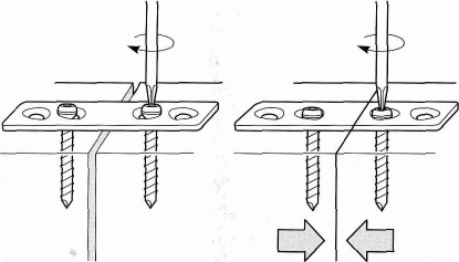
Figure 4. Diagrammatic representation of a small compression plate to illustrate the principle by which compression of the bone interface is achieved. The eccentric pear-shaped holes in the plate cause inward movement of the screw at the final stage of tightening when the head of the screw localizes in the wider part of the pear-shaped hole.
The compression holes in the plate may be positioned one on each side of the fracture (Spiessl, 1972) or both on the same side (Becker, 1974). Because of the tendency for the upper border to open when compression is applied across the fracture at the lower border, it is necessary to apply a tension band at the level of the alveolus before tightening the screws (Figs. 5 and 6). This can be in the form of an arch bar ligatured to the teeth or as a separate plate with screws penetrating the outer cortex only. Schilli (1977) designed a plate with oblique lateral holes which ensured that the compressing force was in part directed towards the upper border so that when the plate was tightened into place there was less tendency for the fracture line to gape. Raveh et al. (1987) employed a special adjustable temporary plate at the upper border which enabled them to achieve precise reduction of the fracture prior to application of the definitive lower border dynamic compression plate. They found no application for the eccentric dynamic compression plate in their large series of cases and reported malocclusion post-reduction in only 0.5 per cent of patients. Other workers have either placed the patient in temporary intermaxillary fixation or employed special adjustable bone-holding forceps to ensure accurate fracture reduction prior to application of the compression plate.
It has become increasingly apparent that compression plates offer no material advantage to the patient in the treatment of facial bone fractures. The operative approach tends to be lengthy and requires considerable expertise to produce consistent results. This is especially so if an intra-oral approach is used or if preliminary intermaxillary fixation is applied. Compression plating becomes more difficult when there is any obliquity of the fracture because of the depth of penetration of the screws. Problems also arise when there is comminution at the lower border, which is not always apparent from conventional radiographs. Perhaps the chief disadvantage, however, is the actual bulk volume of the fixation plate, which necessitates later removal in a high proportion of patients. This, of course, means subjecting the patient to a further general anaesthetic not otherwise indicated. For all of these reasons the majority of surgeons now use non-compression miniplates exclusively.
Miniplates
Roberts (1964) used cobalt-chrome alloy meta-carpal plates up to 1 inch (2.5 cm) in length to treat a series of mandibular fractures. These were applied to the outer cortical plate after reduction of the fracture, by means of 7 mm long screws 1.5 mm in diameter. Roberts later designed plates specifically for mandibular plating. Battersby (1966) subsequently reviewed a series of 350 cases treated in this way over a 12-year period and demonstrated satisfactory fixation. The plates were, however, employed as an alternative to transosseous wiring and most patients needed to be placed into intermaxillary fixation as well. Michelet and Moll (1971) described the use of similar small cobalt-chrome alloy plates of various lengths, and subsequently Michelet et al. (1973) reported results in 300 cases. These chrome-cobalt alloy plates were difficult to adapt and were not widely adopted. Champy et al. (1978) introduced a miniplate system customized for use in mandibular fractures. Originally fashioned in stainless steel, similar plates have now become widely available made from titanium (Figs. 89 and 94).
Champy and his co-workers argued that compression plates were unnecessary because there was a natural line of compression along the lower border of the mandible. They further claimed that compression exerted a stress-shielding effect which was detrimental to ultimate mandibular strength. Non-compression miniplates with screw fixation confined to the outer cortex allow the operator to place plates both immediately sub-apically as well as at the lower border. The stress distribution after fracture of the body of the mandible has been investigated using stressed bars of epoxy resin to simulate the fractured mandible. On the basis of these studies it is suggested that fractures at the angle can be secured with a single plate as near to the upper border as the dental anatomy permits. In the canine region two plates are ideally required, one juxta-alveolar and one at the lower border. All plates can be inserted by an intra-oral approach without the need for intermaxillary fixation (Fig. 7).
|
|
|
|


Figure 5. Diagram illustrating the chief problem with a compression plate, (a) Compression near the lower border opens up the fracture at the alveolar margin, (b) A tension band previously applied to the teeth prevents the distorting effect of the lower border compression plate, (c) A similar effect is achieved by prior application of a small cortical non-compression plate above the level of the inferior.

Figure 6. Radiographs showing the use of a compression plate at the lower border in conjunction with a non-compression cortical plate above the level of the inferior dental canal. (Case treated by Professor J. Prein and reproduced with his kind permission).
|
|
|
| ||
|
(a) |
(b) |
(c) | ||
|
|
| |||
|
(d) |
(e) | |||


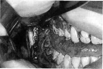

Figure 7. (a) The post-traumatic occlusion of a patient with a bilateral fracture of the body of the mandible. The 'step' deformity in the left mandible is clearly illustrated, (b) Postero-anterior radiograph showing fractures in the right molar and left canine regions of the mandible, (c) Operative photograph showing a non-compression miniplate applied across the fracture in the right retromolar region, (d) Operative view of the left canine region. The fracture was reduced with the aid of a temporary wire around the teeth. Two cortical miniplates have been inserted taking care to avoid damage to the mental nerve, which is shown emerging from the mental foramen.
Miniplates of this design are now widely used and reported results are encouraging.
The operative time involved is no more than that required for transosseous wiring. Cawood (1985) compared 50 cases treated by conventional intermaxillary fixation with 50 cases treated by miniplates. The plated series had a higher incidence of residual malocclusion (8%) but there were no cases of delayed union compared with 6 per cent in the control group. Of the plated cases 6 per cent became infected compared with 4 per cent of the controls and 3.8 per cent in a comparable group treated in an independent hospital department.
Postoperative infection of miniplates appears to vary considerably from unit to unit. Wald et al. (1988) have reviewed the literature and report complications as high as 30 per cent in some series. The risk of infection from various plating systems has been reviewed by Ellis (1999) .
Miniplate osteosynthesis can be used in virtually all types of mandibular body fracture.
Plates can be inserted via an intra-oral approach using special cheek retractors and protective sleeves passed through the soft tissues of the cheek. It is only necessary to reflect periosteum from the outer plate of bone, which is an advantage when compared with transosseous wiring. A few surgeons favour the use of simultaneous intermaxillary fixation when using miniplates (Mommaerts and Engelke, 1986). The plates can usually be left in permanently without causing trouble (Haug, 1996), but on theoretical grounds Cawood (1985) recommends removal because of the continuing effect on the functional forces within the bones. There is also now firm evidence that corrosion and local dispersal of titanium does take place but this is only detectable by ultramicroscopic techniques (Kim et al., 1997). The significance in the long term cannot yet be evaluated, which is regarded by some as an indication for removal of all plates (Haers et at., 1999).
Lag screws
A few oblique fractures of the mandible can be rigidly immobilized by inserting two or more screws whose thread engages only the inner plate of bone. The hole drilled in the outer cortex is made to a slightly larger diameter than the threaded part of the screw. When tightened the head of the screw engages in the outer plate and the oblique fracture is compressed. At least two such lag screws are necessary to achieve rigid immobilization (Fig. 8). The technique has been well reviewed by Leonard (1987).
|
|
|
|
(a) |
(b) |

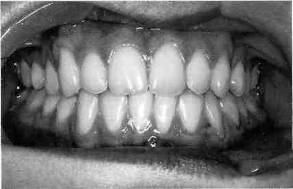
Figure 7. (continued) (e) Postoperative radiograph. The fractures have been satisfactorily reduced and immobilized by non-compression miniplates. (f) Immediate postoperative photograph of the occlusion. The temporary wire ligature around the teeth on each side of the left mandibular body fracture has not yet been removed.
Resorbable plates and screws
The advantage of a small bone plate fashioned in a biocompatible resorbable material has been clear for some time and a number of materials have been investigated. In order to be useful for mandibular fractures these plates need to be both adaptable to the complex surface of the bone and strong enough to stabilize the reduced fracture until bone healing has advanced to the stage of stable union. The mechanical requirements for the fixation of fractures of the upper and mid-facial skeleton are less but the plates need to be smaller than those which can be applied to the mandible. So far the most promising materials appear to be polylactides, or more specifically self-reinforced poly (L-lactide) homopolymer (PLLA) and poly (L/D-lactide) stereocopolymers with an l/d molar ratio of up to 85:15 (Haers et al., 1999).

Figure 8. Diagram to illustrate the principle of the lag screw in an oblique fracture. The deeper section of the screw hole is accurately tapped whereas the superficial section allows free movement of the screw. As the screw is tightened the fracture is reduced and compressed.

Figure 9. Clinical photograph of a self-reinforced PDLLA resorbable miniplate used for the fixation of a mandibular fracture. (Case treated by Professor C. Lindqvist and reproduced with his kind permission).
Plates fashioned in this material have been used successfully in orthognathic surgery and craniofacial surgery and have had limited clinical evaluation for mandibular fractures (Bessho et al, 1997). The material is biocompatible with a somewhat lengthy period of degradation which may not end in total absorption but which does not appear to be a problem clinically (Suuronen et al., 1998). Sufficiently strong plates for the management of mandibular fractures with good overall bone contact can now be constructed in sizes which compare favourably with titanium. They can be bent without heating and after bending the plate will retain its shape. It is fair to say that full clinical evaluation is not yet complete but intermediate-term results are encouraging (Fig. 9).
Indications for application of miniplates
Although there are some who would advocate plate osteosynthesis in all cases of mandibular fracture, there are some fractures which benefit particularly. These are:
Fractures in an edentulous part of the body of the mandible.
Concomitant fractures of the body and condyle when early mobilization is indicated.
When there are associated fractures of the other parts of the facial skeleton.
Patients in whom intermaxillary fixation is contraindicated.
Fractures associated with closed head injury.
6. Fractures in which there are continuity defects.
Fractures in which non-union or malunion has occurred.
Reconstruction of the facial skeleton
Effective reduction and fixation of the facial skeleton can be carried out at any time within the first 2 weeks of injury. There may indeed be considerable advantages in delaying treatment of comminuted mid-face fractures, which are more difficult both to assess and reduce in the presence of the considerable facial oedema which is often an early feature.
There are a few fracture patterns which demand early reduction even if fixation has to be compromised in the short term. These are:
Mobile, painful, nearly always multiple, mandibular fractures which interfere with swallowing and increase restlessness in a patient with a significant head injury.
Facial fractures associated with continuing haemorrhage into the airway.
Upper mid-face fractures accompanied by profuse cerebrospinal fluid leakage.
Impaction of the mandibular condyle into the middle cranial fossa.
Dento-alveolar fractures with subluxed or fractured teeth.
The principles which determine the sequence of reduction and fixation of multiple fractures of the facial skeleton are based on the surgical anatomy. The mid-face is represented by a complex of bones which have to be contained in their correct positions within that outer frame, and finally the projecting nasal complex must be reassembled on a firm foundation. The techniques of reduction and fixation of the individual components of the face will be described in later chapters, but the sequence of reduction and fixation needs to be understood from the outset.
Transfixation with Kirschner wires
Kirschner wires (K-wires) are widely used in orthopaedic practice and are therefore usually available in most hospitals. In rare emergency situations these wires can be used to provide temporary stabilization of a fractured mandible. The fracture is held in a reduced position and one or more wires drilled through the fragments so that part of the wire passes through undamaged bone each side of the fracture. The method is versatile and can be applied with appropriate ingenuity to fractures in any part of the mandible whether there be teeth present or not (Vero, 1968). In the context of fractures of the tooth-bearing area it is of little use when more conventional methods are available.
Shuker (1985) described an ingenious use of a single K-wire for the rapid immobilization of a comminuted fracture of the body of the mandible such as might occur after a missile injury. A horseshoe-shaped 2 mm K-wire is adapted to the mandibular arch and then each end is inserted into two holes drilled from an intra-oral approach into the anterior border of each ramus. The horseshoe-shaped wire lies on the buccal side of the displaced mandibular arch and the individual segments with their contained teeth are ligatured to this semi-rigid frame.
Methods of immobilization
The fact that there is no uniformly accepted method of immobilizing edentulous fractures is indicative of the fact that no completely satisfactory method has yet been devised. There is no doubt, however, that the traditional treatment by means of Gunning-type splints has been largely superseded in recent years by methods which employ some form of direct or indirect skeletal fixation. In older patients intermaxillary fixation is even less desirable than in younger age groups. Nutritional requirements become difficult to maintain and oral candidiasis commonly affects the oral mucosa causing considerable discomfort during the active treatment period. The methods of treatment currently available are listed below:
1. Direct osteosynthesis:
(a) bone plates;
(b) transosseous wiring;
(c) circumferential wiring or straps;
(d) transfixation with Kirschner wires;
(e) fixation using cortico-cancellous bone graft.
2. Indirect skeletal fixation:
(a) pin fixation;
(b) bone clamps.
3. Intermaxillary fixation using Gunning-type splints:
(a) used alone;
(b) combined with other methods.
Direct osteosynthesis
Bone plates are particularly useful for displaced fractures of the edentulous mandible, particularly those at the angle. They allow the fracture to be stabilized without immobilization of the jaw as a whole. The patient is, as a result, more comfortable during the period of healing of the fracture. The main mandibular plating systems described earlier in the chapter are in general applicable to edentulous fractures. Unlike the dentate mandible there is a significant risk of non-union in the edentulous state and it could be argued that compression plates might have a theoretical advantage. However, the reduced depth of bone in the edentulous mandible favours the use of non-compression miniplates rather than the bulkier compression plates in that the former are less likely to interfere with the edge of a future denture. Bone plates are easier to apply in the edentulous state than when teeth are present as there is no need to achieve the same degree of precision in the reduction of the fracture. Any discrepancy in the eventual occlusion of the pre-existing dentures is more easily corrected than when natural teeth are involved (Fig. 10).
It is easier to apply bone plates to the edentulous mandible than it is to insert transosseous wires. There must be fairly liberal exposure of the fracture site with extensive elevation of the periosteum, but that exposure can be confined to one surface of the bone and the overall peri-osteal attachment is often disturbed less than when inserting a number of transosseous wires. Both compression and non-compression systems require an adequate blood supply to achieve uncomplicated bony union and elevation of periosteum in the thinner mandible seriously compromises the blood supply to the fracture site. It has been suggested that in these circumstances plates should be applied with an intervening layer of attached periosteum, but in practice this is difficult to accomplish.
Plates related to the denture-bearing part of the mandible are much more likely to require removal at a later date than those used in the ramus or in dentate fracture sites. Nevertheless some form of plating is currently the preferred method of fixation for the majority of edentulous mandibular body fractures. Resorbable plates would appear to have an advantage but, at their present stage of development, degradation takes place over a period of 2 years or more during which new dentures are likely to be needed. A metal plate removed electively is a better option.
|
|
|
|
(a) |
(b) |


Figure 10. (a) Panoral tomogram of a bilateral fracture of an edentulous mandible with considerable displacement of the fragments, (b) The situation 6 months after treatment. A miniplate was used for fixation on the right and transosseous wiring on the left. No IMF was employed and the healing on each side is comparable.
6. MATERIALS FOR SELF-CONTROL:
A. Questions for self-checking: 1. X-ray foto, a photo and discharge from a hospital from histories of the victims with bones fractures. 2. Materials and the equipment for preparation for operative treatment of fractures of jaws.
B. Tasks for self-checking: 1. To the victim with a fracture of the maxilla by Le Fort ІІ type operative intervention with fastening fragments by spokes by kirshner entered through a skin in a cross-section direction at a level of zygomatic bones is carried out. Name a method of an osteosynthesis by authors. (The answer: by Makienko).
2. At the fighter who is injured with a debris of shell, is diagnosed a ballistic fracture of the mandible with defect of a bone in the chin region more than 3 sem. What method of fixing of fragments of the mandible is shown? (The answer: the osteosynthesis by Rudko and by Bernadsky with apparatus method).3. Patient of 42 years is hospitalized with fracture of the maxilla on type II by Le Fort. Choose the device for treatment of this patient. (The answer: by Zbarsh).
C. Materials for the test control. Test tasks with single right answer (α = ІІ):1. Name the indication for application of device by Zbarzh. A. Fracture of the maxilla by Le Fort I, ІІ, III, by Geren, by Vasmund. B. Fracture of the mandible at absence of the teeth on jaws or at defect of a body of a jaw. C. Fracture of the branch of mandible or process condilaris. D. Fracture of the mandible within the limits of a dental lines or the angle of the mandible.E. Fracture of an alveolar or coronal process of the mandible. (Correct answer: А.).
2. What kinds of fixing concern to a direct type of osteosynthesis at fractures of the maxilla? A. Imposition of spokes by Kirshner, spokes by Makienko. B. Imposition of pin devices. C. Imposition of circular ligature. D. Impositionof a bone seam and a metal frameworks. E. Imposition of wire, banded tape or cast arch bar. (Correct answer: А).
D. Educational task of 3-rd level (atypical tasks):1. Patient С., 28 years, has addressed in 3 days after a trauma. Complaints to a painful swelling in a site of a body of the mandible at the left. Palpation of the body of the mandible at the left side is painful, it is marked mobile оf fragments in a site of 45 teeth. On the X-ray in lateral left projections the crackor fracture line in a site of 45 teeth. The diagnosis is exposed: a fracture of a body of the mandible at the left in a site of 45 teeth. What kinds of surgical treatment should be applied? (The answer: not a direct osteosynthesis).
2.Patient С., 58 years, has addressed in 5 days after a trauma with complaints to a painful swelling in a site of a body of the mandible on the right. Palpation of the body of the mandible at the left side is painful, it is marked mobile of fragments in a site of 35 teeth. On the X-ray in the left lateral projections revealed crack of the bone in the site near the roots of 35 teeth. The diagnosis is exposed: fracture of the body of the mandible on the right side near roots of 35 teeth. What complication is authentic in the given clinical case? (The answer: a suppuration of a bone wound).
3. The patient T. 75 years, has arrived in hospital with the diagnosis: fracture of the angular region of mandible on the right side with displacement. Objectively: asymmetry of the face due to a posttraumatic edema, at palpation is marked mobile of fragments in a site of the fracture, a pain. In the anamnesis - epilepsy since a birth. What method of treatment do you offer? (The answer: the osteosynthesis).
Theme № 17 Combine damages of maxillofacial area. Cranial - maxillofacial damages. Features of clinical current, diagnostics, complications, features of treatment at stages of medical evacuation. Achievement of native scientists, members of chair.
ACTUALITY OF THEME. Knowledge of a clinical picture, features of diagnostics of combine damages of maxillofacial area and their complications, methods of treatment which will allow the doctor - stomatologist to render medical aid by the maxillofacial wounded in a wartime and in conditions of extreme situations.
2. СONCRETE AIMS: 2.1. To analyze a concept about combine damages of maxillofacial area. 2.2. To explain the mechanism of a symptom of mutual burdening. 2.3. To offer the etiological and pathogenetic scheme of a symptom of mutual burdening. 2.4. To classify cranial - maxillofacial damages and a brain traumas. 2.5. To treat the data of X-ray pictures, tomograms and other results of victims with a combine trauma in maxillofacial surgery. 2.6. To draw schemas of stages of medical evacuation. 2.7. To analyze possible complications of combine traumas in maxillofacial region. 2.8. To make algorithm of first aid, examination and treatment at stages of medical evacuation of victims with cranial - maxillofacial damages and a traumatic illness.
3. BASE KNOWLEDGE, SKILLS, THE SKILLS NECESSARY FOR STUDYING OF THE THEME (interdisciplinary integration).
|
Names of the previous disciplines |
The got skills |
|
1. Normal anatomy. |
To be guided in the basic questions of a structure of maxillofacial area. |
|
2. Topographical anatomy and operative surgery. |
To know topographical anatomy of maxillofacial area. Principles of operative access to different departments of maxillofacial area. To define a topical diagnosis of traumatic damages of maxillofacial area. |
|
3. Pathological physiology. |
To represent the aetiology of combine traumas of maxillofacial area and traumatic illness. |
|
4. Neurosurgery. |
To know classification, clinic, diagnostics and the general principles of treatment of the brain trauma. To make the plan of examination and treatment of victims with damage of the face and skull and brain. |
|
5. Clinical pharmacology. |
To know pharmacology products which are prescribe at сombine damages of maxillofacial area. |
|
6. General surgery. |
To be able to make the plan of inspection of the patient with traumatic damage and to fill up the medical documentation. |
4. TASKs FOR INDividual WORK DURING PREPARATION TO Lesson.
4.1. List of basic terms, parameters, characteristic, which a student must master at preparation to lesson:
|
The term |
Definition |
|
1. Cranial - maxillofacial damages. |
This simultaneous damage of the soft tissue, a facial part of skull and a brain. |
|
2. Соmbine damage. |
This action of one injuring agent on two and more areas simultaneously. |
|
3. Traumatic illness. |
Pathological process which develops as a result of traumatic damage. |
4.2. Theoretical questions to the lesson: 1. To define a concept "a cranial - maxillofacial trauma". 2. A classification of combine damages of maxillofacial area. 3. Definition and a pathogenesis of a traumatic illness. 4. Clinic of combine damages of maxillofacial area. 5. Features of the urgent aid the victims with a combine trauma of maxillofacial area. 6. Features of treatment of victims with a combine trauma of maxillofacial area.7. Preventive maintenance of complications at combine damages of maxillofacial area.
4.3. Practical works (task) which are carried out on the lesson: 1. Training from rendering the urgent aid for victims with a cranial - maxillofacial damages and a соmbine trauma in maxillofacial surgery. 2. Drawing up of algorithm of treatment at соmbine traumas in maxillofacial surgery.
5. TABLE OF CONTENTS OF THEME:
Craniofacial fractures
In 1977 Matras and Kuderna pointed out that the rising number of road traffic accidents had added to the incidence of unusual combinations of facial fractures. Among these the frontomax-illary fracture as described by Pape (1969) is the most severe. Such fractures often extend from the anterior base of the skull to the mandible, thus involving the facial skeleton in the upper, lower and mid-facial regions. Cantore et al. (1979) reported that of 387 acute head injuries admitted to their neurosurgical unit during a 3-year period, 8 per cent had cranio-orbito-facial fractures.
There is both an increase in the incidence of these craniofacial fractures together with an improved chance of survival. Extensive disruption of the anterior cranial fossa, often involving the posterior wall of the frontal sinuses, means that reconstruction must be undertaken in a hospital equipped for craniofacial surgery. No maxillofacial injury can be treated in isolation from a coexistent cranial component, not only because of the need to treat the cranial injury but also because the displacement of the frontal bones, particularly the orbital roofs, prevents correct repositioning of the facial skeleton.
Fractures of the mid-face
The imaging required to obtain a detailed picture of a mid-facial fracture needs to be balanced against the benefits to actual treatment. In the tooth-bearing part of the maxillae separation at the Le Fort I level is often a clinical diagnosis. Intraoral films may, however, be most valuable in localizing alveolar fractures or a midline split of the palate, injuries which materially influence a treatment plan.
For higher level Le Fort II and III fractures, it is important to determine the overall fracture pattern and particularly the degree of cranial involvement. When there is comminution and displacement in the naso-ethmoid region or extensive damage to the orbital integrity, the clinician needs the detailed information provided by CT in order to plan the reconstruction. It is very important to obtain accurate reduction and fixation of these fractures at the acute stage when optimal results can be achieved.
The upper facial skeleton
The so-called upper third of the facial skeleton is chiefly the frontal bone making up the superior orbital margin and orbital roof. The base of the skull extends backwards and is angled downwards at approximately 45° where the frontal bone articulates with the sphenoid. The mid-facial complex articulates with this sloping plane and the cribriform plate of the ethmoid extends upwards to make contact with the meninges of the brain and transmit the olfactory nerves. The frontal bone, the body and greater and lesser wings of the sphenoid are not usually fractured. In fact, they are protected to a considerable extent by the cushioning effect achieved as the fracturing force crushes the comparatively weak bones comprising the middle third of the facial skeleton. When fractures of the cranial component of the facial skeleton do occur there are important consequences:
The brain may have sustained direct injury.
The brain may be at risk from indirect injury secondary to bleeding at the fracture site.
A fracture may involve the posterior wall of the frontal sinus, the orbital roof or the cribriform plate, which in turn may be associated with a breach of the dura materand leakage of cerebrospinal fluid.
Displacement, particularly in a caudal direction, will interfere with reduction of the facial bones as a whole.
The mid-facial skeleton
The mid-facial skeleton is defined as an area bounded superiorly by a line drawn across the skull from the frontozygomatic suture across the frontonasal and frontomaxillary sutures to the frontozygomatic suture on the opposite side, and inferiorly by the occlusal plane of the upper teeth, or, if the patient is edentulous, by the upper alveolar ridge. It extends backwards as far as the pterygoid plates of the sphenoid, which are usually involved in any severe fracture.
This area of the facial skeleton is made up of the following bones:
two maxillae;
two zygomatic bones;
two zygomatic processes of the temporal bones;
two palatine bones;
two nasal bones;
two lacrimal bones;
vomer;
ethmoid and its attached conchae;
two inferior conchae;
pterygoid plates of the sphenoid.
Physical characteristics of the mid-facial skeleton
The mid-facial skeleton is made up of a considerable number of bones which are rarely, if ever, fractured in isolation (Fig. 1).
The composite structure of this complex of bones is so ordered that it will with stand the forces of mastication from below and provide protection in certain areas for vital structures, notably the eye. If the comparatively thin areas of bone comprising the outer walls of the maxillary sinuses are removed from the maxillae, it will be seen that the mid-facial skeleton consists of a series of bone struts passing upwards from the upper teeth to the bone of the skull. The forces of mastication are thus distributed round the fragile area of the nose and para-nasal sinuses to the base of the skull (Fig. 2).
This type of structure is able to withstand considerable force from below but the bones are easily fractured by relatively trivial forces applied from other directions. Nahum (1975) conducted experiments in which fracture of the middle third occurred with forces between one-fifth and one-third of those required to produce simple fracture of the mandible. The facial bones as a whole have a very low tolerance to impact forces. The nasal bones are least resistant, followed by the zygomatic arch, while the maxilla itself is very sensitive to horizontal impacts.
Because of the relative fragility of the mid-facial skeleton, it acts as a cushion for trauma directed towards the cranium from an anterior or anterolateral direction. It is analogous to a 'matchbox' sitting below and in front of a hard shell containing the brain, and differs quite markedly from the rigid projection of the mandible below (Fig. 3). These physical differences are extremely important for survival after head injury. An impact directly applied to the cranium may be sufficient to cause severe brain injury or death. This same force applied to the mid-facial skeleton is cushioned sufficiently so that it may not even lead to loss of consciousness, though causing considerable damage to the bones and soft tissue of the face. If, however, the mandible alone withstands the impact, the cushioning effect is reduced and brain injury will result in a manner comparable with a boxer's knockout punch.
Involvement of the brain and cranial nerves
Comminution of the ethmoid occurs with Le Fort II and III fractures and some severe fractures of the nasal complex. This may lead to a dural tear in the region of the cribriform plate of the ethmoid resulting in cerebrospinal rhinorrhoea (Fig. 4). A dural tear may also occur adjacent to fractures involving the posterior wall of the frontal sinus. Cerebrospinal fluid may also escape into the soft tissues via coincident fractures of the orbital roof without appearing in the nasal cavity. More rarely, a profuse cerebrospinal fluid rhinorrhoea occurs as a result of a fracture which passes through the base of the sphenoid, communicating with the sphenoidal sinus and via a crack in the roof of this structure with the middle cranial fossa.
Damage to the infra-orbital and zygomatic nerves may occur with zygomatic and Le Fort II fractures either unilaterally or bilaterally. This gives rise to anaesthesia or paraesthesia of the skin of the cheek and upper lip. Full recovery of sensation may be delayed for up to two years. The anterior, middle and posterior superior alveolar nerves are frequently damaged leading to anaesthesia of the upper teeth and gingiva.
Cranial nerves within the orbit may sustain damage in zygomatic, Le Fort II and III fractures. The sixth cranial nerve is most frequently involved but sometimes the contents of the superior orbital fissure are all damaged, in which case ophthalmoplegia, dilation of the pupil and anaesthesia within the distribution of the ophthalmic branch of the fifth cranial nerve are severally noted. Rarely the orbital apex is fractured with resultant damage to the optic nerve and blindness.

Figure 1. Anatomical specimen showing the disarticulated bones of the skull exploded and mounted to demonstrate their complex interrelationship. Note that the bones of the mid-facial skeleton are all comparatively fragile. They comprise, from above downwards, the ethmoid, flanked by the two lacrimal bones. Next are the paired nasal bones, palatine bones, maxillae and inferior conchae with the zygomatic bone laterally on each side. The vomer is missing. Courtesy of the Anatomical Museum, Royal College of Surgeons of England.

Figure 2. Diagram showing the directions of distribution of the forces of mastication within the skeleton of the mid-face.

Figure 3. Diagrammatic representation of the strength of the bones of the skull and face. The 'matchbox' structure of the mid-facial skeleton cushions the effect of impact force B. Impact force A is transmitted directly to the brain producing the most severe injury. Impact force C is transmitted indirectly to the cranial base via the rigid structure of the mandible (represented here as a bent baseball.

Figure 4. The cribriform plate of the ethmoid. This fragile bone is fractured in Le Fort II and III type fractures and also in severe injuries of the naso-ethmoidal complex.
Involvement of the orbit
The globe of the eye and the optic nerve are remarkably well protected by the physical structure and arrangement of the bones of the orbit. The prominence of the zygomatic bone acts as a protection for the globe from all impinging objects other than very small projectiles. The optic foramen is a ring of compact bone and in high level or Le Fort III injuries, fractures almost invariably pass around it. Rupture of the globe or tearing of the optic nerve is fortunately therefore rarely found with other than the most severe middle third fractures.
Fractures involving the orbit may give rise to alteration in the position of the globe of the eye. The level of the globe is normally maintained by the suspensory ligament of Lockwood, which passes from its medial attachment on the lacrimal bone to be inserted laterally into Whitnall's tubercle situated on the inner aspect of the zygomatic bone just below the frontozygomatic suture. Zygomatic and Le Fort III fractures commonly result in separation at this suture line with a resulting drop in the level of the globe of the eye.
If the bone comprising the floor of the orbit is fractured, orbital contents may herniated through into the maxillary sinus below. The resulting entrapment of these tissues may result in restriction of movement of the inferior rectus and inferior oblique muscles although, anatomically, the muscles themselves are not actually ensnared by fragments of bone. Alternatively, the muscles may become bound down adjacent to the damaged bone by subsequent fibrous tissue formation. In either event, temporary or permanent diplopia results due to prevention of the upward and outward rotation of the eye. The lateral rectus muscle may be partially paralysed by haematoma formation or sixth nerve neuropraxia adjacent to a fracture line resulting in lateral diplopia. Diplopia results mainly from interference with the activity of the ocular muscles rather than physical displacement of the globe. Extreme displacement of the latter will, however, result in such distortion of the visual axis that the muscles will be unable to compensate and double vision will be inevitable.
Orbital contents may also herniate through the thin lamina papyracea of the ethmoid bone on the medial wall of the orbit. Any significant increase in the orbital volume occurring as a result of fracture of the walls of the orbit will lead to enophthalmos. Enophthalmos, particularly if accompanied by inferior displacement of the lateral canthal attachment, may give rise to pseudo-ptosis, a physical sign traditionally referred to as 'hooding of the eye'.
Le Fort II and III fractures and severe nasal complex injuries may involve the naso-lacrimal duct with resulting epiphora. This complication is not noticed at the time of injury, but may become apparent later.
Detachment of the medial canthal attachment of the eye may occur in severe naso-ethmoidal injuries, giving rise to traumatic telecanthus.
Haemorrhage within the orbit is a frequent occurrence in facial injuries. In the absence of actual damage to the globe of the eye it is not usually significant. However, haemorrhage within the muscle cone of the eye (retrobulbar haemorrhage) can very rarely lead to loss of vision. Ord (1981) conducted a comprehensive review of this complication. It is now thought that blindness occurs as a result of spasm of the short posterior ciliary arteries, causing ischaemia of the optic nerve head over a limited but critical area.
The paranasal sinuses
In zygomatic complex and Le Fort I, II and III fractures, the maxillary sinuses are involved. The thin bony walls of the sinuses are often grossly comminuted with bleeding into the cavity and not infrequently inward herniation of the buccal pad of fat. This results in one or more of these paranasal sinuses appearing opaque on radiological examination. Apart from the routine reduction and repositioning of the fracture, no other special treatment is required and the radiological appearance of the sinuses will clear after about 6 weeks. Kreidler and Koch (1975) carried out an endo-scopic study of the maxillary sinus after mid-facial fractures in 25 patients and found 35 per cent had chronic mucosal changes, but all were free of symptoms. No similar studies appear to have been carried out to confirm these observations.
Uncommonly, air may escape into the soft tissues of the face following a fracture which extends into one or other paranasal air sinus. This usually affects the flaccid tissues of the eyelids and leads to 'surgical emphysema'. This in turn gives rise to the physical sign of 'crepitation' of the soft tissues when palpated. Whenever air gains entry into soft tissue planes it is contaminated and adds to the risk of subsequent infection. Air within the cranial cavity and meninges or in the mediastinum is particularly dangerous and can rarely occur as a complication of facial trauma.
Important blood vessels
The third part of the maxillary artery and its terminal branches are closely associated with the lines of fracture in Le Fort I, II and III-type injuries. Occasionally the artery or its greater palatine branch is torn in the region of the pterygomaxillary fissure or pterygopalatine canal resulting in severe life-threatening haemorrhage into the nasopharynx. Packing of the nose via the anterior nares, whilst usually sufficing in more minor nasal haemorrhage, will be ineffectual in this event. A post-nasal pack must be inserted, which will apply direct pressure to the bleeding point without embarrassing the airway through the mouth. It is necessary to retain this pack for 24 hours and to replace it if necessary. Such a pack is a potent source of infection and is not well tolerated by the patient. Adequate reduction of the fracture will fortunately prevent further bleeding in most cases.
Multiple fractures
The same association of direct with indirect violence may give rise to multiple fractures. The most common multiple fracture is that caused by a fall on the mid-point of the chin resulting in fractures of the symphysis and both condyles. These fractures are commonly seen in epileptics, elderly patients who lose consciousness as a result of general disease, and occasionally in soldiers who faint on parade, from which the fracture combination derives its name of 'guardsman's fracture'.
Oikarinen and Malmstrom (1969), in a series of 600 mandibular fractures, found 49.1 per cent were single, 39.9 per cent had two fractures, 9.4 per cent had three fractures, 1.2 per cent had four fractures and 0.4 per cent had more than four fractures.
Comminuted fractures
Comminution of a fracture site is almost invariably the result of considerable direct violence at the site of fracture, as is commonly the case in war missile injuries (Fig. 5). In civilian practice this degree of comminution is most common in the symphysis and parasymphyseal regions. Such fractures require special management and should therefore be considered in a category of their own. It is not unknown for severe missile injuries to cause comminution of the whole of the mandible from one condylar neck to the other.

Figure 5. Lateral oblique radiograph showing comminuted fracture of the left body of the mandible following a medium-velocity missile injury
Cerebrospinal fluid rhinorrhoea
The possibility of cerebrospinal fluid rhinorrhoea should be considered in all Le Fort II and III fractures. In these types of fracture the escape of cerebrospinal fluid into the nose is the result of a dural tear associated with fracture of the cribriform plate of the ethmoid. In most of these instances the cerebrospinal fluid leak arrests within the first few days, either spontaneously or as the result of reduction and fixation of the fracture.
Whenever there is a tear in the dura mater there is a risk that the patient will develop meningitis either in the early days after injury or even many years later. This fact was first established by Lewin and Cairns (1951) in what has become a classical study. As a follow-up to this work, Lewin (1954) analysed 308 cases of non-missile-head injuries with associated fracture of the paranasal sinuses. Of these 84, or 72.3 per cent, developed cerebrospinal fluid rhinorrhoea, which usually occurred within the first few days, but in a number of cases was delayed in onset until the 14th day or later. Of these 84 patients with cerebrospinal fluid rhinorrhoea, 16 developed meningitis at some stage and six of them died. The incidence of late meningitis was virtually eliminated by operative repair of the dura mater.
Since the publication of these papers, argument has persisted concerning the indications for elective repair of the dura mater in cases of cerebrospinal fluid rhinorrhoea. Major fractures of the central mid-face treated in maxillofacial centres are associated with cerebrospinal fluid rhinorrhoea in about 20-25 per cent of cases (Dawson and Fordyce, 1953; Rowe and Killey, 1968). The cerebrospinal fluid leak in these and similar series lasts for about 7 days on average before it either arrests spontaneously or stops after operative reduction of the fracture (Leopard, 1971). Maxillofacial surgeons have not reported meningitis as other than a rare complication in such cases, although it is possible that patients presenting at a later date have been treated elsewhere. Nevertheless, where long-term follow-up of mid-face fractures has been reported (Dawson, 1962), there have been no cases of subsequent meningitis.
The reason appears to be that neurosurgeons are reporting a different type of case; that in which the injury is predominantly to the head.
Summary of management of rhinorrhoea
In most maxillofacial injuries cerebrospinal fluid rhinorrhoea is assumed to be present when there is a combination of the righttype of fracture with a persistent thin discharge of fluid from the nose. Confirmation of the diagnosis by laboratory investigations, CT or MRI scans requires sophisticated equipment.
Reposition and immobilization of the fractures seems to occlude the cerebro spinal fluid fistula safely in the majority of cases.
Neurosurgical advice and possible operative repair of the dura mater should be considered in the following cases:
Combined frontomaxillary-orbital fractures, particularly those involving the posterior wall of the frontal sinus.
Those mid-face fractures in which the connection with the cranial cavity is so large that an aerocele develops.
Cases in which the cerebrospinal fluid leak persists for more than 14 days or cases in which the rhinorrhoea is of late and persistent onset.
In view of the fact that central nervous system capillaries are usually impermeable to large molecules, a blood-brain barrier exists. Antibiotics will, however, reach adequate concentrations adjacent to any dural tear. The value of prophylactic antibiotics in the prevention of meningitis in those patients with actual or suspected cerebrospinal rhinorrhoea is doubtful. However, all patients with maxillofacial injuries of sufficient severity to be associated with a dural tear and CSF rhinorrhoea, should be given prophylactic antibiotics as part of the general management of the facial injury.
Multiple and comminuted fractures
The physical signs of multiple and comminuted fractures depend on the site and number of the fractures. Multiple and comminuted fractures result from extreme direct violence and are usually associated with severe soft-tissue injury. The precise pattern of bony injury may be impossible to determine from the clinical examination. When there is unexpected mobility of what at first sight appears to be a single fracture, a second fracture on the same side should be suspected. In general comminuted fractures of the ramus, angle and molar regions are not associated with gross displacement of the fragments. However, comminution of the symphysis allows the lateral segments to collapse and presents a much more serious problem of management.
Complications from head injuries
Most patients with facial bone fractures associated with a period of loss of consciousness suffer to a greater or lesser extent from the post-concussional syndrome, which consists of headache, dizziness, insomnia, diplopia, intolerance to noise, changes in disposition, intellectual impairment and intolerance to alcohol. Usually these distressing symptoms eventually resolve, but may become aggravated and protracted if litigation for compensation is impending.
An aerocele or a cerebral abscess may develop within a few weeks of the accident. Meningitis may occur as an early or a very late complication, and occasionally epilepsy develops.
6. MATERIALS FOR SELF-CHECKING:
А. Questions for self-checking: 1. Collections of conditional tasks on subjects of cranial and maxillofacial damages. 2. Photos of victims with combone trauma in maxillofacial surgery.
B. Tasks for self-checking:1. In clinic of maxillofacial surgery is hospitalized the victim of 26 years with complaints to a pain in a site of the angle of the mandible at the left side, a headache, anausea. Last night he was beating down of unknown persons, lost consciousness, unitary vomitting took place. Objectively: the face is asymmetric due to a swelling on a site of the left angle of the mandible, a swelling painful and opening of a mouth up to 2sm, infringement of a bite. What preliminary diagnosis? (The answer: the left angular fracture and the closed craniocereberal trauma.)
2. In maxillofacial department is hospitalized the patient with the diagnosis: bilateral fracture of the mandible from one side the angle fracture and a mental from another side and with the closed craniocereberal trauma. What consultation of the expert is necessary first of all? (The answer: the neurosurgeon).
3. The patient of 39 years, complains of a headache, a crunch in ears during movements of a head. A trauma has received in a site of amid-face zone of the face. A consciousness lost, there was a numerous vomitting. Clinical picture is a deformation the nose, the bilateral symptoms of "glasses" over 12 hours, a symptom of "step" on external edge of the orbit. From the nose кровянистое with blood, (containing some blood) and oily (butter(y)) allocation. The bite is open, maxilla is not mobile. Define the preliminary diagnosis of the patient. (The answer: a fracture of maxilla by Le Fort III (the top type) and the open craniocereberal trauma).
C. Material for the test control. Test tasks with one right answer (α = ІІ):1. In the victim with damage of maxillofacial area from the anamnesis of disease it is known about infringement of consciousness, loss of memory, a headache, dizziness, a nausea and vomitting. It testifies about: A. Development of complications of a trauma of maxillofacial area. B. Combine trauma of maxillofacial area. C. Lowering of a resistance of the organism of the patient. D. Damage of the neck. E. Damage of the organs of the belly. (Correct answer: В.).
2. The connected trauma of the face and a brain count: A. Damage of the face and the brain which is caused by one-stage action injuring to the agent on an obverse skull or on an obverse and brain skull. B. Damage of the face and a damages of the brain, a skull with the secondary injured object.C. Damage which is caused by one-stage action of different injuring agents. D. Damage of the face and a brain that has become complicated by inflammatory process. E. Damage of a brain skull that is caused by frustration of other systems including the facial region. (Correct answer: А).
3. What is not a symptom of the closed craniocereberal trauma?A. A nausea. B. A vomitting. C. A giddiness (dizziness). D. Loss of consciousness. E. A bronchospasm. (Correct answer: Е).
D. Educational tasks of 3th levels (atypical tasks): 1. The surgeon - stomatologist is hospitalized the patientof 28 years after road accident in a heavy condition: the consciousness is confused, with vomiting and excitation. After examination with a general surgeon the preliminary diagnosis has been established: the closed craniocereberal trauma, a fracture of the right humeral bone, the left beam bone, a fracture of right VII rib, bilateral mental fracture of the mandible. What are the prime measures of the urgent aid? (The answer: antishock actions, preventions of asphyxia). 2. What type of asphyxia may be? (The answer: dislocation type). 3. Whether there is a necessity of urgent consultation of adjacent experts? (The answer: the anaesthesiologist).
Theme № 18 The combined lesions of tissues of maxillofacial area. Features of clinical current, diagnostics, complication, features of treatment at stages of medical evacuation. Radiation sickness: clinic, diagnostics, treatment
The topicality of the theme. Knowledge of a clinical picture, features of diagnostics of combine damages of maxillofacial area and their complications, methods of treatment which will allow the doctor - stomatologist to render medical aid by the maxillofacial wounded in a wartime and in conditions of extreme situations.
2. SPECIFIC GOALS: 1. To analyze concept "the combined (contrive, scheme) lesions". 2. To explain occurrence of the combined lesions of maxillofacial area. 3. To offer the new approach for accordance the help at the combined lesions. 4. To classify the combined lesions of maxillofacial area. 5. To treat the periods of radiation sickness. 6. To analyze consequences of the nuclear weapon factors on the organism of the victims. 7. To make the plan of diagnostics and algorithm of the help for the victims with the combined lesions of maxillofacial area and radiation sickness at stages of medical evacuation.
3. BASE KNOWLEDGE, SKILLS, THE SKILLS NECESSARY FOR STUDYING OF THE THEME (interdisciplinary integration).
|
Names of the previous disciplines |
The got skills |
|
1. Normal anatomy. |
To be guided in the basic questions of a structure of maxillofacial area. |
|
2. Topographical anatomy and operative surgery. |
To know topographical anatomy of maxillofacial area. Principles of operative access to different departments of maxillofacial area. To define a topical diagnosis of traumatic damages of maxillofacial area. |
|
3. Pathological physiology. |
To represent the aetiology of combine traumas of maxillofacial area and traumatic illness. |
|
4. Neurosurgery. |
To know classification, clinic, diagnostics and the general principles of treatment of the brain trauma. To make the plan of examination and treatment of victims with damage of the face and skull and brain. |
|
5. Clinical pharmacology. |
To know pharmacology products which are prescribe at сombine damages of maxillofacial area. |
|
6. General surgery. |
To be able to make the plan of inspection of the patient with traumatic damage and to fill up the medical documentation. |
4. TASKs FOR INDividual WORK DURING PREPARATION TO Lesson.
List of basic terms, parameters, characteristic, which a student must master at preparation to lesson:
|
A term |
A definition |
|
1.Mechanicaland radiating defeats. |
It is a mechanical trauma and a radiation injury or pollution by radioactive substances and a wound. |
|
2.Mechanicaland chemical defeats. |
It is a mechanical trauma and defeat by poison gases. |
|
3. Radiation sickness. |
Pathological process which develops in an organism under action of a radioactive irradiation. |
4.2. Theoretical questions to the lesson: 1. Mechanisms of injuring of a penetrating radiation.2. Pathogenesis of a symptom of mutual aggravation at mechanical and radiating wounds of maxillofacial area.3. Features of medical aid for the maxillofacial wounded with mechanical and radiating wounds of maxillofacial area.4. Mechanisms of action of poisonous substances. 5. Pathogenesis of a symptom of mutual aggravation at mechanical and radiating wounds of maxillofacial area.6. Features of medical aid by the maxillofacial wounded with mechanical and radiating damages of maxillofacial area. 7. Features of surgical treatment of maxillofacial wounded with the combined damages.
4.3. Practical works (task) which are executed on lesson:1. Professional training from the first aid by the victim with the combined trauma in maxillofacial area. 2. Professional training for medical complex treatment of victims with the combined defeats in maxillofacial area.
5. TABLE OF CONTENTS OF THEME:
Trauma survival
Facial injuries are but one component of a spectrum of morbidity and mortality resulting from trauma. World Health Organisation mortality data show that 5 per cent of all deaths worldwide are caused by trauma, and at least 1 per cent of gross national product is consumed in even the poorest countries in the treatment of injuries. The management of a patient with a facial injury has to be seen in the context of the treatment of injuries in general, and the first priority is obviously ensuring survival. The emergency treatment of even the simplest maxillofacial injury involves securing an airway and an assessment of cervical spine and head injuries, all of which are of vital importance in the general management of a patient whose injuries do not necessarily include the face and jaws. It is pertinent therefore to give a brief outline of the modern approach to managing general acute trauma as a preliminary to a more detailed discussion of the immediate treatment of maxillofacial injuries.
Following trauma there are three recognized peaks of mortality. The first occurs within seconds of injury as a result of irreversible brain or major cardiovascular damage. The second peak occurs between a few minutes after injury and about 1 hour later. It is during this 'golden hour' that modern methods of resuscitation have shown dividends in improved survival. A third peak is found some days or weeks after injury as a result of multi-organ failure despite good medical management. In recent years attention has been directed to the second of these peak periods, mainly as a result of a system of Advanced Trauma Life Support (ATLS), originally introduced and widely taught by the American College of Surgeons Committee of Trauma. Their aggressive interventionist approach to trauma management has also reduced mortality in the third group as active resuscitation leads to less late organ failure.
Organization of trauma services
Triage
Triage is a confusing word. It was originally used to describe the process of sorting goods according to quality, but has been adopted into the vocabulary of warfare to describe the classification of patients according to medical need. In trauma services generally, triage retains its military connotation and is important at three levels:
field triage;
inter-hospital triage;
mass casualty triage.
Triage decisions are crucial in determining individual patient survival and for this reason need to be made at the highest possible level of medical expertise.
Pre-hospital care
Active and appropriate pre-hospital care delivered by fully trained paramedical personnel improves survival during the vital first hour after injury. This means firstly that trained personnel have to arrive at the scene of the accident as rapidly as modern transport permits. They must be trained at the highest level in airway management with cervical spine control, securing intravenous access and initiating fluid resuscitation. Their prime duty is to stabilize the patient prior to rapid transport to a dedicated trauma centre.
Hospital care
A suitable receiving hospital must have senior medical staff organized as a trauma team. Their first duty is further triage to ensure that medical resources are deployed to maximum overall benefit. Accepted resuscitation procedure is in the following order of priority/
Primary survey
A: Airway and cervical spine control
An unconscious patient involved in a road traffic accident or fall has an approximately 10 per cent chance of having sustained a cervical spine injury. Witness statements, where they are able to describe the exact circumstances of the injury, are very important in assessment. Because of the potential catastrophic consequences of this type of injury the neck should be immobilized in a neutral position by a semirigid collar until damage has been excluded.
Early verbal response to a simple question 'Are you all right?' signifies not only that a satisfactory airway is present but also that cerebral function and by implication breathing and ventilation and circulation are adequate. The several techniques available to secure an unobstructed airway are integral to the management of all maxillofacial trauma whatever the degree of severity and are described in detail later.
B: Breathing and ventilation
An adequate airway is an obvious prerequisite for ventilation. Serious chest injuries such as pneumothorax, haemopneumothorax, flail segments and rupture of the diaphragm prevent adequate ventilation and must be recognized early. Cardiac tamponade, which may also accompany serious chest injury, affects cardiac output rather than ventilation. Key signs alone or in combination are:
deviated trachea
absence of breath sounds
dullness to percussion
paradoxical movements
hyper-resonance with a large pneumothorax
muffled heart sounds.
A chest radiograph is essential and will demonstrate where present:
loss of lung markings deviation of the trachea raised hemi-diaphragm fluid levels fracture of ribs.
Emergency treatment will in the majority of cases require chest drainage. Open 'sucking' chest wounds are occluded by a sterile pad and unstable 'flail chest' injuries are managed by endotracheal intubation and intermittent positive-pressure ventilation. Needle decompression of the pericardium relieves cardiac lamponade.
Gastric dilation is a consequence of poly-trauma and constitutes an impediment to respiration and a risk to the airway if vomiting occurs. A wide-bore nasogastric tube is used to decompress and aspirate stomach contents.
C: Circulation
Circulatory deficiency leads to low blood pressure, increasing pulse rate and diminished capillary filling at the periphery.
Fluids for resuscitation
Adequate venous access at two points is essential.
Hypotension should always be assumed to be due to hypovolaemia.
Resuscitation fluid can be crystalloid, colloid or blood.
Recognizable surgical shock will require a blood transfusion, preferably with cross-matched blood or in emergency Group O negative.
Urine output must be monitored as an indicator of cardiac output.
D: Neurological deficit
A rapid assessment of neurological disability is made by noting the patient's response on a four point scale:
Responds Appropriately, is Aware
Responds to Verbal stimuli
Responds to Painful stimuli
Does not respond, Unconscious
In the absence of direct damage to the eye, pupil response must be recorded.
E: Exposure
All trauma patients must be fully exposed, if necessary by cutting away clothing, and the environment accordingly must be warm and protected to ensure the patient suffers no further harm. At some point, unless indicated earlier, the patient must be turned in order that the back and other hidden areas can be properly examined.
Secondary survey
At some point after successful resuscitation of an injured patient a period of stability will be reached when the airway is secure with adequate circulation and vital tissue perfusion maintained. At this point a secondary and detailed survey of the whole body is carried out and repeated at regular intervals until the patient is fully stabilized. The objectives of the secondary survey are:
accurate diagnosis of injuries;
maintenance of a stable state;
determining priorities in treatment;
appropriate specialist referral.
The assessment of maxillofacial injuries will be part of the secondary survey although a maxillofacial surgeon may have become involved at an earlier stage if the airway has been compromised by direct facial trauma. The most important features of the secondary survey are a full assessment of:
head injury;
abdominal injury;
injury to the extremities.
Control of pain
There is surprisingly little pain from maxillo-facial injuries but when present it is important to give adequate analgesia. It is, however, extremely important to avoid giving powerful analgesics, which depress the level of consciousness and respiration. The risk of respiratory obstruction is increased when drugs such as morphine and its derivatives are given to a patient with injuries of the maxillofacial region. Morphine also depresses the cough reflex and so encourages the aspiration of blood into the trachea. In addition it causes constriction of the pupil, which may mask an early sign of rise in intracranial pressure. It is, however, most important to minimize discomfort in the early stages after injury, as a patient is readily exhausted by efforts both to keep his airway clear and to obtain nourishment. Diclofenac, which can be administered rectally, is well tolerated and useful in maxillofacial trauma. Local toilet, support of mobile fractures, posture, availability of suction and administration of intravenous fluids are all of great importance in the early care of the patient.
The majority of patients with mandibular fractures do not appear to suffer much pain, perhaps owing to the frequently associated neuro-praxia of the inferior dental nerve. Some mobile fractures of the body of the mandible are, however, extremely uncomfortable and a potent cause of restlessness in a cerebrally irritated patient. This situation is one of the rare indications for giving priority to immobilization of the mandible in the presence of other serious injury.
Cerebral irritation is often considerable in patients with severe facial bone fractures as a consequence of associated head injury. They may be disorientated and intolerant of interference. In the past there was much debate about how such patients might be safely sedated in order to complete a detailed examination and take appropriate radiographs and impressions. Advances in anaesthesia and intensive care have made these considerations obsolete. It is important first and foremost to find out why the patient is disorientated, which means that after the baseline neurological status has been recorded the patient will need a CT scan. 'Sedation' is now achieved by intubation, anaesthesia and artificial ventilation, which in turn allows detailed examination of the facial region to be completed and CT or MRI scans to be carried out.
Ionizing (or ionising) radiation is radiation composed of particles that individually carry enough energy to liberate an electron from an atom or molecule, ionizing it. Ionizing radiation is generated through nuclear reactions, either artificial or natural, by very high temperature (e.g. the corona of the Sun), or via production of high energy particles in particle accelerators, or due to acceleration of charged particles by the electromagnetic fields produced by natural processes, from lightning to supernova explosions.
When ionizing radiation is emitted by or absorbed by an atom, it can liberate a particle (usually an electron, but sometimes an entire nucleus) from the atom. Such an event can alter chemical bonds and produce ions, usually in ion-pairs, that are especially chemically reactive. This greatly magnifies the chemical and biological damage per unit energy of radiation.
Ionizing radiation includes cosmic rays, alpha, beta and gamma rays, X-rays, and in general any charged particle moving at relativistic speeds. Neutrons are considered ionizing radiation at any speed. Ionizing radiation includes some portion of the ultraviolet spectrum, depending on context. Radiowaves, microwaves, infrared light, and visible light are normally considered non-ionizing radiation, although very high intensity beams of these radiations can produce sufficient heat to exhibit some similar properties to ionizing radiation, by altering chemical bonds and removing electrons from atoms.
Ionizing radiation is ubiquitous in the environment, and comes from naturally occurring radioactive materials and cosmic rays. Common artificial sources are artificially produced radioisotopes, X-ray tubes and particle accelerators. Ionizing radiation is invisible and not directly detectable by human senses, so instruments such as Geiger counters are usually required to detect its presence. In some cases it may lead to secondary emission of visible light upon interaction with matter, such as in Cherenkov radiation and radioluminescence. It has many practical uses in medicine, research, construction, and other areas, but presents a health hazard if used improperly. Exposure to ionizing radiation causes damage to living tissue, and can result in mutation, radiation sickness, cancer, and death.
Biological effects.
Ionizing radiation is generally harmful and potentially lethal to living things but can have health benefits in radiation therapy for the treatment of cancer and thyrotoxicosis. Its most common impact is the induction of cancer with a latent period of years or decades after exposure. High doses can cause visually dramatic radiation burns, and/or rapid fatality through acute radiation syndrome. Controlled doses are used for medical imaging and radiotherapy. Some scientists suspect that low doses may have a mild hormetic effect that can improve health.
Some effects of ionizing radiation on human health are stochastic, meaning that their probability of occurrence increases with dose, while the severity is independent of dose. Radiation-induced cancer, teratogenesis, cognitive decline, and heart disease are all examples of stochastic effects. Other conditions such as radiation burns, acute radiation syndrome, chronic radiation syndrome, and radiation-induced thyroiditis are deterministic, meaning they reliably occur above a threshold dose, and their severity increases with dose. Deterministic effects are not necessarily more or less serious than stochastic effects; either can ultimately lead to a temporary nuisance or a fatality.
Acute radiation syndrome (ARS), also known as radiation poisoning, radiation sickness or radiation toxicity, is a constellation of health effects which present within 24 hours of exposure to high amounts of ionizing radiation. They may last for several months. The terms refer to acute medical problems rather than ones that develop after a prolonged period.
The onset and type of symptoms depends on the radiation exposure. Relatively smaller doses result in gastrointestinal effects such as nausea and vomiting and symptoms related to falling blood counts such as infection and bleeding. Relatively larger doses can result in neurological effects and rapid death. Treatment of acute radiation syndrome is generally supportive with blood transfusions and antibiotics.
Similar symptoms may appear months to years after exposure as chronic radiation syndrome when the dose rate is too low to cause the acute form. Radiation exposure can also increase the probability of developing some other diseases, mainly different types of cancers. These diseases are sometimes referred to as radiation sickness, but they are never included in the term acute radiation syndrome.
Signs and symptoms
Classically acute radiation syndrome is divided into three main presentations: hematopoietic, gastrointestinal and neurological/vascular. These symptoms may or may not be preceded by a prodrome. The speed of onset of symptoms is related to radiation exposure, with greater doses resulting in a shorter delay in symptom onset. These presentations presume whole-body exposure and many of them are markers which are not valid if the entire body has not been exposed. Each syndrome requires that the tissue showing the syndrome itself be exposed. The hematopoetic syndrome requires exposure of the areas of bone marrow actively forming blood elements (i.e., the pelvis and sternum in adults). The neurovascular symptoms require exposure of the brain. The gastrointestinal syndrome is not seen if the stomach and intestines are not exposed to radiation.
Hematopoietic. This syndrome is marked by a drop in the number of blood cells, called aplastic anemia. This may result in infections due to low white blood cells, bleeding due to low platelets, and anemia due to low red blood cells.[1] These changes can be detected by blood tests after receiving a whole-body acute dose as low as 0.25 Gy, though they might never be felt by the patient if the dose is below 1 Gy. Conventional trauma and burns resulting from a bomb blast are complicated by the poor wound healing caused by hematopoietic syndrome, increasing mortality.
Gastrointestinal. This syndrome often follows absorbed doses of 6–30 Gy (600–3000 rad). Nausea, vomiting, loss of appetite, and abdominal pain are usually seen within two hours. Vomiting in this time-frame is a marker for whole body exposures that are in the fatal range above 4 Gy. Without exotic treatment such as bone marrow transplant, death with this dose is common. The death is generally more due to infection than gastrointestinal dysfunction.
Neurovascular. This syndrome typically occurs at absorbed doses greater than 30 Gy (3000 rad), though it may occur at 10 Gy (1000 rad). It presents with neurological symptoms such as dizziness, headache, or decreased level of consciousness, occurring within minutes to a few hours, and with an absence of vomiting. It is invariably fatal.
The prodrome (early symptoms) of ARS typically includes nausea and vomiting, headaches, fatigue, fever and short period of skin reddening. These symptoms may occur at radiation doses as low as 35 rad (0.35 Gy). These symptoms are common to many illnesses and may not, by themselves, indicate acute radiation sickness.
|
Phase |
Symptom |
Whole-body absorbed dose (Gy) | ||||
|
1–2Gy |
2–6Gy |
6–8Gy |
8–30Gy |
Greater Than 30Gy | ||
|
Immediate |
Nausea and vomiting |
5–50% |
50–100% |
75–100% |
90–100% |
100% |
|
Time of onset |
2–6h |
1–2h |
10–60 min |
< 10 min |
Minutes | |
|
Duration |
< 24h |
24–48h |
< 48h |
< 48h |
N/A (patients die in < 48h) | |
|
Diarrhea |
None |
None to mild (<10%) |
Heavy (>10%) |
Heavy (>95%) |
Heavy (100%) | |
|
Time of onset |
— |
3–8h |
1–3h |
< 1h |
< 1h | |
|
Headache |
Slight |
Mild to moderate (50%) |
Moderate (80%) |
Severe (80–90%) |
Severe (100%) | |
|
Time of onset |
— |
4–24h |
3–4h |
1–2h |
< 1h | |
|
Fever |
None |
Moderate increase (10-100%) |
Moderate to severe (100%) |
Severe (100%) |
Severe (100%) | |
|
Time of onset |
— |
1–3h |
< 1h |
< 1h |
< 1h | |
|
CNS function |
No impairment |
Cognitive impairment 6–20 h |
Cognitive impairment > 24h |
Rapid incapacitation |
Seizures, Tremor, Ataxia, Lethargy | |
|
Latent period |
|
28–31 days |
7–28 days |
< 7 days |
none |
none |
|
Illness |
|
Mild to moderate Leukopenia Fatigue Weakness |
Moderate to severe Leukopenia Purpura Hemorrhage Infections Epilation after 3 Gy |
Severe leukopenia High fever Diarrhea Vomiting Dizziness and disorientation Hypotension Electrolyte disturbance |
Nausea Vomiting Severe diarrhea High fever Electrolyte disturbance Shock |
N/A (patients die in < 48h) |
|
Mortality |
Without care |
0–5% |
5–100% |
95–100% |
100% |
100% |
|
With care |
0–5% |
5–50% |
50–100% |
100% |
100% | |
|
Death |
6–8 wks |
4–6 wks |
2–4 wks |
2 days–2 wks |
1–2 days | |
Skin changes
Cutaneous radiation syndrome (CRS) refers to the skin symptoms of radiation exposure. Within a few hours after irradiation, a transient and inconsistent redness (associated with itching) can occur. Then, a latent phase may occur and last from a few days up to several weeks, when intense reddening, blistering, and ulceration of the irradiated site are visible. In most cases, healing occurs by regenerative means; however, very large skin doses can cause permanent hair loss, damaged sebaceous and sweat glands, atrophy, fibrosis, decreased or increased skin pigmentation, and ulceration or necrosis of the exposed tissue. Notably, as seen at Chernobyl, when skin is irradiated with high energy beta particles, moist desquamation and similar early effects can heal, only to be followed by the collapse of the dermal vascular system after two months, resulting in the loss of the full thickness of the exposed skin. This effect had been demonstrated previously with pig skin using high energy beta sources at the Churchil Hospital Research Institute, in Oxford.
Pathophysiology
The most commonly used predictor of acute radiation symptoms is the whole-body absorbed dose. Several related quantities, such as the equivalent dose, effective dose, and committed dose, are used to gauge long-term stochastic biological effects such as cancer incidence, but they are not designed to evaluate acute radiation syndrome. To help avoid confusion between these quantities, absorbed dose is measured in units of gray (Gy) or rad, while the others are measured in sievert (Sv) or rem. 1 rad = 0.01 Gy
In most of the acute exposure scenarios that lead to radiation sickness, the bulk of the radiation is external whole-body gamma, in which case the absorbed, equivalent and effective doses are all equal. There are exceptions, such as the Therac-25 accidents and the 1958 Cecil Kelley criticality accident, where the absorbed doses in Gy or rad are the only useful quantities.
Radiotherapy treatments are typically prescribed in terms of the local absorbed dose, which might be 60 Gy or higher. The dose is fractionated (about 2 Gy per day for curative treatment), which allows for the normal tissues to undergo **repair**, allowing it to tolerate a higher dose than would otherwise be expected. The dose to the targeted tissue mass must be averaged over the entire body mass, most of which receives negligible radiation, to arrive at a whole-body absorbed dose that can be compared to the table above.
Diagnosis is typically made based on a history of significant radiation exposure and suitable clinical findings. An absolute lymphocyte count can give a rough estimate of radiation exposure. Time from exposure to vomiting can also give estimates of exposure levels if they are less than 1000 rad.
Distance
Increasing distance from the radiation source reduces the dose according to the inverse-square law for a point source. Distance can sometimes be effectively increased by means as simple as handling a source with forceps rather than fingers. This could reduce erythema to the fingers, but the extra few cm distance from the body will give little protection from acute radiation syndrome.
Shielding
Matter attenuates radiation in most cases, so placing any mass (e.g. lead, dirt, sandbags, vehicles) between humans and the source will reduce the radiation dose. This is not always the case, however; care should be taken when constructing shielding for a specific purpose. For example, although high atomic number materials are very effective in shielding photons, using them to shield beta particles may cause higher radiation exposure due to the production of bremsstrahlung x-rays, and hence low atomic number materials are recommended. Also, using material with a high neutron activation cross section to shield neutrons will result in the shielding material itself becoming radioactive and hence more dangerous than if it were not present.
Reduction of incorporation into the human body
Where radioactive contamination is present, a gas mask, dust mask, or good hygiene practices may offer protection, depending on the nature of the contaminant. Potassium iodide (KI) tablets can reduce the risk of cancer in some situations, but they do not prevent acute radiation syndrome.
Fractionation of dose
If an intentional dose is broken up into a number of smaller doses, with time allowed for recovery between irradiations, the same total dose causes less cell death. Even without interruptions, a reduction in dose rate below 0.1 Gy/h also tends to reduce cell death. This technique is routinely used in radiotherapy.
The human body contains many types of cells and a human can be killed by the loss of a single type of cells in a vital organ. For many short term radiation deaths (3 days to 30 days), the loss of two important types of cells that are constantly being regenerated causes death. The loss of cells forming blood cells (bone marrow) and the cells in the digestive system (microvilli which form part of the wall of the intestines) is fatal.
6. MATERIALS FOR SELF-CONTROL:
А. Questions for self-checking: 1. A primary medical card. 2. A schema for the first medical aid. 3. A schema for the treatment of victims at stages of medical evacuation.
B. Tasks for self-checking:1. At a stage of the specialized aid the wounded has arrived with combined damage of soft tissues of the face, a facial skeleton and with the big doze of the irradiation. With the purpose of preventive maintenance of complications, displays of a syndrome of mutual aggravation, in what favorable terms it is necessary to make the primary surgical treatment of the given patient? (The answer: at the first 3 hour from the moment of a trauma).
2. A doctor examined the victim. In a cheek and in submandibular regions he has a wound with the size 10х8 sm. The wound with the specific smell of garlic is allocated. Surrounding soft tissue with attributes of a burn. The damaged tissues are covered with a scab of grayish color (as if smoke). As a result of what action of chemical substances the victim has received a trauma? (The answer: the hydrocyanic acid.).
C. Material for the test control. Test tasks with one right answer (α =П):1. What parameters influence on biological effect at radioactive radiation? A. A kind of radiation and size of a dose. B. A kind of radiation and a condition of reactance of an organism. C. Atmospheric conditions and size of a dose. D. Capacity of a doze and the area of the defeat. E. Size of a doze and capacity of a dose. (Correct answer: Е).
2. A combined trauma is: A. A radial or a burn illness. B. A combination of different clinical displays of damages. C. Recur defeat with different intensity of displays. D. Defeat of one anatomic region, one organ or all organism with different agents. E. Defeat of several neighbo(u)ring anatomic regions or organs with one agent. (Correct answer: D).
3. The periods of radiation sickness: A. The period of a shock, the period of the acute toxemia, the period of convalescence. B. The latent period, the period of (high point) height, the period of a convalescence. C. The acute period, a subacute period, the chronic period, the period of an aggravation. D. The latent period, the period of a height, the chronic period, the convalescence period. E. The period of primary reactions, the latent period, the height period, the period of convalescence. (Correct answer: Е).
Theme № 19. Thermal damages and a FROST BITES of the face in a peace time, in extreme conditions, their consequences. Treatment, preventive maintenance, complications, opportunities of plastic surgery. BURNS illness at damages of the face: clinic, diagnostics, treatment.
1. ACTUALITY OF THEME The knowledge of a clinical picture, features of diagnostics of burns and frostbites in maxillofacial area. A knowledge for the doctor necessary for the practice and to render medical aid for wounded in all stages of medical evacuation.
2. СONCRETE AIMS: 2.1. To analyze the etiology of burns and frost bites in maxillofacial region. 2.2. To explain the mechanism of occurrence of burns and frost bites. 2.3. The decision of questions of aetiology of a burn disease. 2.4. To classify burns and frost bites. 2.5. To treat the data of additional methods of diagnostic. 2.6. To draw schemas of depths of burns depending on a degree. 2.7. To analyse possible complications of burns and frost bites in maxillofacial region.2.8. To make the plan of first aid at thermal damages and their treatments at stages of medical evacuation.
3. BASE KNOWLEDGE, SKILLS, THE SKILLS NECESSARY FOR STUDYING OF THE THEME (interdisciplinary integration).
|
Names of the previous disciplines |
The received skills |
|
1. Topographical anatomy. |
To define localization of damage and possible complications. |
|
2. Histology. |
To know a histologic structure of tissues of maxillofacial area. To define localization of thermal trauma according to layers of a skin. |
|
3. Internal illnesses. |
To make the examination of the patient. |
|
4. General surgery and field surgery. |
Definition of concept "burn" and "frostbite", clinical attributes of these damages and methods of their diagnostics volume. To define a character of trauma, to examine the wounded, to define the plan of medical aid and a plan of evacuation of the wounded. |
|
5. Special military preparation. |
To know principles of the organization of medical aid by the wounded at stages of medical evacuation. A primary medical card of the wounded. |
|
6. Medicine of accidents. |
To know the organization of medical aid by the maxillofacial wounded in structure of a civil defence. To organize the first medical, pre-medical and first medical assistance by the wounded at stages of medical evacuation |
4. TASKs FOR INDividual WORK DURING PREPARATION TO Lesson.
4.1. List of basic terms, parameters, characteristic, which a student must master at preparation to lesson:
|
The term |
Definition |
|
1. A burn. |
Thermal trauma. |
|
2. A frostbite. |
A trauma of low temperature. |
|
3. Burns illness. |
Pathological process as a result of a thermal trauma. |
4.2. Theoretical questions to the lesson:1. Aetiology of burns. 2. Aetiology of frost bites. 3. Pathological changes in tissues at burns and frost bites. 4. Classifications of burns. 5. Clinical features of thermal burns of maxillofacial area. 6. Clinical features of chemical burns of maxillofacial area. 7. Methods of definition of depths of burns.8. Methods of definition of the area of burns. 9. Features of granting of medical aid by the maxillofacial wounded with thermal burns. 10. Features of granting of medical aid by the maxillofacial wounded with chemical burns. 11. A pathogenesis of a symptom mutual aggravation at burns and frost bites in maxillofacial area. 12. Principles of surgical tactic of burns wounds of maxillofacial area.
4.3. Practical works (task): 1. Practical training to rendering first aid for the victims with burns of the face. 2. Practical training to make the plan of examination and treatment for victims with burns and frostbite.
5. TABLE OF CONTENTS OF THEME:
Burns are the damage to tissues caused by their exposure to thermal, chemical, electrical or radiation energy.
Classification
A) Causes:
thermal;
chemical;
electrical;
radiation.
B) Dept of damage:
degree 1 – damage of the epidermis only.
degree 2 –damage of the epithelium up to the basal layer
degree 3 – damage of the dermis.
3a –epithelial necrosis with partial involvement of the basal layer, hair follicles, sweat and sebaceous glands are intact.
3 b – complete necroses of the dermis, basal layer and part of the subcutaneous layer.
degree 4 – complete necrosis of the skin and underlying tissues.
The severity of burns depends on the area and depth of damage.
Assessment of the area of burns facilitates the adequacy of the therapy. The methods currently used to calculate the area involved:
1. A “rule of nine”
According to this rule, the body surface regions are divided into areas that are multiples of 9% . Each of the following body regions comprises 9% of total surface burn area:
head and neck – 9%,
upper limb – 9% , anterior part of the trunk – 18%,
back – 18% ,
lower limb – 18% ( thigh – 9%, leg and foot – 9%);
external genitals – 1%.
2. A “rule of palm”
If the damaged areas are not so extensive and scattered on different parts of the body, the rule of palm is applied to determine the areas of deep burns on the basis of superficial ones. The size of an adult palm is about 1% of the body surface area.
Special tables with graduations made according to the body surface areas (cm2) can also be used:
the face 500cm2 or 3,1%;
scalp-the hairy part of the head – 480 cm* or 3,0%;
chest and abdomen – 2990 cm2 or 18,0%;
the hand – 360 cm2 or 2,25%;
the back – 2560 cm2 or 16,0% .
Assessment of the depth of burns.
The classification of burns into superficial and deep (degrees 3 b, 4) is primarily based on the skin’s capability of regenerating through epithelization in superficial burns cap.
Within the first few hours or even days following injury, it is difficult to assess the depth of burns. The evaluation of skin sensation is used.
In superficial burns, pain sensation at the affected areas is intact or somewhat reduced.
In deep burns, unaffected areas below the affected ones become oedematous. The method of infrared thermography can also be used to determine the depth of burns (the areas with deep burns emit heat at a lesser degree that normal ones). The depth of burns can be established on 7-14 days following the injury.
Evaluation of severity of burns
In adults, the rule of 100 can be used (the age in general and burns area in %):
60 – good prognosis;
61-80 – relatively good prognosis;
81-100 – doubtful prognosis;
101 – poor prognosis.
|
Names |
Layers involved |
Appearance |
Texture |
Sensation |
Time to healing |
Complications |
|
First degree |
Epidermis |
Redness (erythema) |
Dry |
Painful |
1wk or less |
Increased risk to develop skin cancerlater in life |
|
Second degree (superficial partial thickness) |
Extends into superficial (papillary) dermis |
Red with clear blister. Blanches with pressure |
Moist |
Painful |
2-3wks |
Local infection/cellulitis |
|
Second degree (deep partial thickness) |
Extends into deep (reticular) dermis |
Red-and-white with bloody blisters. Less blanching. |
Moist |
Painful |
Weeks - may progress to third degree |
Scarring, contractures (may require excision and skin grafting) |
|
Third degree (full thickness) |
Extends through entire dermis |
Stiff and white/brown |
Dry, leathery |
Painless |
Requires excision |
Scarring, contractures, amputation |
|
Fourth degree |
Extends through skin, subcutaneous tissueand into underlying muscle and bone |
Black; charred with eschar |
Dry |
Painless |
Requires excision |
Amputation, significant functional impairment, possible gangrene, and in some cases death. |
Burn disease is a constellation of clinical signs that result from superficial burns (degrees 2-3a) with a burns area of above 15% body surface and in deep burns of more than 10% body surface.
The four periods of the disease are identified:
burn shock;
acute burn toxaemia;
septicaemia;
recovery.
First aid in burns should aim at terminating of burning process and cooling the burnet area.
Cooling is achieved with cold water, ice packs, and snow is to be continued for at least 10-15 minutes. After the pain has subsided, aseptic dressing should be applied locally, and analgesics and non – steroidal inflammatory drags, warm tea and mineral water are given to the patient. During this period, topical treatment (i.e. therapeutic bandages) should be avoided.
Apart from analgetics, the patient is given neuroleptic and antihistamines prior to transportation that should be before an hour if the patient is to be transported for a long distance, he/she has to be given intravenous infusion of plasma substitutes and solutions of electrolytes, oxygen therapy and general anaesthesia, large amount of alkaline drinks and cardio- vascular agents.
Local treatment
The two topical (closed and open) methods are used for burns. First, primary the wound toileting is done. The skin around the burnt areas is cleansed with swabs soaked in 0,25% ammonium, 3-4% boric acid, benzene or warm soapy water, with subsequent application of alcohol. Pieces of clothing, foreign bodies, peeling epidermis are removed from the wound; large blisters are opened to drain their contents, minor ones being left alone. Fibrin deposits are usually left intact since it is under these where regeneration takes place. Excessively dirty burnt areas are cleansed with 3% hydrogen peroxide. Sterile gauze or tissues are used to dry the burnt surface.
As a rule, the primary wound toileting is done after 1-2 ml of promedol or omnopon have been injected subcutaneously.
The closed method (bandaging or covering with dressing material) is the most commonly used and has a number of advantages as follows:
isolation of the wound;
provision of optimum conditions for the application of topical agents;
the possibility of active movement of patients with extensive burns during transportation.
Its pitfalls are the following:
labour intensiveness;
the expenditure of large amounts of dressing material;
painful change of dressing.
The open method avoids these disadvantages. In addition, it promotes formation of the thick eschar on the burnt surface, which is treated by free flow of air over the area, ultraviolet rays or the use of agents that dry it and coagulate protein. It is difficult, however, to implement this method when dealing with patients with deep and wide areas of burns as it requires the use of special equipment (e.g. chambers, cage with electric lamps).
Moreover, there is always a high risk of wound infection (e.g. nosocomial).
When treated by the open method, superficial (degrees 2-3a) burns tend to spontaneously healing. The open method is indicated for facial, genital or perineal burns. The open method requires the use of ointments containing antibiotics (5 and 10% synthomycin emulsions) and antiseptics (0,5% furacilin, 10% sulphacyl) three to four times a day. Suppurated wounds should be dressed. If granulation is found in the areas of deep burns treated with the open method, the closed method should be added.
Each of these methods has its specific indications. At the same time, they can be combined, whenever necessary.
In burns degree 2 it takes 7-12 days for the epithelium to form, while in 3a degree 3 to 4 weeks.
In deep burns, eschar, either as wet or dry necrosis, forms for 3-7 days.
Surgical treatment involves several operations:
early necrectomy,
autodermaplasty,
amputation,
reconstructive operations
Chemical burns
They are caused by concentrated solutions of acids and alkali (base), which leads to necrosis of the skin and mucosal membranes that may extend to deeper lays.
Acids cause- dry or coagulation necrosis, while alkali cause -wet or colliquative necrosis.
Electric burns
High-voltage electric current can cause electric burns at the entry and exit sites of the current. These kinds of burns are always deep, and here the underlying tissues are more damaged than the skin itself. All the tissues on the way of the current get necrotic, the major vessels get thrombosed in addition. In view of these the extent of burn is not established by the skin damage, which is limited to about 2-3 cm in diameter, but by the damage caused to the deep lying tissues that come into contact with the current. When major vessels are damaged there can be tissue necrosis, gangrene of an organ.
On the sites of entry and exit of the current “currentsigns” form - burn wounds are the type of “sign”which differs: circular, oval, with a normal diameter of 2-3 cm with the centre drawn in; in lightning treelike type. “Current signs” consist of grey or dark brown coloured eschars with depressed centres and oedema ( edema) of the adjacent tissue. Skin sensivity is decreased. The “figures” of lightning consist of dark grayish brown tree like forms.
After the cardiac and respiration functions have been restored, dry sterile dressing is applied to the burnt areas. All persons after rescue from an electric shock must be sent to the hospital.
In thermal burns as a result of breathing in hot air or gaseous substances or smoke there can be burns of the respiratory tract.
Frost bite
Frostbite is the medical condition where localized damage is caused to skin and other tissues due to freezing. Frostbite is most likely to happen in body parts farthest from the heart and those with large exposed areas. The initial stages of frostbite are sometimes called "frost nip".
Dept of damage:
degree 1 – blood circulatory disorders and the development of reactive inflammations;
degree 2 – damage to the epithelium up till the germinal layer which is intact;
degree 3 – complete skin necrosis and partial necrosis of the subcutaneous layer;
degree 4 – skin necrosis and necrosis of deep lying tissues.
According to the disease period (period of frost bite):
latent (pre-reactive) period;
reactive period.
Degrees 1 and 2 are superficial, while degrees 3 and 4 are deep.
In first degree frost bite there is blood circulation disorders without necrotic changes in the tissues. Full recovery is usually evident on days 5-7.
In second degree - the superficial layers of skin are damaged, the germinal layer is intact. Skin damage is fully healed within 1-2 weeks.
In third degree - skin regeneration is impossible, and after the eschar has fallen off a skin defect forms, which is covered by granulation tissue and unless skin grafting is done to cover the defect, the wound heals with the formation of a scar.
In fourth degree - a dry or wet gangrene of the affected organ occurs.
Patient with 1-degree complain of pain occasionally burning and unbearable during the warming period. As the patient warms, skin pallor turns into hyperaemia and becomes warm to touch, tissue oedema is minimal, limited to the damaged areas and do not progress. All types of sensation and movement are intact.
Patients with 2-degree complain of itching, burning sensation, tension in the tissues, which persist for several days. Blister formations, which commonly appears in the first days, occasionally on the second day, and rarely on the third-fifth day, is a characteristic sign. Blister are filled with transparent contents, when there are opened a red or pink papilla layer of the skin that iss occasionally covered with fibrin shows. When the bare layer at the base of the blister is touched the patient experiences severe pain. Skin oedema spreads beyond the damaged area.
In 3-degree, pain is more severe and long lasting; there is a history of staying in the cold for long. The skin in the reactive period is violet bluish and cold to touch. During the first days or even hours, all types of sensation are lost. When the blister are opened violet-bluish surface of the blister base that is not sensitive to skin prick or irritation by gauze swabs soaked with alcohol is found. Subsequently dry or wet skin necrosis sets in; and when they peel off granulation tissue forms.
The 4-degree is unlikely to be distinguished from that of the third degree. The damaged skin looks pale or bluish. All types of sensation are lost and cold to touch. Blisters can appear in the first hours and are friable, filled with haemorrhagic dark contents. Oedema develops very fast 1-2 or a few hours after warming. Subsequently dry or wet gangrene develops. After a week, oedema subsides and the demarcation line appears (intact side and necrotic areas).
Treatment:
first aid;
infusion therapy;
detoxication;
immune stimulators;
antibacterial therapy;
surgical treatment (necrotomy, necrectomy, amputation of the damaged segment, plastic and reconstruction surgeries – skin transplant on the granulated wound, restoration of cosmetic defects);
local treatment.
Classification
There are several classifications for tissue damage caused by extreme cold including:
Frostnip is a superficial cooling of tissues without cellular destruction.
Chilblains are superficial ulcers of the skin that occur when a predisposed individual is repeatedly exposed to cold
Frostbite involves tissue destruction.
Signs and symptoms
At or below 0 °C (32 °F), blood vessels close to the skin start to constrict, and blood is shunted away from the extremities via the action of glomus bodies. The same response may also be a result of exposure to high winds. This constriction helps to preserve core body temperature. In extreme cold, or when the body is exposed to cold for long periods, this protective strategy can reduce blood flow in some areas of the body to dangerously low levels. This lack of blood leads to the eventual freezing and death of skin tissue in the affected areas. There are four degrees of frostbite. Each of these degrees has varying degrees of pain.
Causes
Inadequate blood circulation when the ambient temperature is below freezing leads to frostbite. This can be because the body is constricting circulation to extremities on its own to preserve core temperature and fight hypothermia. In this scenario the same factors than can lead to hypothermia (extreme cold, inadequate clothing, wet clothes, wind chill) can contribute to frostbite. Or poor circulation can be due to other factors such as tight clothing or boots, cramped positions, fatigue, certain medications, smoking, alcohol use, or diseases that affect the blood vessels, such as diabetes.
Exposure to liquid nitrogen and other cryogenic liquids can cause frostbite as well as prolonged contact with Aerosol Sprays (see deodorant burn).
6. MATERIALS FOR SELF-CONTROL:
А. Questions for self-checking: 1. Schema of a structure of a skin for definition of depth of a thermal trauma. 2. A photo of victims with burns and frostbite of the face.3. Schemas of inspection the victims who have suffered with burns.
B. Tasks for self-checking:1. The wounded has received a burn of the face and a neck during explosion of a napalm bomb. Objectively: in a site of the nose and wings, a superciliary arches and lips he has a deep necrosis of soft tissues. The surfaces epidermis is exfoliate and lays like fold. Put the preliminary diagnosis. (The answer: a napalm burns of the face and a neck, III degrees).
2.The delivered wounded in two hours after a trauma from the center of napalm defeat. Bandages on the face, on the neck and the hands. The consciousness confusing. A voice hoarse, silent. Breath frequently, loudly. On what prime measures the victim demands? (The answer: the antishock therapy).
3. The military man burnt by a flame. He has hyperemia and edema of the skin of the face. It is revealed different size the bubbles filled with a transparent liquid. What degree of the burn of the skin of the face? (The answer: ІІ degree).
C. Material for the test control. Test tasks with single right answer (α =П):1. Burns depending on genesis subdivide (into): A. Radiative and electrical. B. Chemical and thermal. C. Radiative and thermal. D. Electrical both chemical. E. Physical and chemical. (Correct answer: Е)
2. Consequences of the burns of the face ІІ degrees with aseptic current: A. The ugly scars, deformation of lips, eyebrows, ears, wings of a nose. B. Skin changes a little, the peeling and pigmentation is sometimes marked. C. Epithelization of a skin, which rather sensitive at contact and easily injured. D. Hypertrophic scars. E. Keloid scars. (Correct answer: С)
3. That does not concern to the periods of burns disease: A. Burns shock. B. Acute burns toxemia. C. Sepsis. D. The period of recovery (convalescence). E. Chronic burns sepsis. (Correct answer: Е)
D. Educational tasks of 3th levels (atypical tasks): 1. The victim, 45 years, has received a burn of the face and a neck during explosion of the canister with gasoline. Fainted. Objectively: in area of the external nose and wings, superciliary arches, auricles and lips he has deep necrosis. On other sites of burns surfaces he has bubbles. They are filled with a liquid, some where the epidermis exfoliated and lays like fold. The eyelids of both eyes edematous and he can not open it. Put the diagnosis. How you can explain the irregularity of the burn surface? Where the patient should be treated? (The answer: The burn of the face and necks, a degree II, III, IV. Features of maxillofacial area. In the burns center).
2. The victim of 24th years in clinic with a burn of the face. A trauma has received in consequence the action of an open flame. Complains of a pain and burning in the face region. Objectively: hyperemia of the skin of the face, on a chin, on a nose, on a forehead, cheekbones. There are many bubbles with a transparent liquid. What tactics of the doctor in relation to bubbles? (The answer: Do open bubbles).
Theme 20. SEMINAR. REGENERATION OF BONE TISSUES. HEALING OF BONE WOUND. METHODS OF OPTIMIZATION OF BONE REGENERATION. COMPLEX, COMBINE AND THERMAL DAMAGES OF TISSUES OF MAXILLOFACIAL AREA.
1. ACTUALITY OF THEME: one of the important problems of modern oral surgery is a clinic, diagnosis, treatment and prevention of damage to the maxillofacial area, particularly fractures of the maxilla, which is the highest percentage of all the facial and neck injuries. This is associated with a significant increase in the frequency of road accidents, domestic trauma, using of different types of knives and the growth of street crime and tensions in society. The study of the subject will allow the future doctor is clearly focused on issues clinics, diagnostic methods of first aid to victims the trauma of the lower jaw, in the subsequent practice will avoid many complications primarily related to improper conduct of the doctors in the early stages of providing assistance (late diagnosis, poor and part-time emergency aid).
2. CONCRETE AIMS: 1.1. To explain the effects of physiological, pathological and reparative regeneration of bone tissue.. 1.2. To suggest methods and schemes to optimize the regeneration of bone tissue. 1.3. To classify complex, combined and thermal tissue damage maxillofacial area. 1.4 Interpret the basic principles of diagnosis and treatment of tissue injury in maxillofacial area. 1.5. To analyze the results of laboratory and instrumental investigations. 1.6. To make a chronological scheme of achievements of local scientists and department staff regarding scientific developments relevant topics.
3. BASIC KNOWLEDGE, ABILITIES, SKILLS, WHICH are NECESSARY FOR STUDY THEMES (intradisciplinary integration)
|
Previous disciplines |
The received skills |
|
1. Нistology |
Differentiate basic types of bone cells. |
|
2.General surgery. |
Plan of inspection of patient with a traumatic damage. |
|
3. Рhysiology |
Describe the normal processes of formation of callus. |
|
4. Pathological physiology. |
To define etiology and pathogeny of upper jaw fractures. |
|
5. Topographical anatomy and operative surgery. |
To define the borders of the maxillofacial area.
|
|
6. Traumatology |
Master the basic techniques of immobilization and directed regeneration of bone tissue. |
|
7. Radiology |
Determine the required method of examination. |
4. TASKs FOR INDividual WORK DURING PREPARATION TO Lesson.
4.1. List of basic terms, parameters, characteristic, which a student must master at preparation to lesson:
|
Term |
Definition |
|
1. Regeneration |
Restoration of structural elements instead of dead tissue |
|
2. Combustiology |
This branch of medicine that studies severe burn injury and tied them with pathological conditions, including burn shock, and treatment of these conditions. |
4.2. Theoretical questions to lesson:1. What is trauma? 2. Statistics of traumatic injures of maxillofacial area. 3. Classification of injures of bones of maxillofacial area. 4. Classification of injures of soft tissues of maxillofacial area. 5. Basic methods of examination of patients at policlinic. 6. Additional methods of victim’s examination.
4.3. Practical works (task) which are executed on lesson:
1. Conduct palpation of maxillofacial region of a patient with a suspected fracture of the upper jaw. 2. Conduct a test of indirect loads in a patient with a suspected fracture of the lower jaw.
5. TABLE OF CONTENTS OF THEME:
The facial skeleton can be roughly divided into three areas: the lower third or mandible, the upper third, which is formed by the frontal bone, and the middle third, an area extending downwards from the frontal bone to the level of the upper teeth or, if the patient is edentulous, the upper alveolus.
Fractures of the middleface have also been called «upper jaw fractures» or «fractures of the maxilla», but in view of the fact that bones adjacent to the upper jaw are almost invariably involved in such injuries, these terms are not strictly accurate. It is better to use the term «mid-facial». Fractures of the facial skeleton are one component of a spectrum of «maxillofacial injuries» and they are associated with varying degrees of involvement of the overlying soft tissues and such neighbouring structures as the eyes, nasal airways, paranasal sinuses and tongue. They can vary in severity from a simple crack in the upper alveolus to a major disruption of the entire facial skeleton.
Fracture of the mandible worldwide occurs more frequently than any other fracture of the facial skeleton apart from the nose. Fractures of the zygomatic complex are also common and are often associated with facial lacerations. Therefore all doctors working in Accident and Emergency departments should be able to recognize these injuries and be familiar with the basic management. Fractures of the lower jaw or alveolus may present to a dental surgeon in his practice or, albeit rarely, be a complication of a difficult tooth extraction. Therefore the study of the management of facial bone fractures has a real practical application which is not merely relevant to those studying for higher qualifications or pursuing a career in oral and maxillofacial surgery.
There are three types of regeneration: physiological, pathological and reparative. Physiological regeneration of bone occurs throughout life and is characterized by constant renewal of cells and intercellular organic matter.
Reparative or reconstructive, regeneration occurs when the damaged bone and aimed at restoring tsilesnosti and function of bone. On pathological regeneration say in cases where as a result of various causes distortion occurs regenerative process violation variable proliferation and differentiation.
The result is a wound healing bone callus. There are the following types of callus: periosteal (outer) corn, which is mainly formed by periosteum; endosteal (inner) callus formed in the direction of the endosteum; intermediarna corn fills the gap between the compact substance of bone fragments; paraossalna callus formed as a jumper between fragments of bone on hotels fracture.
Burns (combustiones) - lesion of skin thermal, electrical, radiation factors. Burns mainly affects the skin, much less - mucous membranes, subcutaneous fat, others are deeper anatomical lesions (fascia, muscles, tendons, joints). When a tissues of the head and neck wound process proceeds according to the general laws of tissue damage. The most frequently occurring thermal burns due to high temperatures.
Effect of heat, chemicals and radiation energy leads to pathological changes in the tissues - burns. In peacetime burns occur as a result of violations of safety regulations on business or in life. Burns faces and heads up from 12.4% to 24.5%
Burns, depending on the origin, divided into 4 groups: thermal, chemical, electrical, radiation.
The nature of damage to the skin and deeper tissues located examine four degrees of burns:
1 - characterized by intense redness and slight swelling;
2 - create different skin blisters of different sizes with transparent serous fluid;
3a - part of the dermis with epithelial formations sweat, sebaceous glands and hair follicles are not damaged;
3b - total skin necrosis.
4 - necrosis of tissue (tissue charring)
To severe burns include burns III b and IV degree, leaving deforming scars on the face and neck, deep defects and deformities eyebrows, eyelids, ears, nose, lips, chin and second parts of the face.
Characteristics of thermal burns.
Postburns changes of facial tissue, usually lead to serious view of the lower jaw and the second functional and cosmetic defects. Most of all damaged protruding part of the face - nose, ears, lips, eyebrows, zygomatic part of the chin. Thermal burns of all degrees causing the patient a sense of unbearable pitch and a sharp pain that is worse when touching the damaged surface.
After one degree of burns of face skin varies little, sometimes remains pigmentation. Burns faces have 2 degrees of aseptic downstream end at the end of the second week of treatment epithelialization of the skin which is very sensitive when touched and easily injured. In cases of infected blisters or injured with burns surface face have 2 degrees on the spot granulations are always hypertrophic scars. Burns 3a and 3b degrees accompanied by infection. The process of healing is accompanied by the formation of scars after burn injuries, which often turn into keloid. They are usually formed ulcers, cracks. When defeat ever possible complications in the form of conjunctivitis. When burn injuries face time with injuries the second parts of the body, accompanied by changes in the body necessary observations relevant professionals.
Dimensions of surface burn determine the percentage in relation to the entire body surface through "nine rules" and "rules of the palm." "Rule of Nine" should be used with considerable damage to the surface of the body. The calculation to determine the following: the surface of the head and neck - 9%, lower limb - 18%, upper limbs - 9%, front surface of the body 18% of the rear surface of the body - 18%, intermediate and genitals - 1% of the total area body. More accurate results are obtained using methods of Postnikov. Square custody measure applying to the surface of sterile burn
Characterization of chemical burns
Chemical burns caused by the action of inorganic acids (sulfuric, hydrochloric, nitric), meadow (slaked lime, potassium hydroxide and sodium) salts of heavy metals (silver nitrate) on exposed areas of the body or the mucous membranes of the oral cavity, oropharynx, esophagus. The depth of the burn depends on the concentration and temperature of the substance, duration of exposure. The mechanism of acid burns lies in the fact that acid alter biological fluids - colloids cells is dehydration and coagulation of tissue necrosis develops dry. Mechanism of care meadows lies in the fact that alkali form of tissue alkaline albuminaty, saponified fats, wet necrosis develops.
Chemical burns are further classified into four sequential classification. When chemical burns do not form blisters. Burn disease occurs rarely, but is absorption into the bloodstream chemicals and intoxication them and their metabolites.
Frostbite
Frostbitten arise because of the low temperature. On the face of frostbite are most often the nose, ears, fabric zygomatic region cheeks. From the low temperature cartilage suffer even with small skin lesions. May develop to occurred long and lead to deformity of ears or nose. The bones of the facial skeleton when frostbite affected rarely. There frostbite tongue and lips (usually in children) as a consequence of exposure of these tissues with metal in the cold (attempt to lick a metal object).
In case of violation of natural and artificial thermoregulation possible freezing of tissues under conditions of high humidity at moderate temperatures. In the low temperature damage spreads into the tissue rather than on the surface. Frostbitten facial tissue is rarely an indication for hospitalization. W. policlinic practice they are found in half of patients with frostbite. During WWII single face frostbite were 0.69% among those treated for frostbite in hospitals. Severe frostbite face are rare as a result of prolonged contact of the low temperature on the tissues.
6. MATERIALS FOR SELF-CONTROL:
А. Questions for self-checking:
|
The main objectives |
guidelines |
|
To name the main causes of burns and frostbite face peacetime. |
Pay attention to the peculiarities of burns as a result of various etiological factors (physical, chemical) characteristics of low temperatures on the face. |
|
Clinic of burns and frostbites of face peacetime. |
To make the system of main symptom of burns and frostbite of face at peacetime. |
|
Basic principles of diagnosis of burns and frostbite of face at peacetime. |
To develop a chart diagnosis of burns and frostbite of face at peacetime, pay special attention to determine the area of damage and severity of victims. |
|
The first (emergency) aid for burns and frostbite of face at peacetime. |
Clearly direction to terminate the striking factor and antishock measures. To know the methods of first aid and antishock measures. |
|
Treatment of burns and frostbite of face at peacetime. Indications and contraindications to their use. |
Pay special attention to indications and contraindications to the choice of treatment depending on the clinical situation and prevent complications. |
|
Methods for blending soft bandages on the face with burns and frostbite. |
Clearly direction to desmurgy in maxillofacial area. |
B. Tasks for self-control: 1. Define the term "burn." 2. Classification of burns in origin, depth and severity of damage. 3. Determination of the area burns. 4. Determination of the depth and severity of burns. 5. Features of thermal burns of maxillofacial area. 6. Features of chemical burns of maxillofacial area. 7. Definition and pathogenesis of burn disease. 8. Symptoms of periods of burn disease. 9. First aid for burns and frostbite maxillofacial area. 10. Treatment of burns and frostbite maxillofacial area at peacetime.
C. Materials for test control. Test tasks with the single right answer (a=II):
Written in previouse topics
D. Educational tasks of 3th levels (atypical tasks):
Written in previouse topics
Theme 21. MEDICAL CARE AND NUTRITIONS AT VICTIMS WITH TRAUMA OF MAXILLOFACIAL AREA. EXCERSICE AND PHYSICAL THERAPY AT COMPLEX TREATMENT AT MAXILLOFACIAL WOUNDED.
1. . ACTUALITY OF THEME: organization of care and nutrition of maxillo-facial wounded makes conditions for rational wound healing and quicker recovery.
2. СONCRETE AIMS: 1. To analize methods of rehabilitation of maxillofacial wounded. 2. To know general principles of medical care and nutrition of maxillofacial victims. 3. To propose the algorithm of excersice and physical therapy at complex treatment of maxillofacial victims. 4. To classify excersice and physical therapy for patients with maxillofacial trauma. 5. To know general principles of excersice and physical therapys. 6. To drow the scheme of lesson. 7. To analize different methods of excersice and physical therapy.
3. BASIC KNOWLEDGE, ABILITIES, SKILLS, WHICH are NECESSARY FOR STUDY THEMES (intradisciplinary integration).
|
Names of the previous disciplines |
The received skills |
|
1. Physiology. |
To discribe normal functioning of organism. |
|
2. Hygiene |
To define optimal food for maxillofacial wounded. |
|
3. Traumatology. |
To hold general methods of immobilization and regeneration of bone. |
|
4. Physical therapy. |
To hold general skills of physical therapy. |
|
5. Pharmacology. |
To prescribe the scheme of medical stimulation of regeneration of tissues. |
4.5. TYPES OF INDIVIDUAL WORK OF STUDENTS DURING PREPARATION TO LESSON.
4.1. Basic therms, parametrs, characterics whith are nessesery during preparation to lesson.
|
Therm |
Definition |
|
1. Physical therapy |
It is a component of physical rehabilitation, the branch of medicine which studying physiological action of natural and artificial physical factors. |
|
2. Physical rehabilitation |
This is application with the medical and preventive purpose of physical exercises and natural factors in complex process of recovery of health, a physical condition and efficiency of patients. |
|
3. Excersice therapy |
It is a treatment method which consists in application of physical exercises and natural factors of the nature to the sick person with the treatment-and-prophylactic purposes. |
4.2. Theoretical questions for lesson: 1. What are general components of foodstuff and their biological value. 2. Concept and classification of dystrophies. 3. Hyper - and hypovitaminosis. 4. Features of a homeostasis at traumatic damages of tissues of maxillofacial area. 5. General characteristic of a diet No. 15. 6. Features of a food of thematic patients. 7. Principles of various techniques of a food of thematic patients. 8. Features of a diet of patients with damages of tissues of maxillofacial area. 9. Duties of the medical personnel at rehabilitation of thematic patients. 10. Methods of physiotherapeutic treatment. 11. General indications and contraindications for physiotherapeutic treatment. 12. Principles of excersice therapy in traumatology of maxillofacial area.
4.3. Practical skills for lesson: It isn't provided by working training program of this discipline.
5. ORGANIZATION OF EDUCATIONAL MATHERIAL’s Content.
The principles of medical nutrition of maxillofacial wounded are based on knowledge of metabolic disorders, the periods and phases of wound process. The mechanical trauma causes organism response which consists in a metabolic disorder - proteinaceous, carbohydrate, vitamin, mineral and others. In the general exchange (metabolic) reaction of an organism to a trauma distinguish two phases: the first - katabolic, a phase of the strengthened expenditure of fabric resources. It occurs 7-10 days and changes the second phase - anabolic, a phase, restoration of all types of an exchange.
Violation of a proteinaceous exchange are shown by considerable reduction of the general protein of blood - a hypoproteinemia, strengthening nitrogen allocation with urine-azoturia that testifies to disintegration of protein and negative nitrogenous balance. Patients thus, as a rule, lose weight. Critical falling of weight is observed for 3-4 days after a trauma. Loss of weight of a body at patients happens with injuries of a face and jaws as well at the expense of partial malnutrition which is caused by specifics of a trauma. Consequence of proteinaceous insufficiency which results from a trauma, violation of work of fermental systems. The general hypoproteinemia sharply reduces immunobiological forces of an organism that threatens with emergence of bronchopulmonal and other infectious complications.
Violation of energetic exchange at trauma, the first minutes, is defined by an exit in glucose blood from a liver and muscles. In blood is raised content of sugar - a hyperglycemia and its increased allocation decides on urine - a glucosuria.
At a trauma is carried out higher level of a metabolism, in comparison with norm in which active participation take vitamins - ascorbic acid, tiamin, Riboflavinum, a pyridoxine and niacid. The general develops hypovitaminosis.
Violations of a water-salt exchange at the expense of the increased loss of water by an organism are observed at dehydration. In the first days at a considerable trauma the organism can lose to 2000-3000 ml of water, including in exhaled air - 150-300 ml, at the expense of sweating - to 500 ml, with urine - 1000-1500 ml, with excrement - to 200 ml, and patients with a trauma of bodies have oral cavities - also at the expense of considerable salivation. At dehydration viscosity of blood increases, the volume of circulating blood in vessels and blood-groove speed decreases. Such condition can promote oxygen starvation of tissues and violation of function of cages. At a trauma there is a violation of cellular zymogenic processes that leads to potassium transition from cages on extracellular environment therefore its content in serum of blood raises.
Acid-base balance is the most severe constant of the internal environment. This ratio of concentration of ions of hydrogen (H +) to hydroxyl ions (OH-) in biological environments. In norm рН of blood and histic liquids makes 7,35-7,45. рН less than 6,8 and more than 7,8 incompatible with life. Constancy рН in an organism is provided with existence of four so-called "buffer systems": bicarbonate, phosphatic, hemoglobinic and proteinaceous. The last - the most considerable. The bicarbonate system consists of carbonic acid (H2CO3) and bicarbonates which are out of cages (NaHCO3) and in cages (KHCO3). Bicarbonates prevent shift рН in the sour party. If there is a shift in the sour party, speak about acidosis. Opposite on a condition is alkalosis when in an organism surplus a meadow, that is shift рН in the alkaline party is formed.
A food of patients with injuries of a face and jaws is one of considerable problems in the general complex of treatment and measures for leaving.
The maxillofacial trauma complicates a nibble of food and its chewing. Complications at meal and helplessness of the patient grow in process of weight of a trauma, at violation of integrity of bones of a facial skeleton, especially mandible, or damages of tongue, a hard or soft palate, a throat, a mouth floor. It is very important to remember that all this depressing affects first of all the patient because forms at him idea of full physical weakness. Violation of mentality is promoted also by an injury of a brain which is accompanied by consciousness loss. Damages of the large vessels feeding a brain at wound of the face and jaws can be also one of the reasons of unstable mentality. To features of a maxillofacial trauma belongs as well that after rendering the specialized help which consists most often from an immobilization of jaws by means of dental tires and intramaxillary fixation or draft, meal through a mouth becomes complicated.
Patients with a maxillofacial trauma need before each feeding special preparation, and after food – special care.
Types of diets for patients with injurys of face and jaws. The most widespread in a stomatologic hospital of diets are: maxillary №1, maxillary №2 and the general.
The diet maxillary №1, or probe is appointed to patients at whom owing to a considerable trauma all components of the acceptance report of food are broken that usually happens at a trauma which is accompanied by damage of tongue, the palatine, a mouth floor with the expressed hypostasis of tissues. In this case food introductions in a mouth not only it is inexpedient, but also it is dangerous because its aspiration is possible. The cooked food (diet No. 15) is passed through a meat grinder and a dense sieve, filtered to exclude existence of substances which are difficult crushed (vegetable cellulose, meat streaks, etc.).
The diet maxillary №2 is appointed to patients who have normal function of swallowing and sucking. The food at this type of a diet is necessary to pappi condition.
Thus, the purpose of maxillary diets is providing the patient with a full-fledged enteroalimentation when as a result of a trauma the chewing act is violated, but peroral meal is possible by means of a poilnik or a spoon (a diet maxillary №2) or when all components of the act of feeding are broken, including swallowing, a food through a mouth is replaced broach (a diet maxillary the first or probe). Transfer from a maxillary diet on a table d'hote in the post-traumatic period is carried out on the basis of clinical data.
The general diet d'hote (No. 15) is appointed to patient who doesn't need a special food. It is physiologically full-fledged diet with use of all range of products which meet standards of hospital. So, all products and dishes with use of usual, various culinary processing are resolved.
Methods of feeding of patients
1. By means of drinking bowl with a rubber tube 15 - 20 cm long.
2. By means of a broach which enter either into a stomach, or into a gullet.
3. Nutritious enemas apply only when carrying out other methods of enteralny feeding is impossible.
4. Paranteral feeding by introduction of nutrients intravenously, hypodermically, intramuscularly, as drops (proteinaceous hydrolyzates, fatty emulsions, solutions of glucose, isotonic solutions of salts).
Diet of patients with a maxillofacial trauma
Food reception not less than 4 times a day:
- breakfast – not later than the 9th o'clock in the morning;
- dinner – 13 – 14 hours;
- dinner – 18 hours;
- the second (easy) dinner for 1 – 1,5 hour to a dream.
On caloric content this diet distribute as follows:
- breakfast – 30%;
- dinner – 40%;
- dinner – 20 – 25%;
- the second dinner – 5 – 8%.
Physical methods of treatment of injures of soft tissues of maxillofacial area
|
№ |
Disease |
Method of treatment |
Duration of procedure (minutes and biodoses) |
Quantity of procedures |
Interval of procedures |
Notes |
|
1 |
2 |
3 |
4 |
5 |
6 |
7 |
|
1. 2.
3.
4. |
Contusion Hematoma
Wound
Burn |
1. Diadynamic therapy. 2. Solux.
3. UHF – therapy or
4. Microwave therapy.
5. Paraffin-ozocerite applications.
6. Local UVI.
7. General UVI.
8. Electrophoresis of iodine, ronidase, adrenaline, Novocain, lincomycin.
9. Induct-therapy.
10. Aeroion-therapy. 11. D’arzonvalization.
12. Local cold (bobble-ice). 13. Massage (carefully!). |
1 – 5 10 – 30
5 – 20
6 – 7
20 – 30
3 – 8 biodoses
3 – 4 biodoses 20 – 30
10 – 20
10 – 15 5
15 – 20 |
3 – 5 10 – 25
8 – 15
5 – 10
8 – 15
3 – 8
10 – 15
10 – 15
10 – 12
15 – 20 5 – 10
3 – 6
3 – 5 |
Every day Every day
Every day
Every day
Every day
On alternate days
Every day
Every day
Every day
Every day Every day
First 2 – 3 hours First 2 – 3 hours |
At burns twice a day. At hematomas doesn’t use.
At wounds – applications from 2 to 24 hours. 12-16 biodoses, 2–3 procedures treatment of hematomas from 5–6 day.
Iodine-ronidase – at hematomas; Novocain and adrenaline – at burns; lincomycine or chlor thetracycline – at treatment of wounds. At treatment of hematomes and burns. At wound treatment. At flaccid granulations and ample purulent strips of wound. At hematomas and contuaions. From 5 – 7 days at treatment of hematomas. |

Physical methods of treatment at damages of teeth and jaws
|
№ |
Disease |
Method of treatment |
Duration of procedure (minutes and biodoses) |
Quantity of procedures |
Interval of procedures |
Notes |
|
1 |
2 |
3 |
4 |
5 |
6 |
7 |
|
1.
2.
|
Dislocations and fractures of teeth.
Fracturs of jaws. |
1. UHF – therapy or
2. Microwave therapy.
3. Electrophoresis of iodine, Novocain
4. Aeroion-therapy. 5. Local and general UVI. 6. General hydrochloric-caniferous both. 7. Electrophoresis of calcium and phosphor. 8. Diadynamic therapy.
9. Excersice therapy
10. Paraffin-ozocerite applications.
11. Ultrasound. 12. Solux. (after 10 procedures) |
10 – 12
5 – 7
15 – 25 6
10 – 15 3 – 4 biodoses 4 – 5
20 – 30
20 – 30
30 – 40
3 – 5 10 – 20 |
4 – 10
6 – 10
4 – 25 7 – 10
15 – 20 20 – 25
20
15
15 – 20
15 – 20
5 – 10 5 – 10 |
Every day
Every day
Every day On alternate days
Every day On alternate days Every day
On alternate days Every day On alternate days
Every day On alternate days Every day 2 – 3 times a day |
After immobilization – 1, 2, 3, 4, 5, 6.
Duration of procedure at fracture of jaw is 6 minutes, cours 7 – 10.
At fractures of jaws.
10 – 12 days after trauma.
After 8.
In case of surplus formation of bone cullus 1 – 4, 8, 10, 12. |
|
Note: For treatment of dislocations of mandible on use 1 – 3, 8, 10, 12 procedures + massage of joint’s area 3 – 5 minutes a day. | ||||||
Physical methods of treatment of scurs of face and mucouse membrane of oral cavity
|
№ |
Disease |
Method of treatment |
Duration of procedure (minutes and biodoses) |
Quantity of procedures |
Interval of procedures |
Notes |
|
1 |
2 |
3 |
4 |
5 |
6 |
7 |
|
1.
2. |
Postsurgical scurs
Celloid scurs |
1. Paraffin applications. 2. Ultrasound.
3. Electrophoresis of iodine, lydaza, trypsine.
4. Diadynamic therapy of scur.
5. Diadynamic therapy of simpathic nodle.
6. UHF – therapy or
7. Local UVI. |
30 – 40 5 – 15
20 – 30
10
3
8 – 10
3 – 4 |
10 – 20 10 – 15
20 – 25
5 – 6
5 – 7
7 – 10
10 – 15 |
Every day On alternate days
On alternate days
Every day
On alternate days
Every day
On alternate days |
At second day after suturing. |
|
At scurry contracture of mandible – same procedures (1 – 8) + excersice therapy. | ||||||
6. MATHERIALS FOR SELF-CHECKING:
А. Questions for self-checking:
|
№ |
Tasks |
Directions |
|
1. |
Methods of nutritions of patient |
To cinch methods of enthral and parentheral nutritions. |
|
2. |
Types of diets |
To make mandibular diet №1. To make mandibular diet №2. To prescribe medical drugs for parentheral nutrition. |
|
3. |
Physical methods |
To know physical methods and their directions at organism. |
|
4. |
Types of physical treatment at damages of teeth and jaws. |
To know methods, their duraition, quantity and interval. |
B. Tasks for self-checking: 1. What drug is used for decrease of salivation for patient with damages of salivary glands? (Answer: 1-2 tabs aerone every day, 1 ml, 0,1% solution atropini subcutaneously 4 days).
2. Patient P., 57 years, was admited to resuscitation department of regional hospital. What is the method of nutrition of this patient? (Answer: rectal nutrition).
3. Patient В., 32 years, was admited to maxillofacial department. Diagnosis: closed craniocerebral injury, brain concussion, angular fracture of mandible at 38 toth area, contusion of soft tissues of mental area. What is the movable ragime for this patient? (Answer: absolute bed rest).
C. Matherials for test control. Test tasks with one right answer (α = II): 2.1. Critical weight loss is noted on: А. 1-3 days. В. 3-4 days. С. 5-7 days. D. In a week. Е. At once after trauma. (Correct answer: В).
2.2. What diet is used for maxillar diet? А. № 1. В. № 5. С. № 7. D. № 10. Е. № 15. (Correct answer: Е).
2.3. What is duration of procedure for mechanotherapy? А. 3-5 min., 5 interations at day. В. 5-10 min, 5 interations at day. С. 5-10 min., 6-10 interations at day. D. 10-15 min., 5 interations at day. Е. 10-15 min., 6-10 interations at day. (Correct answer: С).
D. Educational tasks of 3th levels (atypical tasks): 1. The patient of 38 years addressed with complaints on limited mouth opening. From the anamnesis it is known that a month ago it was hospitalized in maxillofacial department concerning odontogenny phlegmon of temporal area and alapanatilane space. What type of treatment during finding of the patient in a hospital was necessary to appoint for prevention of this complication? (Answer: physiotherapeutic treatment and course excersice therapy).
2. In what term physiotherapeutic treatment is appointed at sharp neuritis of a facial nerve? (Answer: in the first days of a disease).
3. Specify a massage site for improvement of a local blood - and a lymph circulation. (Answer: massage of a neck, collar, occipital areas).
Theme № 22 Volume and the order of rendering of the aid for maxillofacial wounded at stages of medical evacuation. Military-medical examination and examination of DISABILITY of patients with maxillofacial trauma in a peace and a wartime.
ACTUALITY OF THEME. The knowledge of principles of medical sorting, volume of medical aid at stages of medical evacuation wounded. To organize rationally a medical aid for maxillofacial wounded. The important component of the knowledge of bases principles of examination of maxillofacial wounded. The knowledge of principles and positions of military-medical examination.
2. СONCRETE AIMS: 2.1. To analyze clinical symptoms of maxillofacial wounded at stages of medical evacuation. 2.2. To explain attributes of disablement (disability; incapacity for work of) maxillofacial wounded in peace and a wartime. 2.3. To offer methods of registration of the medical documentation which explain disablement (disability; incapacity for work of ) of the wounded. 2.4. To classify volume and the order (regime, system) of accordance of the aid for maxillofacial wounded at the stages of medical evacuation. 2.5. To treat (discuss) principles of the organization of a military-medical examination of maxillofacial wounded. 2.6. To analyse criteria of limitation (restriction) of suitability to military service of maxillofacial wounded. 2.7. To know the schema of registration of medical documents of military-medical examination of maxillofacial wounded.
3. BASE KNOWLEDGE, SKILLS, THE SKILLS NECESSARY FOR STUDYING OF THE THEME (interdisciplinary integration).
|
Names of the previous disciplines |
The received skills |
|
1. Special military preparation. |
To validate (draw up) a primary medical card of the wounded, to carry out medical sorting wounded and sick at each stage of medical evacuation. |
|
2. Medicine of accidents. |
To organize the first medical, pre-medical and first medical assistance wounded at stages of medical evacuation. |
|
3. General surgery. |
To define a character of a gunshot wound, to examine the wounded. To determine a turn and the order of medical aid to the wounded, the order and the turn of evacuation of the victims. |
4. TASKs FOR INDividual WORK DURING PREPARATION TO Lesson.
4.1. List of basic terms, parameters, characteristic, which a student must master at preparation to lesson:
|
Тerm |
Definition |
|
1. Medical sorting. |
This distribution of wounded for groups. |
|
2. Volume of medical aid. |
Set of treatment-and-prophylactic actions which are given wounded and sick at each stage of medical evacuation according to fighting and medical conditions. |
4.2. Theoretical questions to the lesson: 1. Concept "medical sorting". 2. Principles of medical sorting maxillofacial wounded. 3. The organization of medical sorting wounded at stages of medical evacuation. 4. Concept "a stage of medical evacuation". 5. The order of medical aid for maxillofacial wounded at each stage of medical evacuation. 6. The order of evacuation of the maxillofacial wounded from a stage of medical aid. 7. To fill in (fill out) a primary medical card of the wounded or the patient. 8. Scoping of medical aid to the maxillofacial wounded at a stage of the qualified medical aid. 9. A determination of the volume and the order of the medical aid to the maxillofacial wounded at a stage of the specialized medical aid. 10. The purposes and tasks of military-medical examination. 11. Features of survey of maxillofacial wounded during military-medical examination. 12. Criteria of definition of the convenience to military service of maxillofacial wounded. 13. The list of diseases which use during military-medical examination. 14. The medical documentation of military-medical examination. 15. Definition of concept – disability (incapacity for work). 16. Kinds of disability. 17. Subjective criteria of invalidity of stomatologic sick and wounded. 18. Objective criteria of invalidity of stomatologic sick and wounded.
4.3. Practical works (task) for the lesson:1. To fix skills of registration of the medical documentation of the surgeon - stomatologist and the medical documentation of examination of disability of the patient. 2. To impose a sling bandage on the mental region.3. To execute manual pressing the general carotid.
5. THE CONTENTS OF THE THEME:
EXPERT EXAMINATION OF TEMPORAL DISABILITY CAUSED BY MAXILLOFACIAL INJURIES IN PEACEFUL TIME
The percentage of maxillofacial injuries among general dental out-patients ranges from 2.2 to 11.8%. Over the last few years this number has been noticeably growing. The percentage of temporal disabilities caused by maxillofacial injuries accounts for 16 to 21% of the total number of temporal disabilities.
BASIC CONCEPTS FOR THE expert examination of the surgical dental patient
The major objectives of the expert examination are:
to assess the type and degree of temporal disability on the basis of a thorough examination of the patient.
to study the causes of morbidity and traumatism.
to determine clinical, labour, community, and social prognosis of the patient or the injured.
To ascertain indications for disability and social aid.
Concept of disability. The criteria for identifying the disability of maxillofacial patients
Disability is verified if a person is unable to perform common work without being detrimental to his health and productive efficiency.
According to characteristics there are two types of disabilities: temporal and permanent which are further subdivided by degrees into complete and partial ones. Temporal disability occurs when the patient is unable to continue his/her professional activities but the disability is reversible through medical treatment.
Complete temporal disability occurs when a patient is completely unable to do any job and is subject to treatment administered by the doctor.
For example, completely temporary disabled patients are those who suffer jaw fractures in their acute period which is characterized by (the symptoms of which are) full-blown (explicit, most evident) pain syndrome and such dysfunctions as: opening the mouth, mastication, speech, deglutition or respiration.
Partial temporal disability is verified when a patient is unable to perform his professional activities but is able to do some other job without exacerbation of his health; a job which ensures rest and permissible force on the injured organ.
To ascertain disability, it is necessary to perform an expert examination of a patient which starts with accurate diagnosis which in turn facilitates labour prognosis.
Labour prognosis can be favourable, unfavourable and doubtful.
Favourable labour prognosis means recovery from disability, resumption of work-status, and returning of the patient to the abandoned or similar job.
Unfavourable labour prognosis is verified when the result of an injury or its complications means the patient can no longer perform his professional activities and has to be transferred to another job appropriate to the state of his health or the patient can no longer do any job.
Doubtful labour prognosis means that at the time of the expert examination there is not enough data to make a decision on clinical outcome of the injury and on the possibility to resume working abilities.
Labour prognosis is closely linked to clinical prognosis, and depends on it, although does not always coincide with it.
The major distinguishing criteria of temporal disability is favourable clinical and labour prognosis which is characterized by complete or partial recovery of jaw malfunctions caused by an injury, and recovery from disability within a relatively short period of time.
To ascertain disability and its period it is necessary:
to thoroughly examine the patient;
to establish the cause of the injury [occupational, non-occupational, in the state of alcoholic intoxication];
to detect anatomical and physiological alterations and dysfunctions;
to perform complex medical procedures;
to check working and living conditions of a patient;
To identify the remoteness of a patient’s home from the patient-care facility.
Rehabilitation from disability caused by jaw fractures is characterized by:
the degree to which the damaged jaw functions can be recovered which is verified by good consolidation of the fractured pieces of bone in the right position;
normal physiological occlusion;
sufficient mobility of temporomandibular joints;
absence of apparent blood- and lymphokinesia disorders;
absence of pains or other complications related to the damage of peripheral nerves of maxillofacial area;
Temporal disability caused by maxillofacial traumatic injuries may be the result of industrial and non-industrial accidents. To identify the cause of temporal disability is one of important tasks of a dental surgeon, because in this case s/he has to deal with both medical and legal issues.
In order to conduct an expert examination it is necessary to determine the view and the cause of injuries which can be followed by both occupational and non-occupational accidents (domestic, transport, street, sport).
The period of disability depends on the type of an injury.
III periods of disability for different types of traumatic injuries.
Mandibular (Lower jaw) fractures account for 60-90% of all fractures of facial bones. Clinical and labour prognoses depend on the type of fracture, the course of recovery process, inflammatory complications, and the patient’s age.
In the case of uncomplicated fractures of the lower jaw in middle-aged patients, primary callus can occur in 2-3 weeks and secondary callus - in 6-8 weeks. Periods of fusion (healing) of mandibular fractures in the region of a branch (process of maxilla, maxillary process) is shorter than in other parts of a jaw. If displacement of bone fragments is not eliminated, the period of immobilization is extended for one more week; in case of developing traumatic osteomyelitis, it is extended for 2 weeks.
Periods of disability of the injured:
|
uncomplicated solitary mandibular fracture in persons who do hard manual labour |
25-28 days 40-43 days
|
|
double mandibular fractures in persons who do hard manual labour |
29-32 days 44-47 days
|
|
multiple mandibular fractures |
30-50 days
|
|
12-16 days
|
Maxillary fractures
This is a severe facial trauma. Clinical and labour prognoses for the injured are serious.
Periods of disability of the injured:
in case of complications |
(до 10 дней) (up to 10 days) |
|
– fractures to the body of maxilla |
– 36-60 days |
|
Le Fort I fractures |
– 56 days |
|
Le Fort II fractures |
– – 65 days |
|
Le Fort III fractures |
– 75 days |
|
the duration of disability for complicated fractures amounts to |
– 120-130 дней |
In case of complicated and multiple fractures of maxilla, disability is determined by a neurologist, otolaringologist, ophthalmologist, or a trauma surgeon.
The period of disability depends on the type of fixation of bone fragments: in case of a surgical intervention – up to 76 days.
Zygomatic arch fractures
Clinical and labour prognoses are favourable. After reposition of bone fragments, outpatient treatment is indicated. Inpatient treatment in hospital is administered to those who are to have surgical fragments fixation or are to undergo maxillary sinusotomy (in case of penetrating injuries).
Periods of disability of the injured:
|
In case of conservative modality of bone fragments fixation |
– 8-10 days |
|
In case of surgical fragments fixation |
– 15-20 days
|
|
when maxillary sinusotomy is applied |
– 18-22 days |
Nasal fractures- prognosis is favourable.
In case of nose deformity medical rehabilitation – plastic surgery - is indicated. Period of disability extends to 8-10 days, in some cases – 3-4 days until breathing is resumed and edema reduced.
Dislocation of temporomandibular joints.
Acute dislocation. Prognosis is favourable. Period of disability extends to 7-8 days, depending on the patient’s job (vocal load) - up to 14-21 days.
Cronical dislocation. Disability occurs during an acute attack of disease and period of disability coincides with that for acute dislocations and vocal load of a patient is taken into consideration.
Soft tissues traumas
In cases of soft tissue bruises and hematomas, disability is determined with regard to the profession of the patient, the degree of his outlook abnormality and dysfuctions - disordered mandibular function, dysphasia (speech disturbance), dysphagia, respiratory impairment. Disability is considered ongoing until functional recovery of maxillary dental system of the patient is achieved; it will take on average up to 7 days.
In case of injuries which are healed by primary intention, the period of disability of the injured depends on the affected site and its lengths, which determines functional and cosmetic disorders (abnormality of appearance, mouth opening, eating, vocal, breathing and sight disorders), which also depends on vocational recommendations (to avoid supercooling, heavy manual labour, wound contamination at work) and on the time to remove stitches - 5-6 days. Those patients whose job involves intensive manual labour, unfavourable weather conditions, dirtyness of a work place; upon removal of stitches are recommended to take an easy job for a month till the end of rehabilitation period.
In case of infected wounds being healed by secondary intention, the period of disability of the injured depends on the severity and extension of inflammation:
|
I – minor fractures |
– 11-13 days |
|
II – moderate fractures |
– 29-31 days |
|
III – severe fractures |
– 30-77 days |
management of temporal disabilities and responsibilities of the attending doctor
The principle person in expert examination of temporal disability is the attending doctor who is allotted adequate rights and responsibilities. Along with diagnosing, surgery and pharmacotherapy, administering regimen and rehabilitation treatment, the attending doctor makes a decision on the necessity of complete or partial relief for the injured with respect to work, and prepares medical documents.
The first medical document of an expert examination of the dental patient are the medical records of the dental patient (from № 043u) or medical records of an inpatient (from № 310) which provides the doctor with data about the disease or injury on the grounds of which they make a conclusion regarding a type, characteristics of the disease or injury, degree of severity, dysfunctions of systems and organs, period and treatment modality.
Data in medical records must be confirmed by records by the patient’s visits to the doctor in an everyday registration sheet of a dentist (from № 037u) and an inpatient’s case history - in an inpatients register of the department and in the register of admitting office.
A document which confirms temporal disability of the patient during his in- or outpatient treatment and which is made on the grounds of hospital records or case history is called a sick leave.
The procedure of granting medical certificates used to be regulated by the Act of Trade Union Organization and the USSA Health Care Commiittee № 1382 of 14.08.1937 with additions and amendments of 13.11.1984.
The regulation currently in use is Law № 455 of 13.11.2001 “On approval of the regulation on granting medical certificates which confirm a citizen’s temporal disability”.
Major clauses of the Regulation
Temporal disability of working citizens is confirmed by a sick leave.
A sick leave is granted to:
Citizens of Ukraine and foreigners who reside in Ukraine and work on the basis of a labour contract at enterprises, institutions, companies and organizations irrespective of the form of ownership.
persons elected to the state authority bodies, local self-governing bodies etc.
The right to grant a sick leave have:
attending doctors who are responsible for case of work at state-owned and municipal medical facilities;
attending doctors who work in inpatient hospitals of prosthetic-and-orthopedic institutions of the Ministry of Labour and Social Policy of Ukraine;
doctor's assistant in areas where doctors are not available, on ships at sea.
The right to grant a sick leave do not have:
doctors who work on an ambulances, at blood transfusion banks, facilities for medical and legal expert examination, recreational hospitals and resort outpatients clinics, holiday homes, tourist centres, prosthodontic departments.
sanitary facilities;
Attending doctors in sanitary institutions of all forms of ownership who provide cosmetic surgery without medical indications.
Granting and extension of documents which confirm the patient’s temporal disability occurs only after the attending doctor have personally examined the patient and made a proper record, which substantiates the temporal disability.
A sick leave in case of the disease or injury is granted on the day of ascertaining the disability for the whole period of temporal disability till it is over or a group of disability is granted by the Medical community commission of experts, except for inpatient treatment. In case of inpatient treatment, a sick leave is issued by the attending doctor and the head of the department for the whole period of treatment. If it is necessary to continue treatment on an outpatient basis, a sick leave may be extended for the period up to 3 calendar days provided it is substantiated in the case history and the abstract of record.
In case of disability caused by a disease or injury the attending doctor in outpatient and polyclinic institutions can personally issue a sick leave for the period of up to 5 consecutive calendar days and its further prolongation depending on the severity of disease up to 10 consecutive calendar days. If the disability lasts more than 10 consecutive calendar days, the prolongation is performed by the attending doctor together with the head of the department for up to 30 consecutive calendar days. Then – the Medical revising commission which follows examination by a commission of experts, periodical examining done at least once in ten days, but no exceeding the period fixed for referring the patient to the Medical community commission of experts.
People who are refused disability are issued a medical certificate in a free form with indication of date and time of his visit to the medical facility.
In case of temporal disability of the patient caused by a disease or injury as the result of alcohol or drug intoxication which is determined in accordance with regulation of Ministry of Health and Ministry of Internal Affairs of Ukraine, the patient is granted a sick leave in which the fact of intoxication is noted. The same record is made in the case history and hospital records.
Temporal disability of university students and students of vocational schools is confirmed by a medical certificate for a student (form № 095/0), approved by the Act of the Ministry of Health of Ukraine № 369 of 29.12.2000.
A free-format medical certificate is granted by a doctor and is to be signed and stamped by the head doctor of the medical facility if the examination is done to determine whether the injury is caused by working conditions or if the individual has a referral by investigating bodies, prosecutor’s office or court: to sick people, unemployed and to individuals referred to be examined by military commissariat, or to an individual on his own initiative.
The violation of the procedure of granting and completing documents of temporal disability is the attending doctor’s responsibility. In case of repeated violations the doctor can be deprived the right to grant certificates of temporal disability.
In the of case an unfavourable labour prognosis, long duration of the disease (more than 4 months), stable or irreversible character, the decision on the necessity of referring the newly-admitted patient to the Medical community commission of experts is made only after consultation with the Medical revising commission or the head doctor to whom an attending doctor together with the head of the department present the patient who in their opinion needs to be sent to an expert commission. At the same time they complete a document "Referral to the Medical community commission of experts" -
form № 088/u - and represent the patient in front of the Medical revising commission.
Activities of the Medical community commission of experts are regulated by the Cabinet of Ministers’ Act № 221 of 04.04.1994 "On approval of the mentioned procedure of organizing and performing medical and community expert examination for disability". The Medical community commission of experts diagnoses disability and gives labour recommendations. The attending doctor in accordance with the decision of the Medical community commission of experts perform regular medical check-up and appoints restorative procedures. This patient’s medical certificate of disability is closed on the day of submission of his documents to the Medical community commission of experts. The date of establishing disability is written on the medical certificate of disability. In the case the patient is not recognized as disabled and is recognized as being capable to work, his medical certificate of disability is closed on the day his examination.
If the patient is recognized as disabled, his medical certificate of disability is extended till his recovery or re-referral to the Medical community commission of experts.
6. MATERIALS FOR SELF-CONTROL:
А. Questions for self-checking:
Stitch the tongue at dislocation type of asphyxia.

Tracheostomy: а) a line of a cut(section) of a skin;
b) a section of fascia;
c) the thyroid gland is displaced down.

Tracheostomy: а) fixing of a trachea; б) introduction трахеотомической of cannula for tracheostomy;
в) a cannula in the trachea.

B. Tasks for self-checking: 1. The military man has received a gunshot wound in a site of the maxilla. What kind of experts is necessary for wounded? (The answer: the maxillofacial surgeon, a throat specialist, the oculist, the neurosurgeon).
C. Material for the test control. Test tasks with one right answer (α =П): 1. Kinds of medical aid in a wartime: A. The mutual aid, the first medical assistance, the specialized help. B. The first medical aid, the pre-medical aid, the medical aid, the qualified medical aid. C. The medical assistant's help, the medical aid, the qualified medical aid, rehabilitation. D. The first medical aid, medical aid, the surgical aid, the specialized aid. E. The first medical aid, the pre-medical aid, the first medical aid, the qualified medical aid, the specialized medical aid. (Correct answer: Е).
2. Where the first medical aid is given: A. On a battlefield. B. On a medical point.C. In a separate medical battalion. D. In separate medical group. (Correct answer: А).
D. Educational task of 3-rd level (atypical tasks):1. The patient of 25 years with complaints: the limited opening of a mouth. A disease connects with a trauma. Objectively: the insignificant edema of soft tissues of a temporal region at the left side, opening of the mouth on 2,0-2,5 sm. The insignificant pain at palpation the TMJ at the left side, at loading on a chin - a pain in the left TMJ. It is not defined a full closing of jaws. What additional methods of inspection is necessary for an establishment of the final diagnosis. (The answer: X-ray foto of the facial bones, X-ray of TMJ by Shooller).
The patient of 22 years, complains on уплощение persons in the bottom third at the left. Opening of a mouth freely, not painfully. Function of mimic muscles is not broken. From the anamnesis it is known, that the patient at children's age has transferred a left-hand osteomyelitis of the mandible. What kind of treatment is shown the given patient for restoration of her function? (The answer: a contour plastic).
The patient of 40 years has a bullet wound of the body of the mandible. Fracture with displacement of fragments and defect of the bone. There are absent molar and premolar in the given area. what method of treatment you will select for restoration ability to work of the patient?
(The answer: a hardware method of treatment).
LITERATURE.
Basic literature:
Banks P. Fractures of the Facial Skeleton / P. Banks, A. Brown. – Edinburg, London, New York, Oxford, Philadelphia, Sydney, Toronto: Churchill Livingstone, 2001. – 200 p.
Coulthard P. Master Dentistry / P. Coulthard, K. Horner, Ph. Sloan, D.E. Theader. – Edinburg, London, New York, Philadelphia, Sydney, Toronto: Churchill Livingstone, 2003. – 251 p.
Fradiskos D. Fradiiskos. Oral Surgery / Fradiskos D. Fradiiskos. – Springer, 2005. – 365 p.
Mahoney P. Ballistic Trauma. A practical guide / P. Mahoney, J. Ryan, A. Brooks, W. Schwal Springer – Verlag London Limited, 2005. – 662 p.
Miloro M. Peterson’s Principles of Oral and Maxillofacial Surgery / M. Miloro, G.E. Ghali, P.E. Larsen, P.D. Waite. – Hamilton, London, Ontario: BC Decker Inc, 2004. – 1461p.
Moore U.J. Principles of Oral and Maxillofacial Surgery / U.J. Moore. – Blacwell Science, 2005. – 273p.
Pedlar J. Oral and Maxillofacial Surgery / J. Pedlar, J. Frame. – Edinburg, London, New York, Philadelphia, Sydney, Toronto: Churchill Livingstone, 2003. – 325 p.
Wray D. General and oral surgery / D. Wray, D. Stenhouse, D. Lee, A. Clark. – Edinburg, London, New York, Philadelphia, Sydney, Toronto: Churchill Livingstone, 2003. – 336 p.
Additional literature:
Mitchell D. An Introduction to Oral and Maxillofacial Surgery / D. Mitchell. – Oxford University Press, Jan, 2006. – 356 p.
Skikevich M.G. The basics of stomatology / M.G. Skikevich, D.S. Aveticov. – Poltava. – ASMI, 2012. – 176 p.
Tkachenko P.I. Propaedeutics of surgical stomatology and inflammatory diseases of maxillofacial region / P.I. Tkachenko, A.I. Pan’kevich, K.Yu. Rezvina. – Poltava. – ASMI, 201. – 283 p.