

2
Evaluation of the Craniomaxillofacial Deformity Patient
Jackson P. Morgan, III and Richard H. Haug
Few procedures are more challenging than the surgical repair of patients with craniomaxillofacial deformities. These deformities are the end results of the effects of trauma, cancer, infection, or congenital anomalies. Experience has shown us that these defects involve both bone and overlying soft tissue. Regardless of the etiology, surgeons must direct their repair toward the correction of the aesthetic defect with the restoration of function. When treating patients who have undergone tumor resection or suffered severe facial trauma, both the surgeon and the patient must understand that the patient most likely will never function or appear as they did prior to their trauma or tumor surgery. Unlike those with facial trauma and maxillofacial tumors, patients who suffer congenital defects are able, for the most part, to have their facial aesthetics and function improved over their preoperative state. Craniomaxillofacial defects are the direct response to trauma, disease, development, and/or the undesirable response to treatment or nontreatment. Undesirable postsurgical results can arise when fractures are misdiagnosed, unrecognized, or the initial surgical treatment is inadequate. When concurrent life-threaten- ing complications interfere with or cause the delay of proper initial treatment, less than optimal results may also occur. No matter how subtle a deformity is, it will more than likely be quite obvious when involving the craniomaxillofacial region.
While much is known about the epidemiology of craniomaxillofacial fractures and congenital defects, it is impossible to truly identify the incidence of craniomaxillofacial defects due to trauma, cancer, or infection; this is because of the large number of local and systemic variables that contribute to the formation of these deformities.
The difficult task for the craniomaxillofacial surgeon is not the surgical correction of the deformity, but the basic understanding of the deformity’s nature. Information obtained in an organized preoperative evaluation is the first step in the diagnosis of craniomaxillofacial deformities.1 The mechanisms that create these deformities can be summarized into three groups: congenital, developmental, and acquired.2
The congenital deformity may be unilateral or bilateral. Examples of these deformities include clefts, craniofacial dysostosis, hemifacial microsomia, as well as deformities associated with branchial arch syndromes, to name just a few. Developmental deformities, on the other hand, are influenced by a multitude of factors such as the involvement of specialized structures, trauma, infection, nutritional deficiencies, endocrine imbalances, and arthritis. Congenital anomalies that involve specialized structures such as hemifacial microsomia with associated facial nerve defects are frequent occurrences. Trauma’s role in developmental deformities usually occurs early in life, interrupting and limiting normal development. Infection, such as with trauma, has the potential to cause developmental deformities if it occurs early in life. If it occurs during adulthood, it will most likely lead to an acquired deformity such as bone loss from osteomyelitis as compared to excessive scar tissue formation that restricts bony development in the child. Nutritional disorders, such as vitamin D deficiencies, can influence development but are extremely rare. The more common endocrine disorders are capable of causing deformities that involve bone and/or soft tissue. Mandibular prognathism associated with adult growth hormone disorders and exophthalmus and associated ophthalmopathy, which is usually associated with hyperthyroidism, are examples of endocrine-influenced deformities. Finally, arthritic and autoimmune disorders, such as adult and juvenile rheumatoid arthritis, can also influence deformities. These deformities can range from a mandibular asymmetry and malocclusion due to condylar degeneration in the adult to ankylosis and associated micrognathia in juvenile patients.
The purpose of this chapter is to describe an organized and accurate means of comprehensively assessing the craniomaxillofacial deformity patient regardless of the deformity’s etiology. Not only is this essential for the proper diagnosis of underlying problems, but this evaluation will be helpful when communicating between specialists as well as providing a medical-legal document.3
5
6
Initial Assessment
As with all patient interviews and examinations, the information obtained during the initial assessment will be the first insight into the patient’s general health and mental readiness regarding their surgical treatment. This information should be clearly recorded and readily accessible to all those involved with the patient’s treatment. Legally, this is a public document that should be available to other physicians, insurance companies with the patient’s permission, and the court system by subpoena.
The first part of the initial assessment should consist of basic identification information such as the date and time of the examination, name, age, race, marital status, and telephone number of the patient. The informant should also be identified regarding from whom the history was obtained. At this point, the surgeon’s feelings toward the accuracy and reliability of the history and information obtained should be stated. Was the patient or informant confused, cooperative, and was there a language barrier? Psychosocial problems that may pose potential problems regarding the surgery and its final outcome should be identified early in the patient interview if possible.
The patient’s chief complaint should be identified and recorded using the patient’s own words. This should not be his or her diagnosis, but rather their complaint. In the craniomaxillofacial deformity patient, the chief complaint is usually multiple and lengthy. In the adolescent and adult patient, the surgeon should try to identify who is the driving force regarding the chief complaint (i.e., the patient, family members, or friends). This information will again reflect the psychosocial status of the patient and family and should be noted because missed signals at this point may cause problems for the treating surgeon when patients enter treatment with unrealistic or misconceived expectations.4 When indicated, patients should be referred for psychological evaluation and counseling. Also remember that the patient’s perceived needs may be totally different than what the surgeon sees and must be addressed.
A detailed history of the deformity is an important part of the evaluation. Traumatic defects should be investigated to identify the etiology of the initial injury and associated concomitant injury in the acute setting. Acquired medical problems such as blindness, preexisting hardware, and seizures should be documented preoperatively.
Deformities secondary to ablated tumor resection should be investigated to determine the type of tumor resected. Some surgeons feel comfortable using a planned primary reconstructive technique immediately following their ablated tumor resection, while other surgeons prefer the delayed secondary reconstructive approach. Regardless of which reconstructive technique has been used, the surgical correction of deformities in these patients should proceed only after it has been es-
J.P. Morgan, III and R.H. Haug
tablished that there is no recurrence of tumor, which must be verified both clinically and radiographically. A detailed history of radiation therapy must also be known, and therapy should begin as indicated.
Medical/Dental History
A variety of medical conditions are commonly associated with craniomaxillofacial syndromes. In planning for the surgical correction of craniomaxillofacial deformities, medical risk factors that contraindicate general anesthesia and surgical reconstruction must be identified.5 Proper evaluation of the patient’s general health requires a comprehensive review of all medical records and a general physical examination such as done on all patients undergoing elective surgery and general anesthesia. Common disease entities such as diabetes mellitus, asthma, and congenital heart defects, just to name a few, can pose little additional risk when appropriately managed in the preoperative setting. Spine and extremity deformities are often associated with craniomaxillofacial syndrome patients as well as patients with acquired deformities. Situations such as these make intubation procedures difficult and can complicate surgery by limiting and interfering with patient positioning during the procedure. No matter how grotesque a deformity is, surgical correction is still considered an elective procedure in which the risks and benefits must be clearly evaluated. In the record, a statement of the patient’s appraisal of his or her general health should be recorded. Previous examinations and treatments should also be noted. A chronologic summary of all hospital admissions, diagnoses, and previous surgical procedures should be recorded as well. This information is of great value and can greatly affect the surgical outcome. A list of medications that the patient takes regularly should be included along with medications that led to untoward reactions in the past. Any other allergies, sensitivities, and blood product transfusions should also be recorded in this section.
The dental history is important. Periodontal disease may indicate poor oral hygiene and compliance, which may slow healing, predisposing the patient to infection and other postoperative complications. When possible, it is best to preoperatively treat all periodontal disease, periapical pathology, and carious lesions when providing optimal comprehensive treatment.
Patients who exhibit or have a history of temporomandibular joint dysfunction must be closely investigated to establish their current joint status. The temporomandibular joint will be directly or indirectly affected in many patients with craniomaxillofacial deformities. Patients with acquired deformities and no history of temporomandibular joint dysfunction in the past may now demonstrate some form of dysfunction, especially if the acquired deformity is secondary to

2. Evaluation of the Craniomaxillofacial Deformity Patient
TABLE 2.1 Common signs of temporomandibular joint dysfunction.
Joint pain
Preauricular pain
Muscle pain
Joint clicking
Joint crepitus
Tinnitus
Vertigo
Decreased motion/function
Deviation upon opening
Muscular spasm
Persistent headaches
trauma. Common joint signs that must be closely evaluated are shown in Table 2.1.
Much controversy exists regarding when to sequence the treatment of symptomatic temporomandibular joints and craniomaxillofacial deformities. Regardless of when symptomatic joints are managed, it is commonly agreed that the correction of craniomaxillofacial deformities may improve the symptoms or potentially create or aggravate joint symptoms in patients with little or no history when correction of the jaws is required. Therefore, it is imperative to accurately document any joint signs or symptoms preoperatively and whether the joint problems will be addressed with concurrent surgical treatment or separately.6,7
Surgical-orthodontic therapy must be considered when planned procedures include the jaws. Early discussion and review of dental casts, bite registrations, and diagnostic mountings with an orthodontist may initially delay the surgery but will greatly reduce the amount of operating time by uncomplicating diagnosis and eliminating unfavorable postoperative results in most cases.
Finally, the services provided by a maxillofacial prosthodontist when dealing with patients who have large acquired deformities can overcome many problems associated with the crippled craniomaxillofacial patient.
Clinical Evaluation
Over the past two-and-a-half decades, there has been an increasing awareness of the vast variations of anomalies and classic syndromes seen in the patient population today.8 Anthropologists, artists, and facial surgeons have studied normal and abnormal facial relationships extensively.9–14 Radiographs, CT scans, dental study models, and photographic measurements can give accurate information regarding large bony movements but should never be substituted for the facial clinical examination. This examination is the surgeon’s most useful diagnostic tool in treating craniomaxillofacial deformities.15
7
Anatomic Soft Tissue Landmarks
Clinically, the face is easily and readily examined, but to know what to look for and understand this information, certain repeatable landmarks should be analyzed to compare observations regarding the normal and abnormal. These landmarks should be noted in the frontal and lateral views. During evaluation, the patient should be sitting comfortably upright and the head should be in the neutral position. For examination purposes the neutral position is achieved when a line that passes through the tragus and infraorbital rim of the patient is parallel to the floor. This reference point is called the Frankfort horizontal plane (FH).
The following anatomic landmarks in the frontal and lateral view may be absent or distorted in the craniomaxillofacial deformity patient. Trichion (Tr) is the point at the most superior portion of the forehead that meets the midpoint of the hairline. Proceeding inferiorily, the next landmark is the soft tissue glabella (G), the most anterior point of the forehead in the midline between the eyebrows. Soft tissue nasion
(N) is the most posterior point of the contour of the nasal bridge and is formed by the soft tissue overlying the most anterior portion of the frontonasal suture. Orbitale (Or) is the lowest point of the inferior orbital rim. Subnasale (Sn) is the inferior junction of the columella or base of the nose with the upper lip. The superior (Vs) and inferior (Vi) vermilion borders are the junctions between the skin and the mucous membranes on the upper and lower lips. Stomion (St) represents the distance between the upper and lower lips at rest. Stomion superioris (Ss) represents the most inferior portion of the upper lip in the midsagittal plane, in which the stomion inferioris (Si) is the most superior portion of the lower lip in the midsagittal plane. Tragion (Tg) represents the supratragus notch of the ear. Rhinion (Rh) represents the junction between the most inferior extent of the nasal bones where they join the cartilaginous nasal dorsum. Tip-defining point (Tp) is the most anterior portion of the nasal tip. The alar crease (A) represents the most posterior portion of the nasal base on the right and left side. The mentolabial sulcus (MLS) is the deepest depression between the chin and the lower lip. Soft tissue pogonion (Pg) is the most anterior point of the soft tissue chin. Soft tissue menton (M) is the most inferior point of contour on the chin at the midline. Gnathion (Gn) is a point in space formed by the intersection of tangents of pogonion and menton. Finally, the throat point (C) is the intersection of tangents drawn vertically along the anterior neck and horizontally through the soft tissue menton, creating a specific soft tissue point in the neck-mandibular region. These anatomic landmarks are shown in Figure 2.1.
Continuing with the specific anatomic landmarks, four common facial angles are used to evaluate facial relationships in the lateral view. These angles are the nasofrontal angle (NFA), which is formed by tangents following the nasodor-
8
FIGURE 2.1 Anatomic landmarks in the profile and frontal views. FH, Frankfort horizontal plane; Tr, trichion; G, soft tissue glabella; Sn, subnasale; Vs, superior vermilion border; Vi, inferior vermilion border; St, stomion; Ss, stomion superioris; Si, stomion inferioris; Tg,
sum, passing through the soft tissue nasion and a tangent extending from nasion through the soft tissue glabella. The nasolabial angle (NLA) is formed by the intersection of tangents paralleling the columella and parelleling the upper lip passing through the vermilion border. The facial contour angle
a |
|
b |
|
|
|
J.P. Morgan, III and R.H. Haug
tragion; Rh, rhinion; Tp, tip-defining point; A, alar crease; MLS, mentolabial sulcus; Pg, soft tissue pogonion; M, soft tissue menton; Gn, gnathion; C, throat point.
(FCA) is the angle formed by the upper facial plane (glabella to subnasale) and the lower facial plane (subnasale to soft tissue mention). The mentocervical angle (MCA) is formed by a tangent extending from pogonion to gnathion and gnathion through menton.
The most common facial planes are the upper and lower facial plane and the throat plane, or length. The upper facial plane (UFP) follows a line that passes through the soft tissue glabella and subnasale. A line passing from subnasale through soft tissue menton creates the lower facial plane (LFP). Throat length is the distance along a line extending from the throat point (C) through menton. The common facial planes and angles are shown in Figure 2.2a,b.
FIGURE 2.2 (a) Common facial angles used in the profile evaluation. NFA, nasofrontal angle; NLA, nasolabial angle; FCA, facial contour angle; MCA, mentocervical angle. (b) Common facial planes. UFP, upper facial plane; LFP lower facial plane; throat length, the distance between point C and M.
General Asymmetry Assessment
Dating back to ancient civilizations, many attempts have been made to establish a set of standards for facial beauty.13 Mathematicians have also attempted to calculate and quantify facial measurements to distinguish what is beautiful and what is not, but these calculations can be complex and difficult to interpret.14–17 However, it was Leonardo da Vinci who felt that anatomic relationships were more valuable than absolute numerical values and divided the face into equal thirds.18 He noted that these divisions should be relatively equal and symmetric.18 Therefore, the clinical examination should begin with the general assessment of symmetry and deformity in the frontal and profile views.
2. Evaluation of the Craniomaxillofacial Deformity Patient
Frontal View
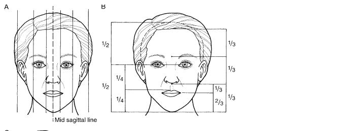
The symmetry assessment is accomplished by dividing the face vertically in half at the midline. This is accomplished by having an assistant hold a silk suture vertically with one hand above the trichion and the other hand below the soft tissue menton with the suture passing through a point between the eyebrows and extending in front of the nasal tip. This allows for the general assessment of rightand left-sided symmetry as well as the relationships between the upper and lower dental midlines. If the deformity or defect is subtle, the frontal profile can be further divided into fifths. Each fifth should approximate one eye’s width beginning at the lateralmost aspect of the ears and extending to the lateral canthus on the right and left sides. Each eye should then be measured from its lateral to medial canthus, and finally, the medial canthal distance should be measured and recorded. This evaluation can also be performed and reviewed at a later date by using a 5 8 frontal photograph. With lines paralleling the midline reference, each fifth should be equal to one eye’s width or the medial canthal distance, thus identifying the region in which subtle asymmetries or deformities are located. During this assessment one should keep in mind that the ideal frontal facial appearance is oval with a width-to-height ratio of three to four.19
Knowing that deformities exist in all three planes of space, the frontal assessment should also be reviewed in relation to horizontal divisions to appreciate the facial balance. This is accomplished by horizontal measurements or lines dividing the face into thirds. The upper third represents the distance between the trichion and soft tissue glabella. The middle third is the space from the soft tissue glabella to subnasale, and the lower
a |
|
b |
|
|
|
9
third is from the subnasale to soft tissue menton. Again, these clinical measurements can be compared and checked with measurements performed on photographs. The lower facial third is also commonly divided into an upper third from the subnasale to stomion and a lower two-thirds from the stomion to soft tissue menton. It should also be noted that upper-facial-third measurements and relations can be misleading due to the varying, and possibly absent, hairlines in some individuals.
The Profile Examination
The profile examination is performed in a similar fashion using the same horizontal landmarks as in the frontal exam. The common facial angles and planes should also be evaluated at this time, assessing the degree of facial convexity or concavity. The Gonzalez-Ulloa line is a reference line that is perpendicular to the Frankfurt horizontal line and passes through the soft tissue nasion. This line helps to establish profiles and the proper chin position.20
At this time all general asymmetries, defects, and deformities should be recorded. Remember that a perfectly symmetric face is an uncommon finding even in the aesthetically beautiful individual. Frontal and profile facial divisions are shown in Figure 2.3a–c.
Cranial Circumference
Absolute measurements of cranial circumference vary with normal adult individuals of the same age and opposite sex. The circumference is approximately 9 mm greater in males
c
FIGURE 2.3 (a) The face is divided into vertical fifths. Each fifth is approximately equal to one eye’s width, beginning at the most lateral aspect of the ear continuing across to the lateral aspect of the opposite ear. (b) Horizontal divisions in the frontal view. The upper third is from trichion to glabella, the middle third is from glabella to subnasale, and the lower third is from subnasale to soft tissue
menton. The lower third can also be subdivided into an upper third and lower two-thirds. The face can also be divided into halves with the distance between the vertex and the midpupillary point being the upper half and the distance from the midpupillary point to menton being the lower half. (c) The facial thirds in the profile view. FH, Frankfort horizontal plane.
10 |
J.P. Morgan, III and R.H. Haug |
than in females of the same age.21,22 In males, cranial growth is rapid during the first 2 years of life with a second growth spurt between ages 12 and 16, whereas females demonstrate their growth spurt between ages 12 and 14 years.21,22 Cranial circumference is not important in adults except when a craniofacial syndrome exists. This measurement is most useful in infants and is a good indication of the size of the intercranial contents as well as of thoracic circumference and body weight.22 The cranial circumference should be measured in centimeters with a measuring tape placed just above the supraorbital rim and encompassing the occiput posteriorly.
Cranial Sutures and Fontanelles
Numerous conditions exist that involve the cranial sutures and fontanelles in infants. This examination should not be overlooked, especially if a syndrome or cranial circumference abnormality is suspected.
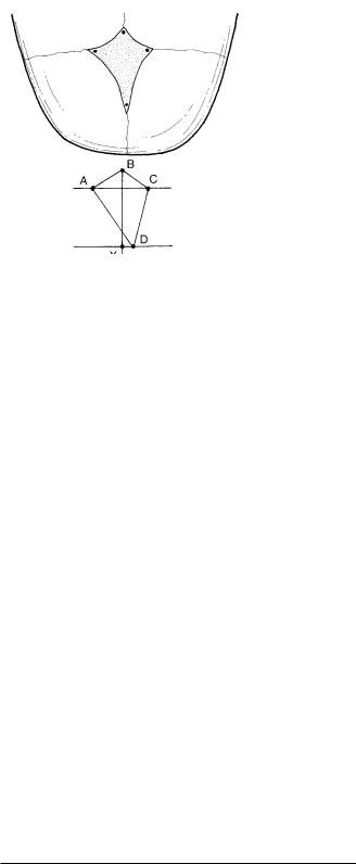
The tension and size of the fontanelles23 are used to estimate intracranial pressure such as that which occurs with meningitis, and it is also used to estimate the degree of brain development. The anterior fontanelle is the largest and is usually obliterated by 2 years of age and replaced by the bregma in the adult skull. During the examination, the area of the fontanelle can be calculated using the formula for the area of a quadrilateral, which is:24
AC BX
Area of ABCD
2
These reference points are made by placing the examiner’s index finger into the right, left, superior, and inferior corners of the fontanelle while using a felt tip pen to mark a point just distal to the examiner’s fingertip.24 The marks are then transferred to a piece of paper by placing the paper directly over the freshly made marks. The points are labeled as in Figure 2.4. Points A and C are connected with a straight line. Then a line parallel to line AC that passes through point D is drawn. A perpendicular line is drawn from line D extending through point B.24 The area is then calculated using the aforementioned formula for the area of a quadrilateral, and compared to the mean values shown in Table 2.2.
Cranial deformities are uncommon and occur when cranial sutures close prematurely. Scaphocephaly occurs when the sagittal suture closes too soon causing the skull to become narrow and elongated. Turrincephaly occurs when the coronal and lambdoid sutures prematurely close giving the skull a tower-like appearance. When the skull becomes even more pointed this condition is called acrocephaly. Complicating matters further, plagiocephaly is caused by an asymmetric premature closure of the coronal or lambdoid sutures resulting in a plethora of asymmetries. The area of the fontanelles and the closure of sutures should be noted and recorded when appropriate.
FIGURE 2.4 The examiner’s index finger being placed into the right, left, superior, and inferior corners of the anterior fontanelle, demonstrating the technique for examining and determining the area of a fontanelle. Each mark is made with a felt tip pen and transferred to a separate piece of paper by gently pressing the paper on top of the freshly made marks. Points A, B, C, and D are labeled, creating a quadrilateral. The area is then calculated using the formula.
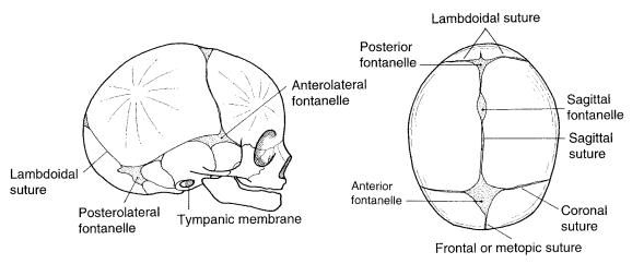
Of the fontanelles, the anterior is the best indicator of brain growth. A small frontal fontanelle for a specific age may indicate abnormally slow brain growth. A third fontanelle, when present, is approximately 2 cm anterior to the posterior fontanelle and occurs in approximately 10% of normal infants and 60% of Down’s syndrome infants.25,26 Figure 2.5a,b shows the fontanelles and their connecting sutures.
Forehead
The forehead composes the upper third of the face, extending from trichion to soft tissue glabella and laterally to the supraorbital rims.
The majority of patients who require surgical correction of the bony forehead usually suffer from craniostenosis, the effects of trauma, or ablative tumor resection. Although the forehead rarely requires surgical correction in normal adults and is commonly overlooked, it does provide important landmarks that are used to evaluate deformities and aesthetics of the rest of the face. In the profile examination, the forehead should exhibit a slight convexity as it extends from trichion to the soft tissue glabella.
TABLE 2.2 Mean areas of the infant’s anterior fontanelle (mm2).
Age |
Mean (mm2) |
Preterm (28–32 weeks) |
113 |
Preterm (33–36 weeks) |
162 |
Term (37–42 weeks) |
220 |
Small-for-dates |
540 |
|
|
11
b
12
patient’s visual acuity should be established. In most cases, this can easily be done using a Snelling chart or a Rosenbaum pocket chart. When a patient is not able to read the largest letter on a Snelling chart, which reveals a visual acuity of 20/400, the examiner should then try to identify the greatest distance at which the patient can count fingers (CF).28 If the patient cannot see the examiner’s fingers, one should try to establish at what distance the patient can note hand motion (HM) by the examiner.28 If HM cannot be established, one must determine if light perception (LP) or no light perception (NLP) exists.28 Other tests that must be considered are determination of extraocular movements, visual fields, and color perception.
Examination of the pupils should not be overlooked and is best performed in a darkened room using the bright light of an ophthalmoscope. The size, shape, and reactivity of each pupil should be evaluated. At this time, a funduscopic examination of each eye should be done evaluating the optic media, disc, and any abnormal pathology. When abnormalities in vision or the ocular examination are noted, a detailed evaluation by an ophthalmologist is recommended.
The Orbit
The clinical evaluation of the interocular distance must be assessed by an actual measurement because clinically the appearance of the distance between the eyes is greatly influenced by the overall height and width of the face, glabellar
J.P. Morgan, III and R.H. Haug
TABLE 2.3 Mean palpebral widths and lengths in Caucasians.
|
Width (mm) |
|
|
Length (mm) |
|
|
|
|
|||
|
|
|
|
|
|
Age (yrs) |
Range |
Mean |
Range |
Mean |
|
|
|
|
|
|
|
1 |
8.0–8.5 |
8.2 |
|
18–21 |
19 |
2–10 |
8.5–9.0 |
8.7 |
|
19–29 |
25 |
11–Adult |
8.0–11.2 |
9.0 |
|
23–33 |
28 |
|
|
|
|
|
|
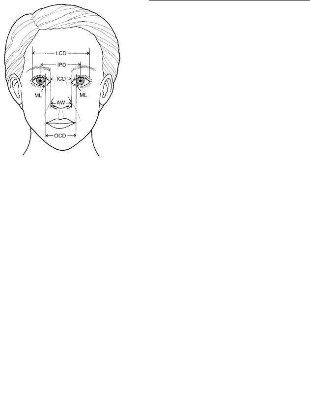
prominence or absence, the shape of the nasal bridge, or the presence or absence of epicanthal folds. Many formulas and methods for evaluating the intercanthal and interpupillary distances appear in the literature.29 A firm distinction between intercanthal distance and interpupillary distance should be made. This is because in patients with anomalies such as Waardenburg syndrome, the outward appearance of ocular hypertelorism is actually a primary telecanthus caused by the lateral displacement of the medial canthus and punctum. Interpupillary and intercanthal measurements are commonly used to assess the position of the orbit and globe.30 The intercanthal distance should be between 30 and 35 mm as compared to the interpupillary distance of 60 to 70 mm.29,30 The interpupillary distance on average should be twice the intercanthal distance and the alar-to-alar nasal base width should be approximately equal to the intercanthal distance in normal Caucasian patients.30
Radiographic measurements can also be used to assess orbital position in children and adults by measuring the distance between the right and left medial orbital walls on an anteroposterior skull radiograph.31 This method has also been used to measure the distance between lateral orbital walls but is shown to have little clinical importance.29 Figure 2.6 demonstrates the relationship between intercanthal and interpupillary measurements as well as their relationship to other facial structures.
If an abnormality is noted in the intercanthal distance, one should also examine the palpebral length and width. In normal infants, the palpebral fissure is extremely narrow and rapidly widens in the first several weeks of life.32,33 In normal infants, children, and adults, measurements of palpebral length will differ between the right and left side 30% of the time.33 Differences greater than 1 mm are usually considered abnormal.33 Table 2.3 demonstrates palpebral lengths and widths.
FIGURE 2.6 The relationship between the intercanthal and interpupillary distances. LCD, lateral canthal distance; IPD, interpupillary distance; ICD, intercanthal distance; AW, alar width; OCD, oral commissure distance; ML, medial limbus tangent to oral commissure.
The Eyebrows
Eyebrow position can be readily evaluated from the frontal view. Abnormalities can obviously be created by soft tissue defects or underlying deformities of the supraorbital rims. The normal eyebrow should begin medially at a point where a vertical line extends up from the medial canthus. It ends laterally at a point along an oblique line that begins at the alar base and extends up through the lateral canthus.34 The medial and lateral extent of the eyebrow should lie on a horizontal line. The eyebrow’s point of maximum height should
2. Evaluation of the Craniomaxillofacial Deformity Patient
be positioned at a point where a vertical line extends up from the lateral limbus of the eye and crosses the brow.34 One must also consider that the integrity of the frontal branch of the facial nerve may also affect brow position. Finally, the brow in men lies on top of the supraorbital rim, while in women it lies above the rim.34
The Eyelids
The upper and lower eyelids should be evaluated for symmetry, shape, and function. The larger and generally more rounded upper eyelid should cover approximately 2 to 3 mm of the iris. The lower eyelid is straight and lies at the margin of the inferior limbus. This assessment should be made with the patient in a primary gaze. No sclera should be noted below the inferior limbus. Excessive anterior position of the globe and/or a poorly supported lower lid will cause excessive sclera to show. Entropion, ectropion, ptosis, elasticity, and function of the lower eyelid should be noted as well as the presence or absence of inferior scleral show.
Globe Position
The anterior, posterior, and superior position of the globe must not be overlooked. The etiology of exorbitism, exophthalmus, and enophthalmus must be identified and noted. Globe position is usually compared to orbital rim projection with the supraorbital rim being approximately 5 to 8 mm anterior to the cornea. The inferior orbital rim should be approximately 2 mm anterior to the cornea. The lateral orbital rims should be approximately 10 to 12 mm posterior to the cornea. These measurements are easily made using a clear ruler and examining the patient from the lateral view with the patient in primary gaze.
Ocular Mobility
Assessment of ocular mobility can be difficult in children and patients who have suffered acute trauma. We suggest that the examiner sit in front of the patient while asking the patient to follow a pen light or the examiner’s fingers. The finger or light should be moved into the six cardinal directions of gaze.35 After the six directions of gaze have been examined, one should ask the patient to follow the light or the examiner’s finger as it is moved toward the nasal bridge. The eyes should converge. This is sustained to within 5 to 8 cm.35 This examination should detect most mobility disorders. If the patient complains of pain or visual disturbances, the exact eye position at which this happens should be documented. If there is a question of entrapment, a forced duction test performed after a local anesthetic is administered will usually differentiate between true entrapment and muscular weakness.
13
The Nose
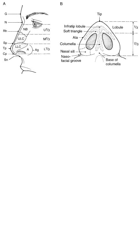
The nose is one of the most aesthetic and functional structures on the face. Its midline position is best examined in the frontal view, and its anterior projection from the profile view. Aesthetically, the nose is not considered as an isolated structure unless it is deformed. The nose is examined in relation to the forehead, orbital rims, eyes, maxilla, lips, and chin. It has been suggested that an aesthetically pleasing nose should flow into the underlying craniomaxillofacial skeleton, represented by smooth interconnecting lines and curves on the topography of the face.36 For traditional examination purposes, the nose is divided into thirds relative to their underlying supporting structures. The proximal third is supported by the nasal bones. The middle third is supported by the upper lateral nasal cartilages. The distal third is supported by the lower lateral cartilages medially and the sesamoid cartilages and dermis laterally.
The nasal septum provides support for both aesthetics and function by separating the bony and cartilaginous vault of the nose. The nasal septum also aids airflow and supports the tip and columella. Owing to trauma, heredity, and developmental changes, the nasal septum is rarely straight.
The mobile portion of the nose includes the membranous septum, columella, and lobule, which contains the tip and alae. The nasal sill and soft triangle make up and support the opening into the nasal vestibule. Figure 2.7a,b shows the common anterior landmarks of the nose.
Congenital, developmental, and acquired deformities of the nose are extremely complex and challenging. Examination of the nose should include inspection from the lateral, frontal, and submental vertex views, as well as a complete intranasal examination.
Because of the vast amount of detailed information concerning the aesthetic evaluation and surgical correction of nasal deformities, the purpose of this section is to provide basic information that describes the normal nose.37–39 When a nasal deformity is identified, obviously a more detailed and specific nasal evaluation is in order. This examination focuses on the characteristics of symmetry, width, projection, and function.
In the frontal view, the nose should be in the midline. A silk suture extending from the glabella to the pogonion should pass through the center of the nasal tip. This divides the nose into equal halves and identifies asymmetries of the nasal bridge and tip. The alar-to-alar width has been described as being approximately 70% of the distance between nasion and the tip-defining point.19,39 This region should be slightly wider in the black and Asian population.19
The area in which the nasal bones and nasal process of the frontal bone blend into the frontal bone makes up the radix, or root, of the nose.39 A normal radix should possess a curvilinear line that begins at the supraorbital ridges and follows the nasal dorsum on the right and left sides of the nose.36,39 Figure 2.8 demonstrates the curvilinear lines of the radix. The
14 |
|
|
J.P. Morgan, III and R.H. Haug |
|
|
|
FIGURE 2.7 (a) Common landmarks and |
a |
|
b |
|
|
|
|
divisions of the nose in the lateral |
|
|
|
|
|
|
|
view. G, soft tissue glabella; N, soft tis- |
|
|
|
sue nasion; Rh, rhinion; Sp, supra-tip- |
|
|
|
break; Tp, tip-defining point; Cp, col- |
|
|
|
umella point; Sn, subnasale; A, ala; |
|
|
|
Ag, alar groove. The nose is divided |
|
|
|
into thirds according to its underlying |
|
|
|
support. The upper third is supported |
|
|
|
by the nasal bones (NB), the middle |
|
|
|
third is supported by the upper lateral |
|
|
|
cartilages (ULC), and the lower third |
|
|
|
is supported by the lower lateral carti- |
|
|
|
lages (LLC). (b) The basilar view of |
|
|
|
the nose and its anatomic landmarks |
|
|
|
and divisions. The lobule should be |
|
|
|
one third of the total height of the base |
|
|
|
of the nose. |
nasal frontal angle (G-N-Tp) should range between 125° and 135°, with the nasal bridge extending approximately 5 to 8 mm anterior to a normally positioned globe.39
The profile view allows one to evaluate nasal length, projection, and rotation of the nasal tip. Projection is defined as the anterior position of the tip relative to the anterior facial plane. Rotation is defined as the inclination of the tip and is indicated by the nasolabial angle. The nasofacial angle assesses the degree of nasal projection and is created by the intersection of facial and nasal planes. For measurement purposes, the angle is represented by a line passing from the soft tissue glabella to the soft tissue nasion and is intersected by a tangent that parallels the nasal dorsum. An angle of between 30° and 35° represents a normal nasal projection.39,40 It has been suggested that nasal projection can easily be assessed where the distance between tip-defining point and subnasial
(Tp-Sn) should equal the distance between subnasale and the vermilion border (Sn-Vs) in a normal nose.41 Situations that alter upper lip length, such as a cleft lip and mentolabial posturing, can make this assessment unpredictable. The nasolabial angle, as described earlier, also evaluates nasal tip projection. Although there are several other techniques used to assess tip projection, the methods that were discussed here are quick, easy, and commonly used.42–44
In the submental vertex view, the nasal base and nostrils are evaluated by merely having the patient tip their head back. A normal base resembles an equilateral triangle.45 The columella should be straight and in the midline. The lobule–nasal base width ratio should be 3:4 in a normal nose. The nostrils should take on a gentle pear shape with the top part of the pear pointing toward the lobule.39,45
When a nasal deformity is present, the nature of the overlying skin should be closely evaluated because superficial scars may distort the mobile portion of the nose and thus make it appear that an underlying defect exists when in reality it does not. Thick skin can make significant bony movements less noticeable and should be considered in the treatment plan, while thin skin may reveal dramatic changes after only subtle bony movements.
The internal nasal exam should identify abnormalities in the septum, turbinates, and/or pathology such as polyps and synechiae. Findings such as these should be documented and investigated as indicated prior to any reconstruction attempts.
The Cheeks
FIGURE 2.8 The topographical curves of the nose. The radix extends from the supraorbital ridges to the lateral dorsal region; the lobularalar rim should be a wide V shape at the tip; the nasolabial junction follows the contour of the upper lip passing through the subnasale extending along the columella.
Subtle deformities that affect malar prominence can be difficult to assess when the overlying skin, underlying bone, and amount of buccal fat mask the true etiology of the deformity. It is agreed that prominent malar bones and arches are generally considered aesthetic and represent a youthful facial
2. Evaluation of the Craniomaxillofacial Deformity Patient
appearance. Normally, the zygomatic arches make up the widest part of the face when viewed frontally. Temporal convexity, buccal fat, and the position of the orbit and auricle influence the interpretation of arch prominence and facial width. When evaluating cheek prominence, one must assess symmetry, projection, and height.
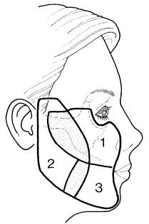
For examination purposes, the cheek can be divided into three regions: suborbital (zone 1), preauricular (zone 2), and the buccal mandibular (zone 3).46 Figure 2.9 shows the zones of the cheek.
Zone 1 extends along the lateral border of the nose medially, the inferior orbital rim and eyelid/cheek junction superiorly, slightly above the gingival sulcus inferiorly, and anterior to the sideburn posteriorly. The underlying bony support in this region is mainly the malar bone and the zygomatic arch. Additional support comes from the anterior maxillary wall and the piriform aperture. Zone 2 extends anteriorly to the anterior border of the masseter muscle and overlaps zone 1 at the malar prominence. Superiorly, it extends above the zygomatic arch to the helix of the ear. Posteriorly, it follows the posterior border of the mandible in the preauricular region and extends all the way to the angle. The inferior border of the mandible makes up its inferior boundary. Bony supporting structures in this region include the zygomatic arch, mandibular ramus, and angle. Other supporting structures in this region include the masseter muscle as well as the parotid gland. The anterior boundary of zone 3 extends from the oral commissure and terminates at the chin midpoint. The superior border meets the inferior border of zone 1, which is su-
FIGURE 2.9 The topographical zones of the cheek. Suborbital (zone 1), preauricular (zone 2), oral buccomandibular (zone 3). The shaded region represents the area of overlap.
15
perior to the gingival sulcus. The posterior border extends back to the masseter muscle and the inferior boundary is made by the remaining inferior border of the mandible. Underlying bony support in this region is made by the mandibular body and symphysis. Significant underlying structures that also provide support and influence the aesthetic appearance of the malar bone are the muscles of facial expression and mastication, which are commonly overlooked.
All three zones overlap at the region of the buccal fat pad. Deformities or defects in any of these regions may affect the overall appearance of the malar bone in zone 1. Thus an apparent malar bone deformity may in reality be normal, while the actual deformity is hidden in zones 2 or 3. Although there are technically three zones for evaluation, the zygomatic arch and malar prominence in zone 1 is where the most attention is directed when evaluating cheek or malar deformities. Close inspection of the other zones must be performed to truly understand the defects’ etiology. Facial nerve palsy, parotid pathology, and the absence of dentoalveolar structures also play a significant role in the interpretation of deformities in this region.
The aesthetic position of the malar region is more dependent on an overall feel for symmetry and balance than an actual measurement. When examining the malar region, the examiner must view the patient from the frontal, profile, oblique, and submental vertex views.47
On frontal view, the examiner must visually inspect and palpate both malar bones and their defects as well as the zygomatic arch and orbital rims for orientation purposes. Deformities in the cheeks, paranasal, and buccal areas must be noted.47 Zygomaticus, the point of maximum prominence of the zygomatic arch, should be identified and compared to the opposite side. Symmetry is of importance here. The most prominent portion of the malar bone should be located approximately 1 cm lateral and 1.5 to 2.0 cm inferior to the normal lateral canthus with the patient in the repose position. Deviations from this point should be documented.
Zone 1 can be further divided into the cheek, paranasal, and buccal areas as described by Zide and Epker to specifically evaluate malar bone position.47 The buccal, cheek, masseter muscle, and intraoral malar buttress region should be palpated to assess the overall thickness of this region. Extraorally, this portion of the cheek should be flat in appearance and should not extend beyond a tangent that extends from the lateral aspect of the malar bone and angle of the mandible.46 Tissue that extends lateral to this line on frontal view is considered to be unaesthetic and abnormal.
The same landmarks should be evaluated when viewing the patient in the profile, oblique, and submental vertex views. When viewing the patient in the profile position for malar deficiencies, one must not overlook globe position and its relation to the supraorbital and infraorbital rims. Exorbitism is a common finding in the non-Caucasian population and usually presents as a malar deficiency.48
16
The Auricle
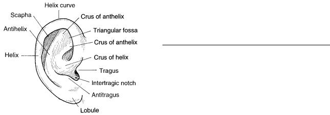
The auricle is an extremely intricate structure made up of convoluted cartilage that is covered by very thin skin except in the lobe region, which is composed of primarily fibrofatty tissue. Figure 2.10 depicts the normal anatomy of the ear. The underlying contour of the cartilage depicts the actual shape of the auricle, making surgical reconstruction difficult. Repair or correction of auricle deformities is one of the greatest challenges a craniomaxillofacial surgeon may be faced with.
The ear is a rich vascular structure that receives its blood supply from the superficial temporal and posterior auricular vessels. The ear has a relatively narrow base when compared to its overall surface area; therefore, any abnormality, previous surgery, or trauma that may involve one of these vessels must be evaluated prior to any reconstruction attempts.
Microtic, constricted, and protruding ear deformities have been shown to have many anatomic and genetic relationships.49 Ear deformities are frequently expressed among families with a history of mandibulofacial dysostosis.50 Studies have also shown that ear deformities may be present in up to 10% of patients or family members of patients with cleft or high-arched palates.50 Possibly up to 25% of patients who present with microtia have family members who demonstrate some evidence of craniofacial microsomia.51 Damage to the stapedial artery causing ischemia has been postulated to be a possible cause of congenital ear deformities as well.52
Congenital ear deformities are evident from birth through adulthood. Traumatic avulsions or loss of ear structure from tumor surgery are dependent on the nature of the injury or location of the tumor and can be acquired at any age. When an ear has been avulsed or amputated and reimplantation attempts have failed, consultation with a maxillofacial prosthodontist is strongly suggested.
The purpose of this section is to give a brief background
J.P. Morgan, III and R.H. Haug
on the etiology of congenital ear deformities and review the shape and position of the normal ear for examination purposes. Generalities and averages will be discussed, and one should remember that normal ears are as distinctive as normal fingerprints.
Although the auricle continues to grow throughout adulthood, it reaches approximately 85% to 90% of its total length by age three and changes very little after the first decade of life.53 The ear grows between 40 and 60 mm until puberty and then continues to enlarge minimally throughout life.53,54 Table 2.4 shows average ear heights and widths associated at various ages for Caucasians.
The width of the ear should be measured from the base of the tragus to the posterior margin of the helical rim. Height is measured from the superior margin of the helical rim to the tip of the earlobe. Ear projection, the amount or degree the ear is elevated off the head, is assessed by measuring the greatest distance the helix is from the mastoid prominence. Although specific numerical values are achieved by measurements, the projection and position of the ear is still considered subjective.55 Actually, ear position should also be related to the position of the external auditory meatus.55 Neck length, cranial vault height, mandibular ramus height, and axial rotation of the auricle all affect the subjective interpretation of ear position.
Preauricular pits, sinuses, appendages, and acquired deformities should be documented. Evaluation of the external auditory canal and tympanic membrane should be performed in a routine fashion in which canal caliber, ossicular function, and integrity of the tympanic membrane should be noted and documented. When external ear deformities exist or when a decrease in hearing acuity is noted, a complete otologic and audiologic evaluation is indicated because middle ear deformities are usually associated with auricle deformities.
If a deformity is present, the surgeon must fully and completely explain the technical limitations involved in the surgical correction or reconstruction of the auricle. The age at which the reconstruction should proceed is determined by both physical and psychologic considerations specific to each individual. Correlation with other necessary facial surgery must be considered.
TABLE 2.4 Average ear widths and heights for males and females.
|
|
Age (yrs) |
Ear width (mm) |
Ear height (mm) |
|
|
0–9 months |
26 |
42.5 |
|
1 |
29.5 |
48.5 |
|
|
4 |
32 |
51.5 |
|
|
8 |
33.5 |
56 |
|
|
10 |
34 |
57.5 |
|
|
16 |
34.5 |
60 |
|
|
18 |
34.5 |
62.5 |
|
|
30–40 |
34.5 |
63.5 |
|
|
50–60 |
34.5 |
66.5 |
|
FIGURE 2.10 The topographical anatomy of the auricle. |
|
70–80 |
35 |
71 |
|
|
|
|
|

2. Evaluation of the Craniomaxillofacial Deformity Patient |
17 |
The Lips
For clinical purposes, the lips should be viewed as they relate to the base of the nose, chin, maxilla, and upper and lower anterior dentition. Much has been written on the physical dimensions of the lips and perioral structures. Unfortunately, most of it is of little clinical importance. With oral competence being the major function of the lips, they are generally viewed as being normal or abnormal by their position and aesthetic value.
The lips should be examined from the facial and profile view where symmetry and balance are of importance. Obvious deformities such as clefts, scars, lesions, and asymmetric regions should be documented. Clefts that involve the lips usually occur once in every 800 to 900 births. The craniofacial and lateralfacial clefts as described by Tessier that can involve the lips are categorized as No. 0 (median craniofacial dysraphia), No. 1 (paramedian craniofacial cleft), No. 2 (similar to No. 1 but more lateral), No. 3 (occlusonasal cleft), No. 4 (occlusofacial cleft I), No. 5 (occlusofacial cleft II), and No. 7 (temporozygomatic cleft).56 Figure 2.11 shows the position of the craniofacial clefts that may involve the lips according to Tessier.
The normal anatomy of the lips should present with two philtral columns along the paramidline of the upper lip. Between the philtral columns, a philtral groove or dimple should be present. Just inferior to the philtral groove should lie the symmetric Cupid’s bow that follows the vermilion border of the upper lip in the midline. The white roll of the upper lip should follow the vermilion border lateral to the Cupid’s bow. The tubercle occupies the mucosal portion of the upper lip, inferior to the Cupid’s bow, and is in the midline. Both the
FIGURE 2.12 The topographical anatomy of the normal lips. Pg, philtral groove; Pc, philtral column; Cb, Cupid’s bow; Tu, tubercle; Wr, white roll; Oc, oral commissure; V, vermilion.
right and left commissures should be symmetric in repose and the vermilion identifies the vermilion border of the lower lip. Figure 2.12 shows the topographical anatomy of normal lips.
Much has been written about the length of the upper lip. It is measured from subnasale to the stomion. On average, it has been shown to be approximately 11 mm in infants, 16 mm at age one, and 20 to 22 mm in the adult (which is reached by 6 years of age).57 Because its borders are poorly defined in many normal individuals, the width of the philtrum is of little concern. The commissure width is measured with the lips in their repose position.58 Table 2.5 shows normal intercommissural widths in Caucasians.
Normal lip fullness is extremely variable, especially in ethnic individuals. Measurements can be made from the middle of the lip to the stomions of the upper or lower lip.
In the repose position, the upper and lower lips should be apart, creating a gap of 3.0 to 3.5 mm. In this position, the amount of upper tooth that is exposed should be approximately 2 to 5 mm from the incisal edge to the bottom of the upper lip. The lower dentition is usually not exposed while the lips are in the reposed position. On full smile, the entire maxillary anterior teeth should be exposed and only 1 to 2 mm of gingival exposure is desirable.59 Abnormal tooth show may be due to jaw or tooth abnormalities, not just lip position.
TABLE 2.5 Mean intercommissural width in Caucasians.
|
Age (yrs) |
|
0–1 |
|
2–3 |
|
8–9 |
|
12–13 |
FIGURE 2.11 The position and numbering of craniofacial clefts that |
14–15 |
involve the lips using Tessier’s classification system. |
16–Adult |
|
Females (mm) |
Males (mm) |
27 |
32 |
30 |
35 |
42 |
44 |
45 |
48 |
47 |
50 |
50 |
52 |
18
FIGURE 2.13 Normal upper and lower lip protrusion. The reference line should pass through the subnasale (Sn) and soft tissue pogonion (Pg). The upper lip should be 3.5 mm anterior to this reference line and the lower lip should be 2.2 mm anterior to the reference line.
On profile view, the upper lip should be fuller than the lower lip. Using a clear plastic ruler, a reference line can be established, which extends from the subnasale to the soft tissue pogonion. This can also be accomplished on a lateral photograph or cephalometric x-ray. The upper lip should be 3.5 mm anterior to the line as compared to the lower lip, which should be 2.2 mm anterior to this line.60 Figure 2.13 shows the protrusion of the normal upper and lower lip.
Finally, the function of the upper and lower lips should be evaluated with the patient in full smile, repose, and the pucker position. Any weaknesses or asymmetries during function may suggest damage to the motor innervation, which is supplied by the buccal and marginal mandibular rami of cranial nerve VII. The locations of these deficiencies should be documented.
Chin-Neck Contour
Generally, the chin and neck contours are evaluated in the frontal and profile views. Clinically, the chin begins at soft tissue menton and extends superiorly into the mentolabial sulcus. The depth of the mentolabial sulcus can give a false interpretation of lower facial height in the frontal view as well as a false interpretation of the protrusion or retrusion of the chin point in the profile view. The correct depth of the mentolabial sulcus is subjective and can be influenced by the actual chin position, lower lip length and position, and the lower anterior dental alveolar structures. Its depth should lie approximately 3 to 4 mm posterior to a line that passes from the vermilion border of the lower lip and extends through soft
J.P. Morgan, III and R.H. Haug
tissue pogonion.61 Excessive anterior flare of the mandibular anterior dentition or mandibular prognathism will usually present with a deficient mentolabial sulcus giving the lower facial third a rather flat appearance. A short lower facial height and a retrognathic mandible will usually be associated with an excessive mentolabial sulcus. The aging face is also associated with an excessive mentolabial sulcus.
Soft tissue chin projection is evaluated in the profile view, in which the distance from the soft tissue pogonion to the Gonzales line (a line extending interiorly from soft tissue nasion and is perpendicular to the Frankfort horizontal plane) is measured. A soft tissue pogonion that falls within 3 mm anterior or posterior to the Gonzales line is considered a normal chin position.
Frontally, the most important clinical aspect of the chin is symmetry and balance. Any abnormality should be documented, remembering that the relative position of the nose, dentition, lips, and neck contour or deformities of these structures will affect the overall appearance and position of the chin.
The neck contour should be evaluated in the frontal, profile, and basilar views. When examining the patient in a frontal and profile position, the patient’s head should be in the neutral position with the facial muscles and lips in the repose position. In the frontal view, a definite line should be easily followed outlining the inferior border of the mandible. Bilateral and symmetric contour concavities should be noted when following the lateral border of the mandibular angle and lateral neck, which should feather out inferiorily and laterally along the trapezius muscle. The sternocleidomastoid muscle should be subtly visible just medial to the mandibular angle and extending inferior and medially as it approaches the sterno-clav- icular region. Any abnormalities in facial width, mandibular (chin) position, and/or excessive laxity of the overlying skin will obviously affect neck contour and appearance. The examiner should palpate the skin in this region to determine its laxity and adherence to underlying structures as well as any hidden mass or defects.
In the profile view, there should be a subtle but definite outline of the inferior border of the mandible as it extends from the chin to the posterior ramus region. The right and left sides should be evaluated and compared where gross asymmetries and defects should be recorded. The chin-neck angle is also evaluated in this position and should be compared to the overall chin projection, lower lip position, and mentolabial sulcus depth. The chin-neck angle is formed by the anterior border of the neck and the submental region extending from point C through soft tissue menton. Normal chin-neck angles are usually between 110° and 120°. Also in the normal neck, one should be able to identify the anterior border and body of the sternocleidomastoid muscle.
Finally, the chin-neck region should be evaluated in the basilar position. One should appreciate symmetry and the amount of redundant tissue in the submental region as well as the skin’s adherence to the underlying structures.
2. Evaluation of the Craniomaxillofacial Deformity Patient
With all of this in mind, the chin-neck area can be classified according to the amount of redundant tissue, platysmal development, and the relative chin position.62 For examination purposes, there are six classes with specific characteristics. Class I presents with a normal chin-neck angle and normal skin tone. Class II shows an increased laxity of the skin with a relatively normal platysma muscle tone. Class III shows a definite accumulation of submental fat. Class IV has obvious banding of the platysma muscle. Class V is seen when the mandible is moderately retrognathic. Class IV presents with an excessively obtuse chin-neck angle and may be due to an inferiorly positioned hyoid bone.62
Oral Cavity and Occlusion
Before any attempts are made to surgically correct any craniomaxillofacial deformity, whether congenital, developmental, or acquired, a complete examination of the oral cavity and occlusion must be performed and not overlooked. While it is easy to focus one’s attention on the very obvious and dramatic aesthetic craniomaxillofacial deformities that a patient may have, the examining surgeon must keep in mind that facial asymmetry and imbalance may be due to poor dentoalveolar structures and relationships. The examination should include a close survey of the lips, labial mucosa, buccal mucosa, mucobuccal fold, hard palate, soft palate and uvula, oropharynx, nasopharynx, tongue, floor of the mouth, muscles of mastication, periodontium, teeth, and occlusion.
While examining the lips and labial mucosa, the overall muscular control of the lips can be evaluated during normal conversation. Visual inspection of the lips will reveal most abnormalities and enlargements. The color and texture of the vermilion border should be noted, and the lips should also be examined for fissuring. Any submucosal nodules or other abnormalities of the lips and labial mucosa can be identified by using bidigital palpation.
The buccal mucosa can easily be evaluated when the patient’s mouth is partially opened using a mouth mirror to retract the cheek laterally. This will allow direct visualization of the area hidden by the maxillary tuberosity. The mucosa of the buccal cheek should be dried using a gauze sponge, and with the aid of bimanual palpation, the parotid gland can be milked, thus evaluating its function and the integrity of Stenson’s duct. Foul-smelling and discolored saliva should be noted.
The mucobuccal fold is usually hidden but should be examined visually and by palpation. This is easily done by retracting the buccal mucosa laterally at its vestibular depth and palpating its depth and alveolar bone using one’s index finger. Contour abnormalities, excessive scar bands, fistulas, and painful regions as well as clefts should be documented.
The hard and soft palate and uvula can be examined by direct vision. The mucosa overlying the hard palate is extremely keratinized and firmly attached. It should be pale pink in color
19
as compared to the soft palate, which sometimes may appear more yellow in color due to its increased amount of adipose tissue and its thin mucosal covering. This region should be palpated and any abnormality should be documented. The patient’s gag reflex should also be noted and appreciated.
The oropharynx and nasopharynx should be inspected by direct and indirect vision. The entire anterior tonsillar pillar should be examined for symmetry and palpated to identify any submucosal masses. Using a gauze sponge to grip the tip of the tongue, retracting it anteriorly, while placing a warmed mouth mirror at its base and having the patient say “ahh,” one can easily and clearly visualize the oropharynx. The nasopharynx can be personally viewed by just rotating this mirror to reflect superiorly. A flexible fiberoptic scope should be used if available. Regardless of the technique used, patient compliance is imperative when examining these regions. The use of topical local anesthetics may decrease the tendency to gag when using the mirror technique and must be used intranasally along with a vasoconstrictive spray when advancing a flexible fiberoptic scope through the nose.
The tongue should be examined. The use of a gauze sponge will aid in retracting the tongue forward, upward, and to the right and left. The entire tongue should be palpated and its shape, size, fissural pattern, color, deformities, and unusual tremors noted.
The floor of the mouth is evaluated by having the patient raise the tongue, enabling the examiner to visually inspect this region. Bimanual palpation of this region is mandatory and is accomplished by placing the index finger along the floor while the other hand supports the submandibular region extraorally. The entire floor should be examined in this fashion. The lingual aspect of the mandible should also be palpated, noting any irregularities. The function and quality of the saliva from the submandibular gland should be evaluated as was performed when examining the parotid gland.
The muscles of mastication should be palpated extraorally and intraorally when possible. Hypertrophy, function, and tenderness should be noted and may indicate possible temporomandibular joint dysfunction.
The periodontium, when visually inspected, is a good indicator of the overall oral hygiene. Poor oral hygiene and inflammation should be noted. The quality, health, and amount of attached gingiva should also be recorded. Selective periodontal probing is recommended in areas where inflammation is noted or where segmental osteotomies may be planned. Patients who present with obvious periodontal disease should be evaluated and treated by a dentist or periodontist to achieve the best gingival health possible prior to any procedure that involves the dentoalveolar structures. This should decrease the chance of postoperative complications such as wound dehiscence and infection.
Finally, the teeth are examined both clinically and radiographically. Missing teeth should be noted. Decayed, symptomatic, and mobile teeth should be restored if at all possible. The occlusion should be examined clinically and by the
20 |
J.P. Morgan, III and R.H. Haug |
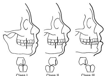
FIGURE 2.14 Angle’s classification of occlusion is based on the position of maxillary and mandibular permanent first molars. Class I is considered normal; the mesiobuccal cusp of the maxillary first permanent molar should rest in the mesiobuccal groove of the mandibular first permanent molar. Class II is considered retrognathic with the mesiobuccal cusp of the maxillary first permanent molar resting anterior to the mesiobuccal groove of the mandibular first permanent molar. Class III is considered prognathic, with the mesiobuccal cusp of the maxillary first permanent molar resting posterior to the mesiobuccal groove of the mandibular first permanent molar.
use of dental study models. Angle’s classification is most commonly used to describe the occlusion. It is based on the position of the maxillary and mandibular first permanent molars. Figure 2.14 shows Angle’s classification of occlusion. The general arch shape and transverse width must also be noted. Other techniques such as transillumination, percussion, and pulp testing should be used as specifically indicated.
Conclusion
When treating craniomaxillofacial deformity patients, it is extremely important that the examining surgeon develop a comprehensive, stepwise system for examining and documenting the complex deformities with which these patients may present. Only through a systematic approach can information and data be collected, reviewed, and interpreted, thus diagnosing the true etiology of the craniomaxillofacial deformity. The examination provides only part of the information that is required to make the diagnosis. Dental study models, radiographs, cephalometric analysis, CT scans, and photo documentation must also be used in conjunction with the clinical examination to both diagnose and document the patient’s preoperative state. These study aids can also be shown to the patient and family members to demonstrate the etiology of their deformity. In today’s litigious society, accurate and organized documentation of the patient’s preoperative state is mandatory, thus providing a complete medical record and good risk management.
Today, third-party payers will often request supplemental information, such as study models, x-rays, and photographs that support your diagnosis and indication for surgery before approval can be granted. Knowing that the information obtained and treatment plan is different for each patient, additional information pertaining to more specific deformities or problems may be needed.
The information obtained in this examination should allow one to accurately diagnose and formulate a treatment that will provide a predictable, functional, and aesthetic result.
References
1.Wood NK. Treatment Planning: A Pragmatic Approach. St. Louis, MO: C.V. Mosby; 1978: ch 2, 29.
2.McCarthy JG. Plastic Surgery, The Face, vol. 2. Philadelphia: W.B. Saunders; 1990: ch 29, 1188.
3.Weed LL. Medical records that guide and teach. N Engl J Med 1968;278(11):593.
4.Peterson LJ, Frost DE. Database Collections. Principles of Oral and Maxillofacial Surgery, vol. 3. Philadelphia: J.B. Lippincott Company; 1992, ch 49.
5.Proffit WR, Epker BN, Ackerman JL. Systematic description of dentofacial deformities: the data base. In: Bell WE, Proffit WR, White RW, eds. Surgical Correction of Dentofacial Deformities. St. Louis, MO: C.V. Mosby; 1980: ch 5, 105.
6.Tucker MR, Thomas PM. Temporomandibular pain and dysfunction in the orthodontic surgical patient: Rationale for evaluation and treatment sequencing. Int J Adult Orthop Orthogn Surg. 1986;1:11.
7.Karabouta I, Mantis C. The TMJ dysfunction syndrome before and after sagittal split osteotomy of the rami. J Oral Maxillofac Surg. 1989;13:185.
8.Gorney M, Harries T. The preoperative and postoperative considerations of natural facial asymmetry. Plast Reconstr Surg. 1974;54:187.
9.Gonzalez-Ulloa M. A quantum method for the appreciation of the morphology of the face. Plast Reconstr Surg. 1964;34:241.
10.Baer MJ. Dimensional changes in the human head and face in the third decade of life. Am J Phys Anthropol. 1956;14:557.
11.Whitaker LA, LaRossa, D, Randall P. Structural goals in craniofacial surgery. Cleft Palate J. 1975;12:23.
12.Farkas LG, Katic MJ, Hreczko Ta, et al. Anthropometric proportions in the upper lip–lower lip–chin area of the lower face in young white adults. Am J Orthol. 1984;86:52.
13.Peck H, Peck S. A concept of facial aesthetics. Angle Orthod. 1970;40:284.
14.Tolleth H. Concepts for the plastic surgeon from art and sculpture. Clin Plast Surg. 1987;14:585.
15.Showfety KJ, Vig PS, Matteson S. A simplified method for taking neutral-head-position cephalograms. Am J Orthod. 1983;83: 495.
16.Romm S. Art, love and facial beauty. Clin Plast Surg. 1987; 14:579.
17.Ricketts RM. Divine proportions in facial esthetics. Clin Plast Surg. 1982;9:401.
18.Larrabee WF. Facial analysis for rhinoplasty. Otolaryngol Clin North Am. 1987;20:653.
2. Evaluation of the Craniomaxillofacial Deformity Patient
19.McGraw-Wall B. Facial analysis. In: Bailey BJ, ed. Head and Neck Surgery—Otolaryngology. Philadelphia: J.B. Lippincott Company; 1993: ch 158, 2070.
20.Gonzalez-Ulloa M. Quantitative principles in cosmetic surgery of the face (profile-plasty). Plast Reconstr Surg. 1962;29:186.
21.Westropp CK, Barber CR. Growth of the skull in young children: I. Standards of head circumference. J Neurol Neurosurg Psychiatry. 1956;29:52.
22.Illingworth RS, Eid EE. The head circumference in infants and other measurements to which it may be related. Acta Pediatr Scand. 1971;333:60.
23.Popich GA, Smith DW. Fontanelles: range of normal size. J Pediatr. 1972;80:749.
24.Davies DP, Ansari BM, Cooke TJ. Anterior fontanelle size in the neonate. Arch Dis Child. 1975;50:81.
25.Chemke J, Robinson A. The third fontanelle. J Pediatr. 1969;75:617.
26.Tan KL. The third fontanelle. Acta Paediatr Scand. 1971;60:329.
27.Ousterhout DK. Feminization of the forehead: contour changing to improve female aesthetics. Plast Reconstr Surg. 1987;79:701.
28.Jelks GW, Jelks EB, Ruff G. Clinical and radiographic evaluation of the orbit. Otolaryngol Clin North Am. 1988;21:13.
29.Laestadius N, Aase JM, Smith DW. Normal canthal and outer orbital dimensions. J Pediatr 1969;74:465.
30.Holt GR, Holt JE. Nasoethmoid complex fractures. Otolaryngol Clin North Am. 1985;18:89.
31.Hansman CF. Growth of interorbital distance and skull thickness as observed in roentgenographic measurements. Radiology. 1966;86:87.
32.Chouke KS. The epicanthus or mongolian folds in caucasian children. Am J Phys Anthropol. 1929;13:255.
33.Fox SA. The palpebral fissure. Am J Opthalmol 1966;62:73.
34.Brennan GH. Correction of the ptotic brow. Otolaryngol Clin North Am. 1980;13:265.
35.Bates B. A Guide to Physical Examination and History Taking.
4th ed. Philadelphia: J.B. Lippincott Company; 1982: ch 7, 170.
36.Sheen JH. Secondary rhinoplasty. Plast Reconstr Surg. 1975; 56:137.
37.Rollin DK, Leslie FG. Rhinoplasty: image and reality. Clin Plast Surg. 1988;15(1):1–10.
38.Larrabee WF. Facial analysis for rhinoplasty. Otolaryngol Clin North Am. 1987;20(4):653.
39.Stella JP, Epker BN. Systematic aesthetic evaluation of the nose for cosmetic surgery. Oral Maxillofac Surg Clin of North Am.
1990;2(2):273.
40.Brown JB, McDowell F. Plastic Surgery of the Nose. St. Louis, MO: C.V. Mosby; 1951: ch 2, 30.
21
41.Simons RL. Nasal tip projection, ptosis and supratip thickening.
J Ear Nose Throat. 1982;61:452.
42.Baum SJ. Introduction. J Ear Nose Throat. 1982;61:426.
43.Powell N, Humphries B. Proportions of the Esthetic Face. New York: Thieme-Stratton; 1984.
44.Crumley RL, Lancer R. Quantitative analysis of nasal tip projection. Laryngoscope. 1988;98:202.
45.Bernstein L. Esthetics in rhinoplasty. Otolaryngol Clin North Am. 1975;8:705.
46.Zide BM. Deformities of the lips and cheeks. In: McCarthy JG, ed. The Face, vol. 2. Philadelphia: W.B. Saunders; 1990: ch 38, 2037.
47.Zide MF, Epker BN. Systematic aesthetic evaluation of the cheeks for cosmetic surgery. Oral Maxillofac Surg Clin North Am. 1990;2(2):351.
48.Block MS, Zide MF. Orbital decompression by midfacial osteotomy. Oral Surg Oral Med Oral Pathol. 1984;57:479–484.
49.Rogers B. Microtia, lop, cup and protruding ears: four directly inherited deformities? Plast Reconstr Surg. 1968;41:208.
50.Rogers B. Berry-Treacher Collins syndrome: a review of 200 cases. Br J Plast Surg. 1964;17:109.
51.Tanzer RC. Total reconstruction of the auricle. The evaluation of a plan of treatment. Plast Reconstr Surg. 1971;47:523.
52.McKenzie J, Craig J. Mandibulo-facial dysostosis (TreacherCollins syndrome). Arch Dis Child 1955;30:391.
53.Lucas WP, Pryor HB. Range and standard deviations of certain physical measurements in healthy children. J Pediatr. 1935;6:533.
54.Rubin LR, Bromberg BE, Walden RH, et al. An anatomic approach to the obtrusive ear. Plast Reconstr Surg. 1962;29:360.
55.Robinow M, Roche AF. Low-set ears. Am J Dis Child. 1973;125:482.
56.Tessier P. Anatomical classification of facial, cranio-facial and latero-facial clefts. J Maxillofac Surg. 1976;4:69.
57.Feingold M, Bossert WH. Normal values for selected physical parameters: An aid to syndrome delineation. Birth Defects. 1974;10(13):1.
58.Cervenka J, Figalovà P, Gorlin RJ. Oral intercommissural distance in children. Am J Dis Child. 1969;117:434.
59.Vig KD, Ellis E. Diagnosis and treatment planning for the sur- gical-orthodontic patient. Clin Plast Surg. 1989;16:645.
60.Burstone CJ. Lip posture and its significance in treatment planning. Am J Orthod. 1967;53:262.
61.Simons RL. Adjunctive measures in rhinoplasty. Otolaryngol Clin North Am. 1975;8:717.
62.Dedo DD. A preoperative classification of the neck for cervicofacial rhytidectomy. Laryngoscope 1980;90:1984.