

- •Preface
- •Acknowledgments
- •Contents
- •Contributors
- •1. Introduction
- •2. Evaluation of the Craniomaxillofacial Deformity Patient
- •3. Craniofacial Deformities: Review of Etiologies, Distribution, and Their Classification
- •4. Etiology of Skeletal Malocclusion
- •5. Etiology, Distribution, and Classification of Craniomaxillofacial Deformities: Traumatic Defects
- •6. Etiology, Distribution, and Classification of Craniomaxillofacial Deformities: Review of Nasal Deformities
- •7. Review of Benign Tumors of the Maxillofacial Region and Considerations for Bone Invasion
- •8. Oral Malignancies: Etiology, Distribution, and Basic Treatment Considerations
- •9. Craniomaxillofacial Bone Infections: Etiologies, Distributions, and Associated Defects
- •11. Craniomaxillofacial Bone Healing, Biomechanics, and Rigid Internal Fixation
- •12. Metal for Craniomaxillofacial Internal Fixation Implants and Its Physiological Implications
- •13. Bioresorbable Materials for Bone Fixation: Review of Biological Concepts and Mechanical Aspects
- •14. Advanced Bone Healing Concepts in Craniomaxillofacial Reconstructive and Corrective Bone Surgery
- •15. The ITI Dental Implant System
- •16. Localized Ridge Augmentation Using Guided Bone Regeneration in Deficient Implant Sites
- •17. The ITI Dental Implant System in Maxillofacial Applications
- •18. Maxillary Sinus Grafting and Osseointegration Surgery
- •19. Computerized Tomography and Its Use for Craniomaxillofacial Dental Implantology
- •20B. Atlas of Cases
- •21A. Prosthodontic Considerations in Dental Implant Restoration
- •21B. Overdenture Case Reports
- •22. AO/ASIF Mandibular Hardware
- •23. Aesthetic Considerations in Reconstructive and Corrective Craniomaxillofacial Bone Surgery
- •24. Considerations for Reconstruction of the Head and Neck Oncologic Patient
- •25. Autogenous Bone Grafts in Maxillofacial Reconstruction
- •26. Current Practice and Future Trends in Craniomaxillofacial Reconstructive and Corrective Microvascular Bone Surgery
- •27. Considerations in the Fixation of Bone Grafts for the Reconstruction of Mandibular Continuity Defects
- •28. Indications and Technical Considerations of Different Fibula Grafts
- •29. Soft Tissue Flaps for Coverage of Craniomaxillofacial Osseous Continuity Defects with or Without Bone Graft and Rigid Fixation
- •30. Mandibular Condyle Reconstruction with Free Costochondral Grafting
- •31. Microsurgical Reconstruction of Large Defects of the Maxilla, Midface, and Cranial Base
- •32. Condylar Prosthesis for the Replacement of the Mandibular Condyle
- •33. Problems Related to Mandibular Condylar Prosthesis
- •34. Reconstruction of Defects of the Mandibular Angle
- •35. Mandibular Body Reconstruction
- •36. Marginal Mandibulectomy
- •37. Reconstruction of Extensive Anterior Defects of the Mandible
- •38. Radiation Therapy and Considerations for Internal Fixation Devices
- •39. Management of Posttraumatic Osteomyelitis of the Mandible
- •40. Bilateral Maxillary Defects: THORP Plate Reconstruction with Removable Prosthesis
- •41. AO/ASIF Craniofacial Fixation System Hardware
- •43. Orbital Reconstruction
- •44. Nasal Reconstruction Using Bone Grafts and Rigid Internal Fixation
- •46. Orthognathic Examination
- •47. Considerations in Planning for Bimaxillary Surgery and the Implications of Rigid Internal Fixation
- •48. Reconstruction of Cleft Lip and Palate Osseous Defects and Deformities
- •49. Maxillary Osteotomies and Considerations for Rigid Internal Fixation
- •50. Mandibular Osteotomies and Considerations for Rigid Internal Fixation
- •51. Genioplasty Techniques and Considerations for Rigid Internal Fixation
- •52. Long-Term Stability of Maxillary and Mandibular Osteotomies with Rigid Internal Fixation
- •53. Le Fort II and Le Fort III Osteotomies for Midface Reconstruction and Considerations for Internal Fixation
- •54. Craniofacial Deformities: Introduction and Principles of Management
- •55. The Effects of Plate and Screw Fixation on the Growing Craniofacial Skeleton
- •56. Calvarial Bone Graft Harvesting Techniques: Considerations for Their Use with Rigid Fixation Techniques in the Craniomaxillofacial Region
- •57. Crouzon Syndrome: Basic Dysmorphology and Staging of Reconstruction
- •58. Hemifacial Microsomia
- •59. Orbital Hypertelorism: Surgical Management
- •60. Surgical Correction of the Apert Craniofacial Deformities
- •Index
39
Management of Posttraumatic Osteomyelitis of the Mandible
Robert M. Kellman and Darin L. Wright
As discussed in a previous chapter, there are several different etiologies for mandibular osteomyelitis. Treatment is to a large extent cause specific since the differing pathophysiologies involved require different approaches. Odontogenic osteomyelitis can often be treated medically with a prolonged course of antibiotics. When surgical debridement is necessary, the treatment will still be medical once debridement has been completed unless a pathologic fracture has occurred. On the other hand, if a fracture is present, treatment should be similar to that described here for posttraumatic osteomyelitis (PTOM).
Osteoradionecrosis (ORN) is a term applied to the specific form of osteomyelitis that develops after exposure of the bone to a treatment course of radiotherapy. This particular problem is addressed in a separate chapter.
The specific entity of PTOM refers to the bone infection that develops after a fracture, whether the fracture has been treated or not. As the name implies, PTOM suggests that an infection has developed in the bone at the site of a fracture. It is more than a soft tissue infection, which can generally be treated successfully by drainage combined with systemic antibiotics and stabilization of a nonfixed fracture. Loose hardware must always be removed from an infected wound, although stable appliances will usually withstand a localized wound infection.
PTOM may develop at the site of an unrepaired mandible fracture. Failure to repair a fracture may be due to poor patient compliance, missed diagnosis, or occasionally the presence of severe, life-threatening injuries. In this situation, it is important to differentiate between a localized infection that will respond to drainage, fixation, and antibiotics and a true PTOM, which will require debridement of osteitic bone.
PTOM may also be seen after inadequately or improperly treated fractures. An improperly applied fixation appliance may become a source of infection at the site of an unstable fracture. Swift removal of the appliance and proper stabilization of the fracture fragments may avert progression of infection in the bone.
Finally, teeth in fracture lines have been implicated by many in the later development of PTOM. This remains quite controversial, and in general, it appears that proper fixation of bony fragments overcomes any tendency toward infection provided by teeth in fracture lines.1–3 The one exception with which most authors will agree is that a preexisting pulp infection in a tooth at the fracture site is highly likely to result in infection and is, therefore, an indication for extraction at the time of fracture repair.4
The management of PTOM has evolved slowly over the past three decades. In addition to intravenous antibiotics, surgery often included debridement and packing of wounds open, allowing healing to take place by secondary intention.5 The advent of transcutaneous suction-irrigation systems has allowed for successful healing using primary closure. The need to stabilize fractures, nonunions, and debridementcreated defects has been recognized, and external fixation has been the mainstay for this.8
Giordano et al. reported on eight cases of PTOM of which four were treated with decortication and packing and four with debridement and primary closure over a transcutaneous suction-irrigation system.5 The latter resulted in less patient discomfort and shorter hospital stays. When needed, stabilization was accomplished using external fixators.
In 1985, Adekeye and Cornah reported on 106 cases of mandibular osteomyelitis. Their recommendations for treatment included debridement, drainage, antibiotic therapy, and fixation of mobile fragments.6 Calhoun et al. advocated the use of judicious debridement, intravenous antibiotics, suction irrigation, and external fixation with the addition of hyperbaric oxygen (HBO) in their study of 60 patients.7
The underlying theme in many authors’ recommendations for treatment of PTOM is that of staged procedures in which the infection is cleared using debridement, suction-irrigation drains to directly apply an antibiotic irrigant, intravenous antibiotics, and fixation of mobile fragments using intermaxillary fixation, external fixators, or both.8,9 Reconstruction of mandibular defects is delayed until all signs of infection have
433
434
been eliminated. Adekeye recommended in 1978 waiting at least 1 month prior to attempting bone grafting for reconstruction.10 Similarly, in 1991 Mercuri advocated a wait of 2 to 3 months prior to reconstructive attempts.11
Not all authors, however, recommend delayed bone grafting. The use of primary bone grafting after debridement of mandibular osteomyelitis was advocated by Obwegeser in 1966.12 However, while some authors have reported on the occasional use of a primary bone graft, success has been variable, and many authors still decry this procedure. Glahn reported on four cases of mandibular osteomyelitis treated using primary bone grafting at the time of debridement.13 He recommended the use of a millipore filter to protect the graft from the surrounding soft tissue inflammation. The only case of the four that failed was one in which this filter was not used. Beckers et al. treated 19 patients with PTOM, and 4 of these patients had bone grafts placed primarily.14 While 2 of the 4 patients developed postoperative infection, both patients went on to osseous union. Obwegeser and Sailer reported on 17 cases of primary reconstruction using rib or iliac bone and stabilization using intermaxillary fixation. Their treatment was successful in 15 patients, with 2 patients requiring removal of necrotic bone.15
Equally controversial is the use of rigid internal fixation (RIF) in the treatment of PTOM. In the late 1960s and early 1970s, the use of rigid internal fixation after bony debridement was introduced for the stabilization of PTOM in orthopedic fractures. While advocated occasionally for PTOM of the mandible, resistance has remained strong. In 1984 Rowe stated that many cases of nonunion in infected fractures could be traced to the use of internal fixation.16 Marx echoed Rowe’s recommendations as recently as 1991 stating that “. . . placement of either an internal reconstruction plate or an immediate bone graft is associated with a high incidence of reinfection and is not recommended.”8
In reviewing reports in the literature, variability in treatments and patient populations make comparative evaluation difficult. The definition of osteomyelitis and particularly acute
R.M. Kellman and D.L. Wright
and chronic osteomyelitis is variable, and criteria for inclusion are quite inconsistent. Most reviews include different types of osteomyelitis including odontogenic, posttraumatic, and osteoradionecrosis as well as less common types in the same series.
In an effort to focus on the particular problem of fixation and PTOM, we have studied the use of rigid internal fixation with or without primary bone grafting in 14 patients. A preliminary report of these patients was presented at the American Academy of Facial Plastic and Reconstructive Surgery meeting in June 1993.17 This reviews the authors’ experience with 14 cases of mandibular osteomyelitis, all of which were associated with persistent nonunions, defects, or both, with a particular focus on the use of RIF and bone grafting. While all cases would fit into the category of chronic osteomyelitis based on the criteria in most reports, the distinction between acute and chronic osteomyelitis is somewhat cloudy and less important than the fact that all patients had failed to resolve on antibiotic therapy of greater than 1 month’s duration and osteitic bone was found at surgery in all cases.
Patients
Fourteen patients with PTOM of the mandible were treated surgically using debridement and RIF with an AO mandibular reconstruction plate between 1983 and 1992. The presumed causes of osteomyelitis included unrecognized or untreated fractures in four patients and identifiable treatment errors in seven patients. The remaining 3 patients were treatment failures after RIF of fractures in which the treatment appeared to have been correct and the cause of failure, therefore, could not be identified. Original treatment sites included six body fractures, five angle fractures, and three parasymphyseal fractures.
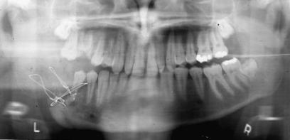
All patients had evidence of infection involving cortical bone and marrow, with bone loss identified radiologically and/or at surgery in all cases (Figure 39.1). All failed to re-
FIGURE 39.1 Panorex of one of the patients included in this series revealing osteomyelitis of an inadequately treated left angle fracture. The fracture is not well reduced, and the surrounding bone is osteopenic.
39. Management of Posttraumatic Osteomyelitis of the Mandible
spond to antibiotic therapy (culture specific or empiric) for 1 month or more. Eleven patients had been treated prior to referral, 7 with courses of antibiotics only and 4 with antibiotics and one or more surgeries in efforts to clear the PTOM.
Prior to surgery, all patients received intravenous antibiotic treatment for 10 to 14 days. Surgery included radical debridement of osteitic bone in all cases without regard for the size of the defect that would result. Defects ranged from 0 to 7 cm after debridement.
Seven patients underwent immediate RIF for stabilization using the AO mandibular reconstruction plate (stainless steel or titanium) at the time of debridement. Five of these patients had bone defects after debridement. Three underwent primary bone grafting at the time of debridement and plate placement and two underwent secondary bone grafting. All grafts consisted of cancellous iliac bone (no cortical component), which was pressed into the defect between the bone ends in the space under the plate. Generous amounts of cancellous bone were used.
Seven patients underwent aggressive surgical debridement without plate placement. In two patients, external fixation was used for stabilization, and in five patients no fixation was used. Secondary plate placement was carried out within 7 to 25 days. At the time of plate placement, five patients had defects grafted using cancellous iliac bone pressed into the space under and around the plate.
Three patients underwent elective plate removal, two of whom had undergone primary placement. At the time of removal, bones showed complete healing and replacement of the grafts with normal-appearing bone. The mandibles were completely stable and tolerated normal masticatory function after plate removal. No plates were removed for any nonelective reasons.
Results
All patients in this series went on to complete bony union without further evidence of infection over a follow-up period of 6 months to 7 years. One patient presented 2 months after secondary repair complaining of pain over the graft site. No clinical evidence of infection was found. Nonetheless, the patient was treated with 4 weeks of intravenous clindamycin using home therapy. No further complaints of pain were made, and clinically normal healing was present at last follow-up. As noted earlier, complete bony union was found in the three patients who underwent plate removal.
Time hospitalized was noted to range from 7 to 43 days (cumulatively, excluding treatment by prior physicians). There was no significant difference between those who had plates placed primarily (range of 7 to 42 days, mean and median of 23 days) and those who had plates placed secondarily (range of 9 to 43 days, mean of 27 days, median of 28 days).
435
Status of dentition, the presence of teeth in the fractures, and how these were handled were randomly distributed. Cigarette smoking is frequently noted in series to be associated with mandibular osteomyelitis, and this proved true in this series as well (9 of 12 in which it was recorded smoked more than one pack of cigarettes per day); however, this association may well reflect other factors such as socioeconomic status, nutrition, hygiene, etc., and certainly, no causal relationship can be concluded.
Bacteria were cultured from debrided bone material in all but one patient. There was no predominant organism, but anaerobes were most common. Biopsies were consistent with osteomyelitis in eight patients and less definitive in three. In the remaining three, the bone was sent for culture, and no histologic evaluation was performed.
Discussion/Recommendations
While this series certainly does not prove conclusively that RIF with primary placement of a bone graft is the best approach for treatment of PTOM, the success rate of 100% in this series suggests that multistaged approaches may be unnecessary. The authors’ current experience includes 17 patients with no cases of nonunion, infection, or bone graft failure. It is the authors’ belief that a primary reason for the high success rate is the aggressive use of surgical debridement and the liberal use of preoperative and postoperative intravenous antibiotics, along with the use of a long plate with numerous fixation points (at least three or four per side) placed at a distance from the infected site. As this technique is evaluated further by more surgeons, failures are, of course, inevitable. It is hoped that failures will be critically assessed to determine if they represent failures of RIF or failures of the particular method of application, particularly if the specific technical points noted here are not part of the technique employed.
The early experience involved initial debridement and subsequent plate placement at a secondary procedure. When necessary, bone grafts were placed at this second procedure (thereby avoiding the need for a third procedure in these patients). Complete success in these cases encouraged the author (RMK) to proceed to primary plate placement at the time of debridement, again placing bone grafts when necessary. Successful use of this approach decreases the number of surgeries from three or four to one, even in the presence of a significant bone defect. Cumbersome suction-irrigation systems and external fixators are avoided as well.
As a result of this experience, our current recommendations for the treatment of PTOM include the following important steps:
1.Local incision and drainage (outpatient) if an abscess is present
2.Intravenous antibiotics (can be given as an outpatient) for 1 to 2 weeks or until external signs of local infection (erythema, swelling, drainage) have resolved or diminished

436
FIGURE 39.2 A patient with osteomyelitis showing resolution of external signs of infection after preoperative antibiotic treatment. Surgical treatment should be delayed until such infection has resolved or has markedly decreased.
R.M. Kellman and D.L. Wright
FIGURE 39.4 Positioning of bone fragments and placement of a mandibular reconstruction plate after completion of debridement.
markedly (Figure 39.2). Antibiotics should be culturespecific when cultures can be obtained; otherwise, clindamycin has been the drug of choice
3.Surgery to include:
a.Aggressive debridement, removing all osteitic and questionable bone, leaving only solid, healthy bone (Figure 39.3). Bone is sent for culture and pathologic examination. Any hardware is removed and cultured.
b.Placement of a long, strong plate for RIF, taking care to properly reposition the bone fragments prior to fixation. At least three but preferably four screws should be placed into each bone fragment. Note that it is critical to place the screws far enough away from the affected area so that they are not in proximity to the debrided edges or any vaguely questionable bone. This frequently entails wide exposure with the use of a 12to 14-hole plate (Figure 39.4).
c.Filling the space under the plate between the bone segments with autologous, freshly harvested, cancellous bone packed tightly into the space (Figures 39.5–39.7). Broadspectrum intravenous antibiotics should be given 1 to 1.5 hours prior to graft harvesting so that the graft bone will be impregnated with antibiotics.
d.Meticulous closure of the soft tissue pocket around the plate and graft, followed by placement of a suction drain and skin closure. The drain is removed after 24 to 48 hours, depending on the amount of drainage.
e.Intravenous antibiotics should be continued for 1 to 4 weeks depending upon the clinical impression at surgery.
f.Later plate removal can be performed after 6 to 12 months (Figure 39.8). The need for removal to prevent stress protection is unclear, but it should be kept in mind. If removal is carried out, it can be done as an outpatient procedure.
FIGURE 39.3 Exposure of a fracture showing sequestration and involucrum. Debridement must be aggressive to remove all infected bone. Debridement should continue until bleeding bone is seen.
FIGURE 39.5 The iliac crest is uncapped for harvesting of cancellous bone. (The cortical cap is replaced after harvesting of the graft.)

39. Management of Posttraumatic Osteomyelitis of the Mandible
FIGURE 39.6 Harvested cancellous bone.
One of the major advantages using this single-stage approach is the reduction in the number of operative procedures necessary to adequately treat PTOM. With the routine availability of outpatient intravenous therapy, the number of days of hospitalization can be reduced, and consequently an overall reduction in the cost of treating patients with PTOM will result. Furthermore, the high success rate should encourage this approach as well.
Finally, the avoidance of cumbersome appliances such as external fixators and suction-irrigation systems should improve patient satisfaction and compliance. External fixators may be a source of secondary infection, and avoiding their use may help minimize complications.
Conclusion
The use of RIF with and without primary bone grafting for the management of PTOM is advocated. This is combined with perioperative intravenous antibiotic therapy. In the au-
437
FIGURE 39.8 Postoperative radiograph demonstrating healing of the graft and complete bony union (same patient as in Figure 39.1 and 39.4).
thors’ series, primary plate placement at the time of surgical debridement was equal in success rate to secondary plate placement after successful primary debridement. Osteomyelitis resolved in 17 cases, without recurrence or failure of hardware. Aggressive surgical debridement and the use of long, solid fixation plates is believed to contribute to the high success rate.
FIGURE 39.7 Another patient showing how cancellous bone is packed tightly into the bone defect under and around the plate.
References
1.Thaller SR, Mabourakh S. Teeth located in the line of mandibular fracture. J Craniofac Surg. 1994;5(1):16–19.
2.Kamboozia AH, Punnia-Moorthy A. The fate of teeth in mandibular fracture lines. A clinical and radiographic followup study. Int J Oral Maxillofac Surg. 1993;22(2):97–101.
3.Berg S, Pape HD. Teeth in the fracture line. Int J Oral Maxillofac Surg. 1992;21(3):145–146.
4.Dierks EJ. Management of associated dental injuries in maxillofacial trauma. Otolaryngol Clin N Am. 1991;24(1):165–179.
5.Giordano AM, Foster CA, Boies LR, Maisel RH. Chronic osteomyelitis following mandibular fractures and its treatment. Arch Otol. 1982;108:30–33.
6.Adekeye EO, Cornah J. Osteomyelitis of the jaws: a review of 141 cases. Br J Oral Maxillofac Surg. 1985;23:24–35.
7.Calhoun KH, Shapiro RD, Stiernberg CM, Calhoun JH, Mader JT. Osteomyelitis of the mandible. Arch Otol Head Neck Surg. 1988;114:1157–1162.
438
8.Marx RE. Chronic osteomyelitis of the jaws. Oral Maxillofac Surg Clin N Am. 1991;3(2):367–381.
9.Koorbusch GF, Fotos P, Goll KT. Retrospective assessment of osteomyelitis: etiology, demographics, risk factors and management in 35 cases. Oral Surg Med Pathol. 1992;74:149– 154.
10.Adekeye EO. Reconstruction of mandibular defects by autogenous bone grafts: a review of 37 cases. J Oral Surg. 1978;36: 125–128.
11.Mercuri LG. Acute osteomyelitis of the jaws. Oral Maxillofac Surg Clin N Am. 1991;3(2):355–365.
12.Obwegeser HL. Simultaneous resection and reconstruction of parts of the mandible via the intraoral route. Oral Surg. 1966;21: 693.
R.M. Kellman and D.L. Wright
13.Glahn M. The surgical treatment of chronic osteomyelitis of the mandible. J Maxillofac Surg. 1974;2:238–241.
14.Beckers HL. Treatment of initially infected mandibular fractures with bone plates. J Oral Surg. 1979;37:310–313.
15.Obwegeser HL, Sailer HF. Experiences with intra-oral partial resection and simultaneous reconstruction in cases of mandibular osteomyelitis. J Maxillofac Surg. 1978;6:34–40.
16.Rowe N. Nonunion of the mandible. In: Mathog RH, ed. Maxillofacial Trauma. Baltimore: Williams & Wilkins; 1984:177–185.
17.Kellman, RM. One stage versus two stage management of posttraumatic mandibular osteomyelitis using the AO mandibular reconstruction plate. Presented at the International meeting of the American Academy of Facial Plastic and Reconstructive Surgery, San Francisco, CA; June, 1993.