

14
Advanced Bone Healing Concepts in Craniomaxillofacial Reconstructive and Corrective Bone Surgery
Tomas Albrektsson, Lars Sennerby, and Anders Tjellström
When working with experimental oral implants at our bio- |
sequent implant loss) relates to the implant stability. Accord- |
materials unit in Gothenburg during the 1960s, Brånemark et |
ing to this theory, implant failure owing to infection and soft |
al.1 observed that there seemed to be a direct bone anchorage |
tissue problems is unlikely if fixture stability is maintained, |
of the metallic devices. At that time proper methods were not |
a notion with some clinical support at least with Brånemark- |
available to verify the presence of direct bone anchorage, |
type implants.11,12 Implant failure would then occur mainly |
which is why the osseointegration of metal implants was not |
because of overload that would result in gradual bone saucer- |
generally recognized until well into the 1980s.2 In Bråne- |
ization. Whether the osseointegration of an oral implant is |
mark’s early concept, osseointegration was a feature of c.p. |
mainly threatened by so-called peri-implantitis13 or over- |
titanium alone and characterized by an almost complete cor- |
load14 remains a controversial issue, and presently it seems |
tical bone encapsulation of the foreign material. This is in |
as though the predominantly quoted failure mode is mainly |
contrast to our present knowledge when, in fact, many look |
related to the background of the investigator (i.e., periodon- |
upon bone anchorage of metallic implants to be a primitive |
tics versus prosthodontics). |
foreign body reaction that is observed to occur with numer- |
This chapter will first discuss oral implants with an em- |
ous metals of varying biocompatibility.3 Furthermore, it is |
phasis on grafting techniques to augment the severely re- |
now understood that in reality the osseointegrated interface |
sorbed maxilla. Our aim is to present the background to and |
consists of a mixture of bone and soft tissue.4 The reason os- |
current results of various ways of augmenting maxillary bone. |
seointegration (which at present is best defined as a stability |
The chapter will end with an overview of extraoral, osseoin- |
concept)5 has survived as a relevant important contemporary |
tegrated implants in the auricular, nasal, and orbital bone bed. |
term is related to the clinical superiority of osseointegrated |
In this part of the chapter we will briefly discuss the clinical |
craniofacial implants compared to soft tissue anchored de- |
outcome in radiated bone and the use of hyperbaric oxygen. |
vices that have a high failure rate. There is substantial evi- |
|
dence that a biocompatible metal such as c.p. titanium shows |
Bone Grafting and Endosseous Implants |
a stronger bony interface response than other metals, which |
|
lends support to the early concept.6 The osseointegrated, clin- |
|
ical implant, which is similar to the experimental version, does |
Reconstruction of the craniomaxillofacial region may require |
not have complete encapsulation in bone. With retrieved c.p. |
a combination of bone grafting and placement of endosseous |
titanium oral implants, an average bony interface of about |
implants for anchorage of extraoral as well as intraoral pros- |
80% has been reported.7 |
theses. The incorporation of the graft and the integration |
However, osseointegration is not dependent only on the |
process of the implants individually are complex healing sit- |
type of biomaterial. Albrektsson et al. summarized8 the then- |
uations that must be successful for an acceptable clinical out- |
current knowledge of six different factors important for os- |
come. Owing to the extent of tissue loss, various types of |
seointegration: biocompatibility, design, and surface condi- |
grafts and different strategies for placement of the implants |
tion of the implant, the state of the host bed, the surgical |
may be used. The implants can be inserted in conjunction with |
technique at insertion, and the loading conditions. With con- |
grafting or after primary healing of the graft. A third option |
trol of these factors Brånemark et al.9 were able to demon- |
is preformed osseointegration surgery, in which implants are |
strate a high percentage of clinical success with osseointe- |
placed at the donor site in the planned graft prior to its trans- |
grated oral implants, as well as with skin-penetrating extraoral |
fer to the recipient bed. |
ones. To some investigators,10 the reason for the good clini- |
By definition, a bone-grafting procedure is either a trans- |
cal results (despite penetration of oral or skin soft tissues that |
plantation or an implantation. The former involves surgical |
theoretically would seem likely to result in infections and sub- |
transfer of living tissue, with or without vascular supply, from |
124
14. Advanced Bone Healing Concepts
a donor site to a recipient bed. The latter comprises the surgical insertion of a nonliving biocompatible material. The use of free autologous bone grafts clearly dominates in the scientific literature, although allogeneic and alloplastic grafts and implants have been used as well. To minimize necrosis of autologous grafts and thereby improve the healing and incorporation process, grafts with an internal blood supply may be considered (i.e., free-vascularized or pedicled grafts).
Basic Principles of the Integration of Grafts
and Implants
Bone healing occurs as a two-step process, with woven bone initially formed rapidly by osteoblasts in a random manner, for instance, to bridge a defect. This immature bone has poor biomechanical properties, owing to its lack of organization and its low mineral content. In the next step, the immature bone will be replaced by lamellar bone via a coupled osteoclastic/osteoblastic activity, known as creeping substitution, which originally was described based on histologic sections by Axhausen15 and for the first time was observed in vivo by Albrektsson.16 Bone-metabolizing units (BMUs; i.e., cutting and filling cones in cortical bone) will result in the formation of secondary osteons or haversian systems in cortical bone and bone-structural units in trabecular bone. Any surgical intervention in bone will provoke such a well-programmed and complex tissue response, which aims at regeneration of the traumatized tissues. In the ultimate healing situation, the repair will proceed until the tissues are completely restored (i.e., all voids are filled with lamellar bone). Frequently however, bone defects heal incompletely and/or with an admixture of bone and fibrous scar tissue, depending on the influence of several factors such as nutrition, pressure, instability, competition from adjacent tissues, etc. If stability, adequate blood supply, and the prevention of soft tissue collapse and ingrowth in a bone defect can be provided, the bone defect will undergo complete healing. In essence, if the conditions are favorable, bone will fill in and be condensed and remodeled in any space and toward any surface. In introducing a graft/ implant in a bone defect, the healing process may be influenced by the graft or implant itself: passively, by its action as a mechanical barrier and by its mobility, and actively, by interaction between the graft/implant surface or molecules released from it and the biological environment. For example, an autologous graft may enhance the healing process owing to the presence of viable cells and bone inductive agents in the graft, while a fresh allograft will have a negative influence, since an immunological reaction will be elicited due to the lack of histocompatibility. In conclusion, the outcome of a grafting/implantation procedure depends on the extent to which the present circumstances allow the newly formed bone from the recipient bed to fill voids and undergo remodeling (Figure 14.1).
125
Healing of Free Autologous Bone Grafts
The healing of a free autologous bone graft is determined by the vascular supply at the recipient bed and to some still unknown extent by the survival of the cells in the graft. The graft is incorporated by the enveloping of a complex of necrotic old bone with viable new bone. Since sufficient nutrition is a prerequisite for any cellular activity, the ability of newly formed vessels to penetrate the graft is crucial to the repair process. This process differs when comparing the healing of cortical bone grafts with that of cancellous bone grafts, due to their three-dimensional structures. Morphologically, cortical bone is built up by densely packed circular, parallel, and interstitial bone lamellae around haversian and Volkmann’s canals. Cancellous bone is porous and appears as a lattice of rods, plates, and arches, generally described as trabeculae, in between which marrow tissue is present. A larger surface of the cancellous bone graft is consequently within reach of cells and vessels from the recipient site as compared to the cortical bone graft. Vascular ingrowth has been demonstrated, in vivo, to occur 30% more rapidly into cancellous, as compared to cortical, bone grafts.16 Furthermore, owing to the large surface area, more bone marrow cells and cells lining the bone surfaces (which possibly can survive and take part in the repair) are present in cancellous bone grafts as compared to cortical bone grafts.
Cancellous Grafts
After the surgical trauma, a hemorrhage is formed around and in the graft. A number of mediators are released from the tissue, as well as from the fluid and cellular components of the blood, which stimulate migration of inflammatory cells, phagocytes, and mesenchymal pluripotential cells by chemotaxis. Depending on the kind of stimuli, the mesenchymal cells proliferate and differentiate into endothelial cells, fibroblasts, and osteoblasts, resulting in the formation of vessels and new connective tissue. The revascularization may occur as a result of end-to-end anastomoses of the host vessels with those of the graft within a few hours after grafting.17 Revascularization of a cancellous bone graft may be completed after a few weeks. Bone formation can occur without the preceding resorption by osteoclasts, which is in contrast to cortical bone grafts. Osteoblasts line the surfaces of the old trabeculae and start to produce osteoid, which sequentially becomes mineralized to immature bone. On radiographs, this can be seen as an increase in radiolucency. Cores of necrotic bone will consequently be entrapped in the newly formed bone. In the final remodeling stage, the immature newly formed bone and the necrotic bone will be resorbed by osteoclasts and replaced with mature lamellar bone by BMUs, and with time, the cancellous bone graft will be totally replaced.
126 |
T. Albrektsson, L. Sennerby, and A. Tjellström |
a |
b |
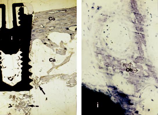
FIGURE 14.1 (a) Ground section of a conical Brånemark System implant (i) removed 4 weeks after insertion in an onlay iliac crest bone graft in the maxilla of a patient. There are no signs of bone formation or resorption in the cortical bone (Co) of the graft. However, bone formation (arrows) is evident around the apex of the implant
Cortical Bone
The initial healing phase of cortical bone grafts is identical to that of cancellous bone grafts. The most apparent difference is in the rate of revascularization, since this takes at least twice as long for cortical bone grafts as for cancellous ones. Complete revascularization usually occurs within 2 months. As stated earlier, it is this difference that is most likely attributed to the structural differences. In cortical bone grafts, vascular penetration is primarily the result of osteoclastic resorption and vascular ingrowth into previously existing Volkmann’s and haversian canals. The resorption of the internal cortical bone proceeds by enlargement of haversian canals followed by the apposition of new bone by osteoclasts (i.e., as creeping substitution). In this way, the repair of cortical grafts results in an admixture of viable and necrotic bone.
Healing of Vascularized Autologous Bone Grafts
If the graft has an internal vascular supply (i.e., free vascularized and pedicled grafts), it does not necessarily become necrotic or require a lengthy process of incorporation. The
in the cancellous and the marrow part of the graft (Ca) near the recipient side. (b) Close-up showing solitary bone formation in the vicinity of the implant (i). Osteoblasts (Ob) are being trapped in a mineralized matrix.
graft will heal with the recipient bone at either end by a process that is analogous to fracture healing.
Healing of Allogeneic and Alloplastic Grafts
Allogeneic grafts are widely used in orthopedic reconstructive surgery. However, fresh allografts are treated to avoid an immunological reaction and rejection of the graft. Such treatments include deep freezing, freeze drying, chemical processing with chloroform-methanol, paracetic acid, hydrogen peroxide, etc. Most of the allogeneic grafts used in oral surgery today are demineralized or mineralized freeze-dried grafts. These materials have, however, poor biomechanical properties and are suitable to fill defects and cavities. The healing process of allogeneic and alloplastic materials follows the same principles as for the incorporation of an autologous graft (i.e., penetration of vessels and bone condensation) provided that an immunological reaction toward the graft can be prevented. However, the bone-forming process is delayed as compared to autologous grafts, which is attributed to the absence of living cells in the graft. Overall, allografts appear to
14. Advanced Bone Healing Concepts
undergo decreased incorporation as compared to autologous grafts.
Factors of Importance for Successful Incorporation and Maintenance of a Graft
Vascular Supply
A free graft placed in a defect within the skeletal envelope is surrounded by highly vascularized bone surfaces, which provides the optimal conditions for revascularization of the graft. This is in contrast to the situation in which the graft extends beyond the skeletal borders so that there is a reduced surface of the host bone, which can provide contact for the graft with vessels and osteogenic cells. Moreover, the situation with such defects is that the degree of spontaneous healing is high, while bone is not likely to form spontaneously beyond the skeletal borders. In the former situation, probably any type of graft can be successfully incorporated. In the latter case, however, greater demands are at hand, and large differences might be found when comparing different types of grafts and surgical procedures. When vascularized grafts are used, the incorporation does not depend entirely on contributions from the local tissue of the recipient bed.
Stability
It is well known that stability of the bone-bone, bone-graft, or bone-implant interface is crucial for bone formation to occur. Mesenchymal cells are sensitive to strain and may differentiate into fibroblasts or chondroblasts if micromovements are present in the healing area. The graft may be stabilized with screws or wires, but it is also the biomechanical properties of the graft itself that can be of importance for its stability.
Biocompatibility
The implantation of any biological or inorganic material in the human body elicits a tissue reaction in response to the surgery and to the material itself. The tissue response to the transplant/implant can either be the result of a specific immunological reaction, as in the case of nonself biological transplants or implants (e.g., allografts, xenografts), or be of a nonspecific character. The specific reaction correlates to the degree of matching of major histocompatibility antigens (transplantation antigens) between donor and recipient. Specifically, an activation of B- and T-lymphocytes results in the production of antibodies and direct actions between T- lymphocytes and the foreign cells, leading to rejection of the transplant. The nonspecific reaction is related to factors other than the antigenic properties of the implanted material. The nonspecific reaction is induced by the interaction between biomolecules, cells, and the surface of the implant, and it is related to the chemical and physical properties of the implant surface. Moreover, it is well known that the macroscopic de-
127
sign, such as the pore size of alloplasts, or the thread design of an implant, as well as the topography of the implant surface can modify the tissue response. From a host-acceptance point of view, it is likely that autologous bone is the preferred grafting material.
Prevention of Soft Tissue Ingrowth
Complete healing of large bone defects cannot be expected, since soft tissue will occupy the defect by ingrowth, collapse, or both. The bone healing can be enhanced in such a situation by using a physical barrier for guided tissue regeneration (GTR). The barrier will hinder ingrowth of soft tissues and seclude the defect so that only bone cells will have access to the defect. It is reasonable to consider that there is some degree of competition between soft tissue and bone tissue formation during the incorporation of a graft. In that respect, it is possible that a porous graft (i.e., a cancellous bone graft) is more prone to soft tissue ingrowth than a nonporous graft (i.e., cortical bone graft). Theoretically, a corticocancellous graft may be better incorporated, when the cancellous layer is oriented toward the defect since the cortical layer will act as a barrier to prevent soft tissue ingrowth into the cancellous part of the graft. Moreover, it may also be important to pack voids between the graft and the recipient bone surfaces with particulate bone grafts. In the literature, some authors have suggested that barrier membranes should be used to enhance the incorporation of bone grafts. However, to date it is not clear if any of the various types of alloplastic membranes are superior to an intact periosteum.
Loading
It is well known that unloaded grafts are usually completely resorbed with time. However, several studies indicate that the graft will maintain its dimensions when implants are inserted and loaded (Figure 14.2).
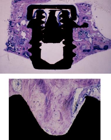
Experimental Studies of Grafts and Implants
Albrektsson16 used a rabbit tibial model in which he had inserted a specially constructed c.p. titanium implant that would enable in vivo visualization of the pending bone graft (Figure 14.3). Following transplantation, repeated inspections were performed at varying times for follow up of the remodelling of the graft structures (Figure 14.4). In this manner it was possible to compare the vascular activity before and after grafting and to monitor the ingrowth of new vessels. In cancellous bone, this was observed to occur at a maximal rate of some 1.2 to 0.4 mm a day, while cutting cones invaded cortical bone at a maximal rate of 30 to 40 m a day.
Donath et al.18 compared the healing of Tübingen implants in vascularized and free iliac crest grafts in a sheep model. Bone segments of identical size were osteotomized in both left and right iliac crests of eight animals. The blood supply to the graft was kept intact on one side, while the vessels were
128
FIGURE 14.2 Ground section showing the bone implant interface of a Brånemark System implant removed 3 years after insertion in an iliac crest onlay graft in the maxilla. Normal lamellar bone with marrow cavities (M) and an apparently direct contact between bone and implant is seen.
cut on the contralateral site. The segments were repositioned and fixed with ostheosynthesis plates and screws, after which one or two Tübingen implants were placed in the grafts. The animals received fluorochromes during a 1- to 12-week healing period. After sacrifice, the histological analysis showed that the implants became osseointegrated in the vascularized graft and that soft tissue encapsulated the free graft. It was concluded that the immediate placement of implants in free bone grafts cannot be recommended, which is in contradistinction to the results presented by other researchers.19,20 Neukam et al.19 demonstrated the integration of Brånemark System implants in onlay grafts inserted into the mandibles of 10 minipigs. In that study, mandibular defects were created 3 months following the removal of the premolars in the minipigs, and free grafts were transplanted from the iliac crest and stabilized with two implants into the defects. The authors observed osteoneogenesis with direct contact between the recipient bone and the implants and between the bone graft and the implants at 3 and 5 months. These authors concluded that their experimental and clinical experiences provided the basis for expecting good long-term results when using onlay osteoplasties and simultaneous insertion of implants. Similar results were presented by Lew et al.,20 who performed a
T. Albrektsson, L. Sennerby, and A. Tjellström
comparison of the integration of Brånemark implants in free corticocancellous block grafts and particulate corticocancellous grafts. The iliac crests in 17 dogs were used as experimental sites. On one side, a block of corticocancellous bone was osteotomized and the cortical bone at one end of the bone block was removed. The same procedure was performed on the other side, but the graft was additionally sectioned into 2- to 3-mm segments. The corticocancellous block graft was placed in the contralateral defect and stabilized with a 20-mm- long implant. An implant was inserted on the other side and the nonburied portion of the implant was covered with bone particles. Light microscopy and microradiography were performed on the specimens, including the implant and the surrounding bone, taken at 1, 2, and 3 months after surgery. Both types of grafts were determined to be viable through the evaluation of fluorescent labels. The bone density appeared to be greater with a higher degree of bone/implant contact calculated by morphometry in the block grafts, as compared to the particulate ones. The authors concluded that the osseointegration of titanium implants developed more rapidly in corticocancellous bone blocks, as compared to the particulate bone grafts.
In a rabbit model, Lundgren et al.21 studied the integration of Brånemark System implants in particulate autologous grafts. One implant was inserted in the tibias of eight rabbits in such a way that five threads were not covered with bone on one side. On the test side, particulate cortical bone grafts from the calvarium were packed over the exposed implant surfaces, and covered with a bioresorbable polylactide barrier. At the control side, only particulate bone grafts were applied over the implant threads. Histology from 12-week-old specimens showed significantly thicker bone at the test side, with osseointegration occurring to the same degree on both the test and control sides. It was concluded that the barrier probably stabilized the grafts and prevented the ingrowth of soft tissue and resorption of the bone particles during the healing period.
The integration of titanium implants in dog alveolar ridges augmented by allogeneic material was evaluated by Pinholt et al.22 Six weeks after the removal of two premolars in all quadrants via block resections, the ridges were augmented with allogeneic demineralized and lyophylized dentin or bone. A total of 32 titanium implants were inserted in the augmented regions of 10 dogs 5.5 months later and followed for another 3.5 months, at which time the animals were killed and specimens retrieved. All implants were encapsulated by a fibrous tissue containing a few multinuclear giant cells and some other inflammatory cells. The graft material was without signs of remineralization, except when there was contact with the recipient bone surfaces.
Clinical Histology of Implants in Bone Grafts
In 1989, Riediger et al.23 presented histology from a patient treated with a vascularized iliac crest graft and Tübingen implants. The patient had been reconstructed following the re-
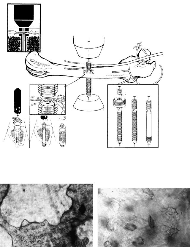
FIGURE 14.3 Albrektsson-constructed titanium implants permitting direct visualization of bone graft remodelling and revascularization. The implants, existing in slightly different designs (bottom right), were inserted through long bones in various animals. Bone and vascular tissue invaded a space that went straight through the body of
the titanium chamber. Grafting was performed when the ingrown bone and vessels were found to be in a steady-state situation (bottom left). In this manner, it became possible to inspect bone tissue before the graft-bone complex was transplanted in an autologous or allologous manner.
a
b
FIGURE 14.4 (a) Low-power view from bone chamber at 4 weeks after autologous transplantation with bone tissue (darker with numerous rounded bodies representing osteocyte lacunae) undergoing resorption. The depicted bone was completely resorbed and replaced
with invading soft tissue (light), when inspected a couple of weeks later. (b) Osteocyte lacunae with visible canaliculae in living bone after autologous transplantation.
129
130
section of a mandibular segment because of a tumor. Three years later the patient had a recurrence, and the graft and implants were removed. The implants were clinically stable at the time of removal, with direct bone contact observed at the apical portion of the implants, while soft tissue and epithelium contact were observed at the coronal portion of the implant.
Nyström et al.24 presented histology of one patient who died 4 months after an onlay grafting procedure and immediate placement of six Brånemark System implants. The grafted bone from the iliac crest demonstrated signs of resorption, but also areas where new bone formation was seen on old trabeculae. There was only a patchy contact between the grafted bone and the implants, with the major part of the interface consisting of soft tissues. Bone condensation into the implant threads was evident in some areas, both in the graft and in the recipient bone.
Histology from a patient who died 8 months after a sinus elevation procedure was reported by GaRey et al.25 Freezedried cortical allografts and resorbable hydroxyapatite had been used in conjunction with immediate placement of “rootformed” implants. One of the two implants studied was totally submerged in bone, and the microscopical examination revealed a bone interface, while the other implant had a minimal amount of bone in the interface. The authors concluded that “eight months would not have been enough healing time prior to loading for this patient.”
Jensen and Sennerby26 used small test implants of c.p. titanium to study osseointegration in patients that had undergone sinus augmentation with radiated mineralized cancellous allografts (RMCA) or autografts and immediate placement of Brånemark System implants. The test implants were inserted through the buccal bone into the grafts and were removed at abutment connection 6 to 14 months later. The histological examination of the specimens revealed a minor degree of bone formation and osseointegration when using allografts. Most of the specimens contained nonviable particles of the allograft. Bone formation was evident only in the cortical passage. However, in one specimen of the particulate cancellous autograft/titanium interface, mature lamellar bone in direct contact with the implant was observed. The study indicated that autologous bone grafts are preferable to allografts.
Clinical Use of Grafts and Implants
Cortical Bone Grafts and Implants
In 1994, Donovan et al.27 reported on the clinical outcome of two techniques using calvarial bone grafts and Brånemark System implants. With the first technique, the grafts were placed as horizontal onlays and inlays for immediate insertion of the implants. After an average follow-up time of 18 months, 98% of the initially inserted 43 implants were still clinically stable. The second technique involved the fixation of calvarian strips as vertical onlay grafts and the de-
T. Albrektsson, L. Sennerby, and A. Tjellström
layed insertion of implants 6 to 8 months later. Using this approach, 86% of the 50 implants inserted were considered to be successful.
Cancellous Bone Grafts and Implants
Breine and Brånemark28 used tibial cancellous bone chips and bone marrow packed around titanium implants inserted in the atrophied maxilla or mandible of 18 patients. The result was not satisfactory as 75% of the implants were lost and a dramatic resorption of the graft was observed during the first years.
Cancellous bone particles have been used for maxillary sinus augmentation and were first described by Boyne and James in 1980.29 In 3 of 14 cases treated, blade implants were inserted 12 weeks after the augmentation procedure. No signs of resorption were evident during the 1 to 4 years the implants were followed.
Jensen30 described the clinical outcome of using cancellous autografts for sinus augmentation and the immediate (n 179) and delayed (n 43) insertion of Brånemark System implants. The lateral wall of the sinus was covered with an e- PTFE barrier to prevent the ingrowth of soft tissue into the augmented sinus cavity. Delayed implant placement improved the overall implant survival rate, 93% versus 81%. More implant failures occurred when less alveolar bone was available inferior to the sinus cavity.
Corticocancellous Bone Grafts and Implants
Preformed Endochondral Grafts and Implants
Brånemark introduced the technique of bone graft preformation on theoretical grounds. The idea was to insert, as a guide, a mold together with fixation screws at the donor site several months before the actual grafting. As the mold was shaped in accordance with the estimated needs at the recipient site, the advantage of the preformed bone graft would be that it had already remodeled before grafting. The grafting procedure would be less traumatic since the separation surgery had been partially performed during the initial surgical procedure. Experimental studies in rabbits confirmed that the preformation procedure indeed produced a highly viable graft.31 One variety of the preformed bone graft was applied clinically by Tjellström et al.32,33 The authors inserted titanium molds into the tibias of humans. The mold had a canal that was preformed as an ossicular bone graft. Bone tissue invaded the canal and 6 months later the molds were removed with a trephine, which upon opening ossicular “selfcast” grafts were found inside. In a total of 11 cases, such preformed ossicular bone was grafted to the middle ear, and when evaluated 5 years later,34 the grafts continued to demonstrate very adequate function.
Breine and Brånemark28 placed implants in a preformed tibial graft 3 to 6 months prior to harvesting the graft and transplanting it to the maxilla. During the follow-up period of 1 to 8 years, more than 50% of the implants had failed. Lindström
14. Advanced Bone Healing Concepts
et al.35 used the preformation technique to obtain large bone grafts to treat defects resulting from the ablation of major mandibular tumors. In addition to molds, fixation screws and “oral implants to be” were inserted at the donor site and allowed to be incorporated in the intended bone graft before disruption of the vascular connections. The actual grafting was performed 4 to 5 months after the first surgical procedure when adequate remodelling of the grafts had occurred. Of the five patients, two died from metastatic disease within a year after surgery, and three others had experienced good graft and implant function for follow-up periods ranging from 5 to 10 years.
Although theoretically advantageous, the preformation principle is time-consuming and troublesome for patients in that it involves a two-stage surgical procedure. Furthermore, it is questionable as to whether the technique really results in an improved outcome compared to routine procedures. Therefore, at the Göteborg University clinic, the procedure was abandoned many years ago.
Other Types of Endochondral Bone Grafts
and Implants
In a study by Keller et al. in 1987,36 9 patients were treated with iliac corticocancellous grafts and immediate placement (5 patients) or delayed placement (4 patients) of titanium implants. Of 28 immediately placed implants 4 (14.3%) were removed owing to clinical mobility, and 5 (23%) of 21 delayed implants failed.
In a Swedish and an international multicenter study, Albrektsson et al.37 reported on 42 implants inserted in grafted mandibles with only one failure over a follow-up period of 1 to 5 years. In the maxilla, 183 implants were inserted in grafted bone and 50 of those failed over a follow-up of 1 to 5 years.
The use of iliac onlay-grafts and immediate insertion of titanium implants in patients with severely resorbed maxillae and mandibles was reported by Neukam et al. in 1989.19 Of 110 implants inserted in 21 patients, 13 failed (11.8%) during an observation period of up to 4 years. The overall survival rate was higher in the upper jaws (95.95%) as compared to the lower jaws (82.0%).
Adell et al.38 reported on the outcome of 124 implants inserted together with iliac onlay grafts in 23 patients who were followed from 1 to 10 years. Seventeen of the patients had stable fixed prostheses, 5 had overdentures, and 1 patient returned to using a complete denture. Of the originally placed fixtures, 8.1% were lost during the healing period until the abutment connection and 73.8% of the implants were still in function after 5 years of loading. The mean marginal bone resorption during the first year in function was 1.49 mm and 0.1 mm/year thereafter. Similar results were presented by Gunne et al.39 using the same technique. In that study, 30 patients divided into a development group (10 patients) and a routine group (20 patients) were followed for 3 years. Of the total 177 implants inserted, 43 failed during the observation
131
period (24.3%). However, the implant survival rate was higher in the routine group (87.5%). The remodeling of the residual ridge was also studied, and it was concluded that a reduction of height and faciopalatal width of the grafted area was observed during the first year.
This technique was applied to the partially edentulous patient by Schliephake et al.40 Fifty-five Brånemark System implants were inserted in 16 patients. Two implants (3.6%) failed because of wound dehiscence and subsequent infection, and another 2 implants were left as “sleepers.” No further implants failed during the 2- to 80-month period of loading.
In 1989, Sailer41 described a new method for augmentation of the atrophied maxilla in conjunction with installation of titanium implants. He would perform a Le Fort-1 osteotomy, placed iliac corticocancellous bone blocks to obturate the floor of the nose and maxillary sinus, and simultaneously inserted titanium implants as a single-step surgical procedure. The maxilla/graft complex was held in position with the use of osteosynthesis screws and plates. None of the 35 implants in 5 patients failed during the follow-up period of 0 to 13 months. Using the same technique, Isaksson reported a survival rate of only 68% of the first 10 cases with 57 implants placed. However, Isaksson42 had a considerably longer follow-up time, 33 to 95 months, as compared to Sailer in which the implants in 2 of the 5 patients had not yet been uncovered.
Intramembranous Grafts and Implants
In a series of publications, Jensen et al.43–45 have described the use of autologous intramembranous corticocancellous bone grafts taken from the chin and immediate insertion of implants for augmentation of the severely resorbed maxilla. The grafts were taken from the chin by the use of specially made instruments that permitted preparation of the implant sites through the tapping stage at the donor site prior to harvesting and transfer to the recipient site. The grafts were used as onlays or sinus grafts or as a combination of the two. In a preliminary report in 1990, they presented the results of 107 implants inserted in 26 patients. The overall implant survival rate was 93.5% after a follow-up of 6 to 26 months. All 7 implants lost had been inserted in a combination of onlay and sinus grafts. Four of the lost implants were removed due to wound dehiscence and exposure of the grafts. An average marginal bone resorption of 1 to 2 mm was observed in this study. The authors suggested that this minor bone resorption was attributed to the intramembranous origin of the graft bone. In 1994, Jensen et al.45 reported the outcome of different procedures when inserting implants in the maxilla. In one group, chin transplants were used as onlays and sinus grafts for immediate placement of implants. Of 152 implants, 17 (11.2%) failed to integrate or lost the integration during the 13to 58-month follow-up period.
A two-stage technique of using intramembraneous retromolar or chin grafts and ITI implants was presented by
132
Krekeler et al.46 The grafts used as buccal onlays and fixated by osteosynthesis screws were allowed to heal for 4 to 6 months prior to installation of the implants. None of the 47 implants installed in 30 patients failed during the 1-year follow-up period. The marginal bone loss was reported to be less than 1 mm.
T. Albrektsson, L. Sennerby, and A. Tjellström
gen implants and 15 IMZ implants where placed in 22 iliac crest grafts. Two of the Tübingen implants were lost during the healing period. In 1991, Riediger and Ehrenfeld49 reported on the use of the same technique for augmentation of the atrophied mandible. Three of 12 Tübingen implants, 2 of 12 IMZ, and none of 8 Brånemark implants failed during the up to 34-month follow-up period.
Allogeneic Grafts and Implants
Allogeneic allografts have mostly been used for sinus lift procedures. Jensen and Greer47 reported on the use of radiated mineralized cancellous allograft and immediate insertion of implants in the augmented maxillary sinus. Of 38 implants, 8 (21.1%) failed during the 2.5-year follow-up. They also used demineralized cancellous allografts for sinus augmentation and immediate placement of implants, and lost 10 (45.5%) of 22 implants.
In 1993, Small et al.48 reported on the use of demineralized freeze-dried cortical bone mixed with 50% hydroxyapatite for maxillary sinus augmentation and immediate placement of cylindrical implants. Of 111 implants placed in 45 sinus grafts in 27 patients, 76 were used for anchorage of dental prostheses. None of the 76 implants failed during a 1- to 5-year period of loading.
Vascularized Grafts and Implants
In a study by Riediger et al.23 a microsurgical technique using vascularized iliac crest grafts and immediate insertion of implants for the replacement of mandibular and maxillary segments, owing to tumor surgery, trauma, or infection, was described. The graft was taken in such a way that the deep circumflex iliac artery and vein could be used for anastomosis with the facial artery and vein. In the 1989 study, 46 Tübin-
a
Craniomaxillofacial Prostheses
There are two major indications for inserting skin-penetrat- ing, craniofacial implants. One is related to the stable fixation of a bone-anchored hearing aid, which is useful for patients with certain types of hearing disorders. The other indication is related to craniofacial reconstruction with the stable anchorage of a facial prostheses. Patients in the latter category may represent congenital malformations, cancersurgery deformities, or facial trauma injuries with ear loss (Figure 14.5).
Skin-Penetrating Implants for External
Hearing Aids
The indication for treatment may not represent the most appropriate example of craniomaxillofacial reconstructive bone surgery, as the primary indication for surgery is a hearing disorder. Nevertheless, the first patients to be treated with skinpenetrating implants received those for the indication related to hearing impairment,50 and we have gained invaluable experience on skin-penetrating implants from treating such patients. In a review of the first 100 patients who were treated with skin-penetrating implants and external hearing aids, Tjellström and Granström51 came to the conclusion that 90%
b
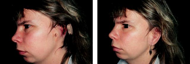
FIGURE 14.5 (a) Patient with a thalidomide embryopathy who had a conventional bone-conducting hearing aid until 1979. She received a bone-anchored hearing aid (BAHA) the same year and showed a clear speech improvement after surgery. Her ability to hear particu-
larly high-frequency sounds was greatly improved. (b) The same patient had bilateral ear atresia. She was operated on both sides in 1992 and received bone-anchored silicone ear replacements (b).
14. Advanced Bone Healing Concepts
of the implants were still stable after a follow-up of between 8 and 16 years. Five percent of the implants had been removed due to trauma, whereas another 5% had lost their integration for other reasons. About 80% of the patients were without or experienced only a single episode of adverse soft tissue reactions, with the majority of those complaints treated without any associated problems with the bone-anchored implants. In a more detailed analysis of soft tissue reactions around skin-penetrating implants, Tjellström52 divided the problems into five categories in which 0 implied no irritation and 4 meant infection leading to removal of percutaneous implant. Of all 1739 observations made in 1989, 92.5% of those were of grade 0, 4.1% of grade 1 (redness), 1.8% of observations were of grade 2 (moist), 1.5% of observations were of grade 3 (granulation), and only 0.1% of all observations were of grade 4. There were similar results for skin-penetrating auricular prostheses and no tendency for increased problems over time. The average score of all observations was found to be 0.14. The author concluded that adverse skin reactions were not a major problem with the type of permanent skin penetrating implants used in the study.
133
than those quoted earlier, altogether 53 implants were placed in the nonirradiated nasal region for an average success rate of 83%. Only 10 implants were placed in the irradiated, nasal region for a success rate of 80%. One difference from the Swedish experience was the much better outcome of 28 Canadian implants inserted in the irradiated orbit, where there was a 96.4% success rate. Of a total of 1365 implants inserted at all 20 participating centers (13 in the United States, 6 in Canada, and 1 in Sweden), 1290 or 94.5% had integrated in the bone.56
a
Skin-Penetrating Implants for Stable Anchorage of Facial Prostheses
The great advantages with skin-penetrating implants in conjunction with facial prostheses are the stability of the devices and the improved aesthetics (Figure 14.6). Psychologically, the patients often regard the implant and prosthesis to be “self” rather than “nonself.” The first patient with skin-pen- etrating fixtures for the anchorage of an auricular prosthesis was treated at the University of Göteborg in 1979.53
When evaluating the outcome of skin-penetrating fixtures, Jacobsson et al.54 came to the conclusion that implant survival in the auricular area was 95.6%, and in the orbital region it was only 67.2%. These statistics were based on 234 auricular and 81 orbital consecutively inserted fixtures followed up from 6 months to more than 5 years. The comparatively poor result in the orbit region is partly explained by previous irradiation in that 16 of 19 mobile orbital implants had been inserted in previously irradiated bone beds. Excluding irradiated cases, the implant survival in the orbit region was much improved in that only 3 of 38 placed implants were lost, for a survival of 92%. The interesting question is whether hyperbaric oxygen treatment will improve the longterm survival of orbital implants inserted into previously irradiated bone beds. Jacobsson et al.54 were the first to suggest criteria for success with respect to skin-penetrating maxillofacial implants. Their criteria were based on the guidelines for oral implants suggested by Albrektsson et al.55 and included implant immobility, soft tissue reactions of 0 and 1 in a minimum of 95% of all observations, and the absence of persistent pain, infections, or paresthesia. Wolfaardt et al.56 compared the Swedish (Göteborg University), Canadian (University of Alberta), and U.S. (University of San Antonio) experiences with skin-penetrating fixtures. In regions other
b
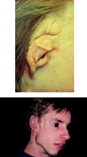
FIGURE 14.6 Patient with hemofacial microtia initially treated by conventional plastic surgery. The figure shows the plastic-surgically created ear after some 40 surgical interventions. This patient was initially operated with implants in 1983 but had then decided that he wanted to keep his surgically made ear as depicted in (a). The resultant prosthesis had therefore quite bulky proportions (b). In 1993, the patient decided that his ear fragments from plastic surgery could be removed and it was then easier to provide him a better-fitting artificial silicone ear on skin-penetrating implants.
134
One central issue with respect to craniofacial, skinpenetrating implants is trying to increase survival rates in previously irradiated bone. Granström et al.57 concluded that even heavily irradiated bones could integrate the implants and bear the load from the prosthesis. There were no major complications such as wound infection, fistulation, or osteoradionecrosis reported after surgery. Nevertheless, there was an increased loss of implants with time after irradiation, particularly in the orbital region. The authors reported that hyperbaric oxygen treatment (HBO) reduced the number of implant losses. In another study,58 the problem of postimplantation irradiation was addressed, in which 32 implants were placed before irradiation. Two of those were removed from the temporal bone in a secondary surgical intervention, and 2 others were lost from the frontal bone region during chemotherapy. Osteoradionecrosis developed in 3 of 11 patients. The authors recommended the subsequent removal of all prostheses,
a
b
T. Albrektsson, L. Sennerby, and A. Tjellström
frameworks, and abutments before irradiation, whereas the fixtures should be allowed to remain in the bone but should be covered with skin or mucosa.
Craniomaxillofacial Implants in Children
The youngest child, to the knowledge of the authors, ever operated with an implant aimed for permanent skin penetration was 6 months old and suffering from Mb Apert. However, the implant was later removed on request of the parents before any second-stage surgery was performed. Histology demonstrated some 47% bone-to-implant contact, and the threaded region was filled out by bone to some 79% (Figure 14.7).
One problem with operating on children is, of course, that their cortex is thin compared to adults, but children have, on the other hand, a better bone-forming capability. The outcome
FIGURE 14.7 (a) We have experience with about 100 implants inserted in children. In children with hearing disorders, we have sometimes operated on patients as young as 3 years, whereas craniofacial disorders are often not operated until the child reaches puberty. The youngest child to receive a bone-anchored implant that we know of
was operated on in a European clinic at the age of 6 months. Indication for surgery was Morbus Apert. However, the implant was later removed on request from the parents. (b) The implant of (a) that was never loaded showed some 47% bone-to-implant direct contact.
14. Advanced Bone Healing Concepts
of 59 consecutively inserted skin-penetrating implants in 30 children was reported by Jacobsson et al.54 Of those implants, 16 were inserted in an equal number of patients with the indication of impaired hearing. In this group we operated on the youngest patients, down to an age of 3 years, as we hoped to establish an improved feedback situation with a bone conduction hearing aid, in turn leading to better social development. The average age of this patient group was 9.3 years compared to 10.6 years for the 14 patients with 43 implants inserted with the indication to anchor a facial prosthesis. The follow-up time of all patients ranged between 1 and 144 months. The skin reactions and the clinical results did not differ significantly from the situation in adults, and there was an average fixture survival rate of 96.6%
Tjellström59 has pointed out that in selecting young patients for auricular prostheses, the most important factor is for the child to be clearly motivated for this type of surgery. Sometimes there is a parental guilt complex in the background instead of a true problem for the patient. It is extremely important that the child understand that the skin has to be carefully cleaned to avoid soft tissue reactions and that the prosthesis will have to be remade every 2 to 3 years.
Stability of Maxillofacial Implants and
Histological Examinations
In an investigation approved by the Göteborg University ethical committee, two implants were inserted in the temporal bone of patients, although only one was necessary to attach the hearing aid. One of these screws was later removed with a torque-gauge analyzer revealing an average removal torque of 42.7 Ncm over an average follow-up of 107 months.60 Yamanaka et al.61 inserted 31 similar implants in the temporal bone. The implants were divided into three groups depending on the time of implantation. Group 1 (n 10), with an average follow-up of 3.4 months, demonstrated an average removal torque of 39.3 Ncm; group 2 (n 7), with an average follow-up of 15.6 months, demonstrated an average removal torque of 67.9 Ncm; and group 3 (n 14), with an average follow-up of 69 months, demonstrated an average removal torque of 96.4 Ncm. Thus there was a clear tendency of an increasing removal torque with increasing time.
Retrieval analyses have confirmed a good bone-interfa- cial response as the reason for the torque needed for implant removal.62 One implant removed for psychological reasons at 9 months after insertion demonstrated a bone mineral contact percentage of 54 and a mean bone area of 84%. In a case in which the reason for implant removal was “insufficient hygiene,” inflammatory cells, macrophages, and osteoclasts were observed, but there was nevertheless an average bone-to-metal contact of 78% and a mean bone area of 90% at 18 months after implant insertion. Another patient who had been irradiated with 90 Gy and had four implants inserted some 5 months later was available for re-
135
trieval analyses post mortem another 2 years later. Bone remodelling was then still active. Mean percentage of bone- to-metal contact was about 40 and mean bone area in the implant threads was 75%. Another patient received irradiation of 50 Gy and cytostatics for the treatment of a partial maxillectomy for squamous cell carcinoma. Two years later, three fixtures were placed in the orbital region. When these implants were removed because of pain and other discomfort 3 years later, it was demonstrated that they had penetrated into the frontal sinus and were covered by a mucous membrane. The bone structure did not appear very normal, and there were numerous empty osteocyte lacunae without any bone to implant direct contact.
Conclusion
An essential aspect of every new clinical procedure is the careful documentation of shortand long-term results so that techniques that truly represent new, advanced modes of treatment can be properly differentiated from questionable procedures that at best are theoretically advantageous. An example of the latter category is the preformed bone graft. New grafting techniques, such as sinus lift procedures, which are now presented as more or less routine at numerous meetings, are still difficult to properly evaluate owing to differences in graft selection and implant types. Therefore, they should be used with some caution at the present time. By contrast, several good papers have been published on the outcome of onlay bone grafts, which at the current level of knowledge are much better documented than other more novel types of grafting procedures. In craniomaxillofacial surgery, a thorough clinical documentation has at all times accompanied the advancement of threaded titanium implants, which is why we now have substantial documentation of the excellent outcome of such implants inserted in the temporal bone, in contrast to the less reliable outcome of the same implant inserted in previously irradiated orbit bone. This has resulted in our trying new techniques, such as hyperbaric oxygen treatment, to improve results of orbital implants. The authors are convinced that when introducing new implant techniques, a meticulous mode of clinical documentation joined with the supervision of university ethical committees should replace what is commonly seen today; specifically, a series of relatively poorly controlled human experiments supervised by numerous clinical entrepreneurs.
References
1.Brånemark P-I, Breine U, Lindström J, et al. Intra-osseous anchorage of dental prostheses I. Experimental studies. Scand J Plast Reconstr Surg. 1969;3:81–93.
2.Albrektsson T. The response of bone to titanium implants. CRC Critical Reviews in Biocompatibility. 1985;1:53–84.
3.Donath K, Laass M, Günzl HJ. The histopathology of different foreign-body reactions in oral soft tissue and bone tissue. Virchows Arch A Pathol Anat. 1992;420:131–137.
136
4.Johansson C, Albrektsson T. Integration of screw implants in the rabbit. A 1-year follow-up of removal of titanium implants.
Int J Oral Maxillofac Implants. 1987;2:69–75.
5.Zarb G, Albrektsson T. Osseointegration—A requiem for the periodontal ligament?—An editorial. Int J Periodont Res Dent. 1991;11:88–91.
6.Johansson C. On tissue reactions to metal implants. Göteborg, Sweden: University of Göteborg; 1991. PhD Thesis.
7.Albrektsson T, Eriksson A, Friberg B, et al. Histologic investigations on 33 retrieved Nobelpharma implants. Clin Mater. 1993;12:1–9.
8.Albrektsson T, Brånemark P-I, Hansson HA, Lindström J. Osseointegrated titanium implants. Requirements for ensuring a long-lasting, direct bone anchorage in man. Acta Orthop Scand. 1981;52:155–170.
9.Brånemark P-I, Zarb G, Albrektsson T, eds. Osseointegration in Clinical Dentistry. Berlin/Chicago: Quintessence; 1985.
10.Ten Cate R. The gingival junction. In: Brånemark PI, Zarb G, Albrektsson T, eds. Tissue Integrated Prostheses. Chicago/ Berlin: Quintessence; 1985:145–153.
11.Apse P. Clinical and microbiological aspects of the periodontal and periimplant sulcus: a cross-sectional study. Toronto, Canada: University of Toronto; 1987. MSc Thesis.
12.Carmichael R, Apse P, Zarb G, McCulloch C. Biological, microbiological, and clinical aspects of the peri-implant mucosa. In Albrektsson T, Zarb G, eds. The Brånemark Osseointegrated Implant. Chicago/Berlin: Quintessence; 1989:39–78.
13.Marinello C. Resolution of experimentally induced periimplantitis. Göteborg, Sweden: Göteborg University; 1995. MSc Thesis.
14.Hoshaw S. Investigation of bone modeling and remodeling at a loaded bone-implant interface. Troy, NY: Rensselaer Polytechnic Institute; 1992. PhD Thesis.
15.Axhausen, G. Die patologisch-anatomischen Grundlagen der Lehre von freien Knochentransplantation beim Menschen und beim Tier. Med Klin. 2 1908; Beiheft 23.
16.Albrektsson, T. Repair of bone grafts. A vital microscopic and histological investigation. Scand J Plast Reconstr Surg. 1980;14:1–12.
17.Albrektsson, T. In vivo studies of bone grafts. The possibility of vascular anastomoses in healing bone. Acta Orthop Scand. 1980;51:9–17.
18.Donath K, Hillman G, Ehrenfeld M, Riediger D. Enossale Einheilung Tübinger Implantate in frei und gefäßgestielt replantierte Beckenkammsegmente. Eine tierexperimentelle Studie. Z Zahnärztl Implantol. 1991;VII:58–61.
19.Neukam FW, Scheller H, Günay H. Experimentelle und klinische Untersuchungen zur Auflagerungsosteoplastik in Kombination mit enossalen Implantaten. Z Zahnärztl Implantol. 1989;V:235–241.
20.Lew D, Marino AA, Startzell JM, Keller JC. A comparative study of osseointegration of titanium implants in corticocancellous block and corticocancellous chip grafts in canine ileum.
J Oral Maxillofac Surg. 1994;52:952–958.
21.Lundgren AK, Sennerby L, Lundgren D, Taylor Å, Nyman S. Bone augmentation at titanium implants using autologous bone grafts and a bioresorabable barrier. An experimental study in the rabbit tibia. Clin Oral Implant Res. 1996;8:82–89.
22.Pinholt EM, Haanaes HR, Donath K, Bang G. Titanium implant insertion into dog alveolar ridges augmented by allogeneic material. Clin Oral Implant Res. 1994;5:213–219.
23.Riediger D, Ehrenfeld M, Donath K. Tübinger Implantate im
T. Albrektsson, L. Sennerby, and A. Tjellström
vaskularisierten Beckenkammtransplantat. Klinische Ergebnisse und morphologische Befunde. Z Zahnärztl Implantol. 1989;V: 137–141.
24.Nyström E, Kahnberg KE, Albrektsson T. Treatment of the severely resorbed maxillae with bone graft and titanium implants. Histologic review of autopsy specimens. Int J Oral Maxillofac Implants. 1993;8:167–172.
25.GaRey DJ, Whittaker JM, James RA, Lozada JL. The histologic evaluation of the implant interface with heterograft and allograft materials. An eight month autopsy report. Part II. J Oral Implant. 1991;XVII:404–408.
26.Jensen OT, Sennerby L. Histological analysis of titanium microimplants placed in conjunction with maxillary sinus floor augmentation. Int J Oral Maxillofac Implants, 1997.
27.Donovan MG, Dickerson NC, Hanson LJ, Gustafson RB. Maxillary and mandibular reconstruction using calvarial bone grafts and Brånemark implants. A preliminary report. J Oral Maxillofac Surg. 1994;52:588–594.
28.Breine U, Brånemark P-I. Reconstruction of alveolar jaw bone.
Scand J Plast Reconstr Surg. 1980;14:23–48.
29.Boyne PJ, James RA. Grafting of the maxillary sinus floor with autogenous marrow and bone. J Oral Surg. 1980;38:613–619.
30.Jensen OT. Guided bone graft augmentation. In: Buser, Dahlin, Schenk, eds. Guided Bone Regeneration in Implant Dentistry.
Chicago: Quintessence; 1994:235–264.
31.Albrektsson T, Brånemark P-I, Eriksson A, Lindström J. The preformed autologous bone graft. An experimental study in rabbits. Scand J Plast Reconstr Surg. 1978;12:215–223.
32.Tjellström A, Lindström J, Albrektsson T, Brånemark P-I, Hallén O. A clinical pilot study on preformed, autologous ossicles
I.Acta Otolaryngol (Stockh). 1978;85:33–39.
33.Tjellström A, Lindström J, Albrektsson T, Brånemark P-I, Hallén O. A clinical pilot study on preformed, autologous ossicles
II.Acta Otolaryngol (Stockh). 1978;85:232–242.
34.Tjellström A, Albrektsson T. Five-year follow-up of preformed, autologus ossicles in tympanoplasty. J Laryngol Otol. 1985;99: 729–733.
35.Lindström J, Brånemark P-I, Albrektsson T. Mandibular reconstruction using the preformed autologus bone graft. Scand J Plast Reconstr Surg. 1981;15:29–38.
36.Keller EE, Van Roekel NB, Desjardins RP, Tolman D. Pros- thetic-surgical reconstruction of the severely resorbed maxilla with iliac bone grafting and tissue-integrated prostheses. Int J Oral Maxillofac Implants. 1987;2:155–164.
37.Albrektsson T, Dahl E, Enbom L, Engevall S, et al. Osseointegrated oral implants. A Swedish multicenter study of 8139 consecutively inserted Nobelpharma implants. J Periodontol. 1988; 59:287–296.
38.Adell R, Lekholm U, Gröndal K, et al. Reconstruction of severely resorbed edentulous maxillae using osseointegrated fixtures in immediate autogenous bone grafts. Int J Oral Maxillofac Implants. 1990;5:233–246.
39.Gunne J, Nyström E, Kahnberg KE. Bone grafts and implants in the treatment of the severely resorbed maxillae: A 3-year fol- low-up of the prosthetic restoration. Int J Prosth. 1995;8:38–45.
40.Schliephake H, Neukam FW, Scheller H, Bothe KJ. Local ridge augmentation using bone grafts and osseintegrated implants in the rehabilitation of partial edentulism. Preliminary results. Int Oral Maxillofac Implants. 1994;9:557–564.
14. Advanced Bone Healing Concepts
41.Sailer HF. A new method of inserting endosseous implants in totally atrophic maxillae. J Craniomaxillofac Surg. 1989;17: 299–305.
42.Isaksson S. Evaluation of three bone grafting techniques for severely resorbed maxillae in conjunction with immediate endosseous implants. Int Oral Maxillofac Implants. 1994;9:679– 688.
43.Jensen J, Krantz-Simonsen E, Sindet-Petersen S. Reconstruction of the severely resorbed maxilla with bone grafting and osseointegrated implants. A preliminary report. J Oral Maxillofac Surg. 1990;48:27–32.
44.Jensen J, Sindet-Petersen S. Autogenous mandibular bone grafts and osseointegrated implants for reconstruction of the severely atrophied maxilla: a preliminary report. J Oral Maxillofac Surg. 1991;49:1277–1287.
45.Jensen J, Sindet-Pedersen S, Oliver AJ. Varying treatment strategies for reconstruction of maxillary atrophy with implants. Results in 98 patients. J Oral Maxillofac Surg. 1994;52:210–216.
46.Krekeler G, ten Bruggenkate C, Osoterbeek HS. Verbesserung des Implantatbettes durch Augmentation mit autologem Knochen. Z Zahnärztl Implantol. 1993;IX:231–236.
47.Jensen OT, Greer R. Immediate placement of osseointegrated implant into maxillary sinus augmented with mineralized cancellous allograft and Gore-Tex. Second stage surgical and histological findings. In: Laney W, Tolman D, eds. Tissue Integration in Oral, Orthopedic and Maxillofacial Reconstruction.
Chicago: Quintessence; 1992:321–333.
48.Small SA, Zinner ID, Panno FV, Shapiro HJ, Stein JI. Augmenting the maxillary sinus for implants. Report of 27 patients.
Int J Oral Maxillofac Implants. 1993;8:523–528.
49.Riediger D, Ehrenfeld M. Mikrochurigische Beckenkamm-
transplantate in Kombination mit enossalen Implantaten. Ein neues Verfahren zur Rehabilitation extrem atrophier Kiefer.
Z Zahnärztl Implantol. 1991;VII:178–183.
50.Tjellström A, Lindström J, Albrektsson T, et al. The boneanchored auricular episthesis. Laryngoscope. 1981;91:811–815.
51.Tjellström A, Granström G. Long-term follow-up with the boneanchored hearing aid: A review of the 100 patients between 1977 and 1985. ENT J. 1994;73:21–23.
137
52.Tjellström A. An analysis of soft tissue reactions around skinpenetrating implants. In: Yanagihara N, Suzuki J, eds. Transplants and Implants in Otology. II. Amsterdam/New York: Kugler; 1992:2–3.
53.Tjellström A, Lindström J, Albrektsson T, et al. Osseointegrated titanium implants in the temporal bone. A clinical study on boneanchored hearing aids. Am J Otol. 1981;2:303–310.
54.Jacobsson M, Tjellström A, Fine L, Andersson H. A retrospective study of osseointegrated skin-penetrating titanium fixtures used for retaining facial prostheses. Int J Oral Maxillofac Implants. 1992;7:523–528.
55.Albrektsson T, Zarb G, Worthington P, Eriksson RA. The longterm efficacy of currently used dental implants: A review and proposed criteria of success. Int J Oral Maxillofac Implants.
1986;1:11–25.
56.Wolfaardt J, Wilkes G, Parel S, Tjellström A. Craniofacial osseointegration: the Canadian experience. Int J Oral Maxillofac Implants. 1993;8:197–204.
57.Granström G, Tjellström A, Brånemark PI, Fornander J. Boneanchored reconstruction of the irradiated head and neck cancer patient. Otolaryngol Head Neck Surg. 1993;108:334–343.
58.Granström G, Tjellström A, Albrektsson T. Postimplantation irradiation for head and neck cancer treatment. Int J Oral Maxillofac Implants. 1993;8:495–500.
59.Tjellström A. The relevance for a child of the BAHA and auricular prosthesis. In: Fior R, Pestalozza G, eds. The Child and the Environment: Present and Future Trends. Amsterdam: Elsevier Science Publishers BV; 1993:251–255.
60.Tjellström A, Jacobsson M, Albrektsson T. Removal torque of osseointegrated craniofacial implants. A clinical study. Int J Oral Maxillofac Implants. 1988;3:287–289.
61.Yamanaka E, Tjellström A, Jacobsson M, Albrektsson T. Longterm observations on removal torque of directly bone-anchored implants in man. In: Yanagihara N, Suzuki J, eds. Transplants and Implants in Otology II. Amsterdam/New York: Kugler; 1992:112–117.
62.Johansson C, Tjellström A, Albrektsson T, Sennerby L. Retrieved extraoral implants. Biomaterials Club 3rd Winter meeting, Val Gardena, Italy. 1993.