- •Preface
- •Acknowledgments
- •Contents
- •Contributors
- •1. Introduction
- •2. Evaluation of the Craniomaxillofacial Deformity Patient
- •3. Craniofacial Deformities: Review of Etiologies, Distribution, and Their Classification
- •4. Etiology of Skeletal Malocclusion
- •5. Etiology, Distribution, and Classification of Craniomaxillofacial Deformities: Traumatic Defects
- •6. Etiology, Distribution, and Classification of Craniomaxillofacial Deformities: Review of Nasal Deformities
- •7. Review of Benign Tumors of the Maxillofacial Region and Considerations for Bone Invasion
- •8. Oral Malignancies: Etiology, Distribution, and Basic Treatment Considerations
- •9. Craniomaxillofacial Bone Infections: Etiologies, Distributions, and Associated Defects
- •11. Craniomaxillofacial Bone Healing, Biomechanics, and Rigid Internal Fixation
- •12. Metal for Craniomaxillofacial Internal Fixation Implants and Its Physiological Implications
- •13. Bioresorbable Materials for Bone Fixation: Review of Biological Concepts and Mechanical Aspects
- •14. Advanced Bone Healing Concepts in Craniomaxillofacial Reconstructive and Corrective Bone Surgery
- •15. The ITI Dental Implant System
- •16. Localized Ridge Augmentation Using Guided Bone Regeneration in Deficient Implant Sites
- •17. The ITI Dental Implant System in Maxillofacial Applications
- •18. Maxillary Sinus Grafting and Osseointegration Surgery
- •19. Computerized Tomography and Its Use for Craniomaxillofacial Dental Implantology
- •20B. Atlas of Cases
- •21A. Prosthodontic Considerations in Dental Implant Restoration
- •21B. Overdenture Case Reports
- •22. AO/ASIF Mandibular Hardware
- •23. Aesthetic Considerations in Reconstructive and Corrective Craniomaxillofacial Bone Surgery
- •24. Considerations for Reconstruction of the Head and Neck Oncologic Patient
- •25. Autogenous Bone Grafts in Maxillofacial Reconstruction
- •26. Current Practice and Future Trends in Craniomaxillofacial Reconstructive and Corrective Microvascular Bone Surgery
- •27. Considerations in the Fixation of Bone Grafts for the Reconstruction of Mandibular Continuity Defects
- •28. Indications and Technical Considerations of Different Fibula Grafts
- •29. Soft Tissue Flaps for Coverage of Craniomaxillofacial Osseous Continuity Defects with or Without Bone Graft and Rigid Fixation
- •30. Mandibular Condyle Reconstruction with Free Costochondral Grafting
- •31. Microsurgical Reconstruction of Large Defects of the Maxilla, Midface, and Cranial Base
- •32. Condylar Prosthesis for the Replacement of the Mandibular Condyle
- •33. Problems Related to Mandibular Condylar Prosthesis
- •34. Reconstruction of Defects of the Mandibular Angle
- •35. Mandibular Body Reconstruction
- •36. Marginal Mandibulectomy
- •37. Reconstruction of Extensive Anterior Defects of the Mandible
- •38. Radiation Therapy and Considerations for Internal Fixation Devices
- •39. Management of Posttraumatic Osteomyelitis of the Mandible
- •40. Bilateral Maxillary Defects: THORP Plate Reconstruction with Removable Prosthesis
- •41. AO/ASIF Craniofacial Fixation System Hardware
- •43. Orbital Reconstruction
- •44. Nasal Reconstruction Using Bone Grafts and Rigid Internal Fixation
- •46. Orthognathic Examination
- •47. Considerations in Planning for Bimaxillary Surgery and the Implications of Rigid Internal Fixation
- •48. Reconstruction of Cleft Lip and Palate Osseous Defects and Deformities
- •49. Maxillary Osteotomies and Considerations for Rigid Internal Fixation
- •50. Mandibular Osteotomies and Considerations for Rigid Internal Fixation
- •51. Genioplasty Techniques and Considerations for Rigid Internal Fixation
- •52. Long-Term Stability of Maxillary and Mandibular Osteotomies with Rigid Internal Fixation
- •53. Le Fort II and Le Fort III Osteotomies for Midface Reconstruction and Considerations for Internal Fixation
- •54. Craniofacial Deformities: Introduction and Principles of Management
- •55. The Effects of Plate and Screw Fixation on the Growing Craniofacial Skeleton
- •56. Calvarial Bone Graft Harvesting Techniques: Considerations for Their Use with Rigid Fixation Techniques in the Craniomaxillofacial Region
- •57. Crouzon Syndrome: Basic Dysmorphology and Staging of Reconstruction
- •58. Hemifacial Microsomia
- •59. Orbital Hypertelorism: Surgical Management
- •60. Surgical Correction of the Apert Craniofacial Deformities
- •Index
21B
Overdenture Case Reports
Alex M. Greenberg
Overdentures have been an excellent solution for the management of the completely edentulous patient.1–4 By using a minimum of two to three implant fixtures, stabilization attachments can be utilized for improved retention of the complete denture. Individual ball type, ERA, spark erosion, clip bar, and modified Dalbo attachments may be used as the retentive element(s). When sufficient implants are in place, overdentures can have retention that nearly replicates the stability of fixed prosthetics in function, while allowing superior cleansability and oral hygiene. Multiple maxillary dental implants5–7 can allow the elimination of palatal coverage, which is poorly tolerated by many patients, and provide patients with better taste sensation and phonetics.
The following are cases that represent maxillary and mandibular overdenture treatment.
Case 1
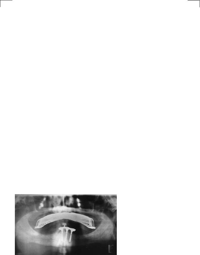
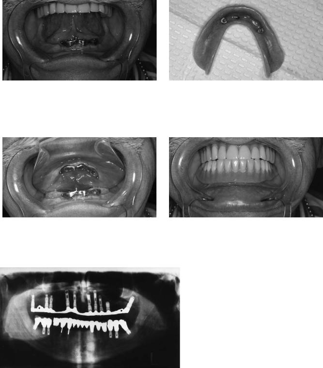
Completely edentulous mandible reconstructed with 3-screw type dental implants and fabrication of a bar with three ball attachments and an overdenture (Figures 21B.1–21B.5). (Dental implant surgery: Alex M. Greenberg, DDS, Oral and Maxillofacial Surgeon, New York, NY. Implant prosthodontics: Ava Thaw, DDS, Prosthodontist, Private Practice, New York, NY.)
FIGURE 21B.1 Panoramic radiograph demonstrating 3-screw type dental implant fixtures of the anterior mandible with overdenture bar.
262
21B. Overdenture Case Reports
FIGURE 21B.2 Frontal view of edentulous mandible with overdenture bar. Note the unfavorable floor of mouth and tongue position relative to the edentulous ridge.
FIGURE 21B.3 Occlusal view of edentulous mandible with overdenture bar.
263
FIGURE 21B.4 View of overdenture base and three o-rings.
FIGURE 21B.5 Mandibular overdenture in occlusion with maxillary fixed bridge.
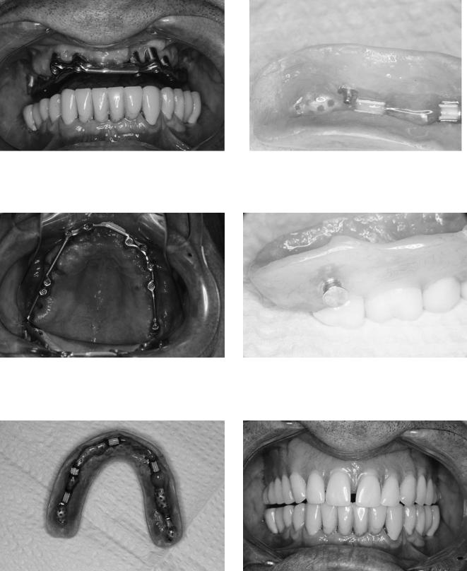
FIGURE 21B.6 Panoramic radiograph demonstrating 10 maxillary dental implant fixtures as well as the overdenture bar and bilateral posterior skirts for push–pull Lew attachments.
264
FIGURE 21B.7 Frontal view of maxillary overdenture bar and occlusal clearance.
FIGURE 21B.8 Occlusal view of maxillary overdenture bar.
A.M. Greenberg
FIGURE 21B.10 Close-up view of overdenture base with Lew attachment pushed into position.
FIGURE 21B.11 Close-up buccal view of Lew attachment push–pull button.
FIGURE 21B.9 View of overdenture base and 5 Hayder nylon clips. Note the absence of palatal coverage.
FIGURE 21B.12 Maxillary overdenture in opposing mandibular fixed bridge.
21B. Overdenture Case Reports
Case 2
Completely edentulous maxilla reconstructed with 10 dental implants, fabrication of continuous bar with posterior bilateral skirts for Lew attachments, 5 Hayder clips, and overdenture (Figures 21B.6–21B.12). (Dental implant surgery: Alex M. Greenberg, DDS, Oral and Maxillofacial Surgeon, New York, NY. Implant Prosthodontics, Joel Hirsch, DDS, Prosthodontist, Private Practice, New York, NY.)
References
1.Branemark P-I, Zarb GA, Albrektsson T. Tissue Integrated Prostheses. Quintessence: Chicago, 1987:283–287.
2.Worthington P, Branemark P-I. Advanced Osseointegration
265
Surgery: Applications in the Maxillofacial Region. Chicago:
Quintessence, 1992:233–247.
3.Misch CE. Contemporary Implant Dentistry. St. Louis: MosbyYearbook, 1993:223–240.
4.Jemt T, Chai J, Harnett J, Heath MR, et al. A 5-year prospective multicenter follow-up report on overdentures supported by osseointegrated implants. Int J Oral Maxillofac Implants. 1996; 11:291–298.
5.Misch LS, Misch CE. Denture satisfaction: a patient’s perspective. Int J Oral Implant. 1991;7:43–48.
6.Floystrand F, Karlsenk, Saxegaard E, Orstavik JS. Effects on retention of reducing the palatal coverage of complete maxillary dentures. Acta Odontol Scand. 1986;44(2):77–83.
7.Lundqvist S. Speech and other oral functions. Clinical and experimental studies with special reference to maxillary rehabilitation on osseointegrated implants. Swed Dent J. (suppl) 1993;91:1–39.
This page intentionally left blank
Section III
Craniomaxillofacial Reconstructive
and Corrective Bone Surgery
This page intentionally left blank