

Учебники / Enlarged Bony Portion of the Eustachian Tube in Oculoauriculovertebral Spectrum
.pdfOtology & Neurotology
24:961–962 © 2003, Otology & Neurotology, Inc.
Imaging Case of the Month
Enlarged Bony Portion of the Eustachian Tube in
Oculoauriculovertebral Spectrum
*Shin-Ichi Haginomori, *Ryuzaburo Nonaka,
*Hideaki Hoshijima, *Masahiko Higashikawa, *Hiroshi Takenaka,
†Yasuo Uesugi, and †Isamu Narabayashi
*Department of Otolaryngology and †Department of Radiology, Osaka Medical College, Takatsuki, Japan
The patulous eustachian tube (ET) has been reported as the cause of cholesteatoma, otitis media, and tinnitus. Almost all reports on the patulous eustachian tube mentioned the patulous lumen in cartilaginous portion of the ET and atrophy of peritubal tissue, especially fat tissue. In contrast, enlarged bony portion of the ET is rare (1) and its pathophysiology remains unknown. Radiological studies are extremely useful to detect this anomaly.
In this case of oculoauriculovertebral spectrum (OVAS), the multi-detector row computed tomography (1-mm collimation, 1-mm interval) reveals the enlarged bony portion of the left ET (Figs. 1 and 2). The width of the bony portion of the ET, which is closed to the junctional portion of the ET (2), is 7 mm and is much wider than that in normal children (1.5 mm) as reported by Suzuki et al (3) (Fig. 1). Huge bony dehiscence is recognized in the carotid canal (Fig. 2). Moreover, underdeveloped vestibule and semicircular canals, complete absence of the cochlea (Fig. 1 and 2), and anomalous ossicles are observed. In the right ear, which has a normal aspect in the middle ear and inner ear, the shape and width (1.3 mm) of the bony portion of the ET are normal (Fig. 3).
Embryologically, the tubotympanic recess and primary tympanic cavity are derived from the expanding terminal end of the endoderm-lined first pharyngeal pouch and probably the second pharyngeal pouch (4). The endoderm of the tubotympanic recess approached the surface that comes in contact with the ectodermal membrane of the first branchial groove. Near the end of the second month, the tubotympanic recess undergoes a bottleneck constriction, then the medial constricted portion lengthens and becomes the ET (4). The bony portion
Address correspondence and reprint requests to Dr. Shin-Ichi Haginomori, Department of Otolaryngology, Osaka Medical College, 2–7 Daigakucho, Takatsuki, Osaka 569–8686, Japan; Email: hagi@poh.osaka-med.ac.jp
FIG. 1. Axial computed tomography shows an enlarged bony portion of the eustachian tube (white asterisk) and complete absence of the cochlea in the left ear.
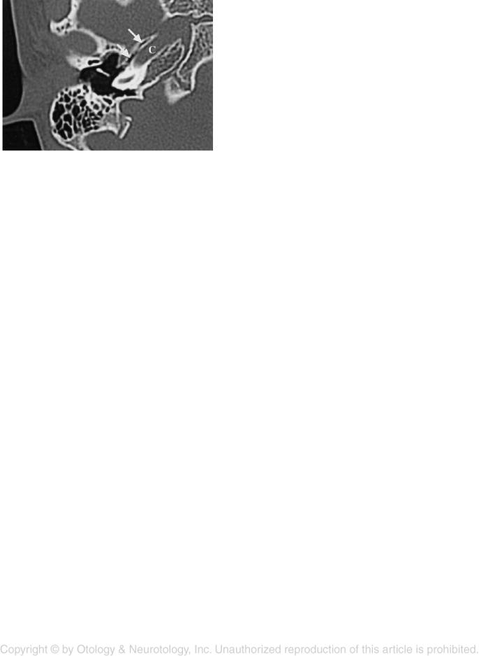
FIG. 2. Axial computed tomography shows a huge bony dehiscence (white arrow) in the carotid canal (C) facing the bony portion of the eustachian tube (E) in the left ear.
961
962 |
S.-I. HAGINOMORI ET AL. |
FIG. 3. Axial computed tomography shows normal shape and width of the bony portion of the eustachian tube (white arrows) in the right ear. C, carotid canal.
of the ET is surrounded by the petrous part of the temporal bone that is first formed in cartilage (4) and the sphenoid bone that is formed by membranous bone development. The anomalies in OVAS (e.g., epibulbar dermoids or lipodermoids, microtia, mandibular hypoplasia, and vertebral anomalies) are considered to be congenital malformations secondary to developmental abnormali-
ties of mostly first and second branchial arch derivatives
(5). Accordingly, there is a possibility that OVAS has abnormalities in the ET. With regard to anomalies in the bony portion of the ET in OVAS, Miura et al (6) reported histopathologically that it was rather narrow with a slit like shape. The enlarged bony portion of the ET in our case is opposite findings to Miura’s report (6). This difference suggests that OVAS does not arise from one etiopathogenetic factor, but from some complex factors.
REFERENCES
1.Tolley NS, Phelps P. Patulous eustachian tube: a radiological perspective. J Laryngol Otol 1990;104:291–3.
2.Sudo M, Sando I, Ikui A, et al. Narrowest (isthmus) portion of eustachian tube: A computer-aided 3-dimensional reconstruction and measurement study. Ann Otol Rhinol Laryngol 1997;106: 583–8.
3.Suzuki C, Balaban C, Sando I, et al. Postnatal development of eustachian tube: A computer-aided 3-dimensional reconstruction and measurement study. Acta Otolaryngol (Stockh) 1998;118: 837–43.
4.Pearson AA. Developmental anatomy of the ear. In: English M, ed. Otolaryngology. New York, NY: Harper and Row, 1978:1–68.
5.Rollnick BR, Kaye CI, Nagatoshi K, et al. Oculoauriculovertebral dysplasia and variants: Phenotypic characteristic of 249 patients. Am J Med Genet 1987;26:361–75.
6.Miura M, Sando I, Takasaki K, et al. Histopathologic study of temporal bone and eustachian tube in oculoauriculovertebral spectrum. Ann Otol Rhinol Laryngol 2001;110:922–7.
Otology & Neurotology, Vol. 24, No. 6, 2003