

Учебники / Diagnosis in Otorhinolaryngology Onerci 2009
.pdf
T. Metin Önerci
Diagnosis in Otorhinolaryngology

T. Metin Önerci
Diagnosis in
Otorhinolaryngology
With 432 Figures and 62 Tables
Prof. T. Metin Önerci
Hacettepe University
Faculty of Medicine
Dept. of Otorhinolaryngology 06100 Ankara
Sihhiya
Turkey metin@tr.net
ISBN: 978-3-642-00498-8 e-ISBN: 978-3-642-00499-5
DOI: 10.1007 / 978-3-642-00499-5
Springer Dordrecht Heidelberg London New York
Library of Congress Control Number: 2009926009
Springer-Verlag Berlin Heidelberg 2009
This work is subject to copyright. All rights are reserved, whether the whole or part of the material is concerned, specifically the rights of translation, reprinting, reuse of illustrations, recitation, broadcasting, reproduction on microfilm or in any other way, and storage in data banks. Duplication of this publication or parts thereof is permitted only under the provisions of the German Copyright Law of September 9, 1965, in its current version, and permission for use must always be obtained from Springer. Violations are liable to prosecution under the German Copyright Law.
The use of general descriptive names, registered names, trademarks, etc. in this publication does not imply, even in the absence of a specific statement, that such names are exempt from the relevant protective laws and regulations and therefore free for general use.
Product liability: The publishers cannot guarantee the accuracy of any information about dosage and application contained in this book. In every individual case the user must check such information by consulting the relevant literature.
Cover design: eStudioCalamar, Figueres/Berlin
Printed on acid-free paper
Springer is part of Springer Science+Business Media (www.springer.com)

Preface
In preparing the material for this book, I took the advice of my students who generously shared their views and opinions with me. I was told that it would be preferable to have images of the various diseases with legends describing the disease. Students would be able to learn and retain the information more successfully if the material was accompanied by pictures and schematic drawings.
Recent advances in technology have made it possible to photograph regions that are difficult to view with the naked eye, such as the ear, nose, throat, nasopharynx, and larynx – all the areas of otorhinolaryngology. Such an illustrated text in this field is important and necessary for teaching purposes.
In this book I tried to compile images of the basic conditions that are commonly seen in general practice and to give the reader a visual survey with a brief description of the condition. I added tables and schematic drawings in order to provide practical information. It is not the purpose of this book to be a comprehensive textbook, since many textbooks are already available with more detailed information of the conditions illustrated here.
This book is primarily intended for medical students, family and general practitioners, and ENT trainees. It may also serve as basic reading material for those in allied specialties. I hope my colleagues find this book useful and it contributes toward their teaching purposes.
Ankara, Turkey |
T. Metin Önerci |

Contents
Chapter 1 Ear |
|
2.5 |
Sinusitis |
72 |
|
|
|
|
|
2.6 |
Complications of Sinusitis |
79 |
|
1.1 |
Ear Anatomy 2 |
|
2.7 |
Nasal Polyposis 87 |
|
|
1.2 |
ENT Examination |
8 |
2.8 |
Nasal Obstruction 92 |
|
|
1.3 |
The Pinna 14 |
|
2.9 |
Septum |
97 |
|
1.4 |
External Ear Canal |
18 |
2.10 |
Epistaxis |
101 |
|
1.5 |
Otitis Media with Effusion 24 |
2.11 |
Traumas |
104 |
|
|
1.6 |
Acute Otitis Media |
28 |
2.12 |
Dacryocystorhinostomy |
110 |
|
1.7 |
Chronic Otitis Media 34 |
2.13 |
Tumors |
112 |
|
|
1.8Facial Nerve Paralysis 41
1.9Complications of Otitis Media 43
1.10 |
Hearing Loss 45 |
Chapter 3 Throat & Neck |
||||||
1.11 |
Otalgia |
48 |
|
|
|
|
|
|
1.12 |
Temporal Bone Fractures 50 |
3.1 |
Acute Tonsillopharyngitis 122 |
|||||
1.13 |
Tinnitus |
52 |
3.2 |
Adenoids |
126 |
|
||
1.14 |
Vertigo |
54 |
3.3 |
Snoring |
129 |
|
||
|
|
|
3.4 |
Temporomandibular |
|
|||
|
|
|
|
Joint |
132 |
|
|
|
Chapter 2 Nose |
3.5 |
Airway Obstructions |
134 |
|||||
|
|
|
3.6 |
Hoarseness |
137 |
|
||
2.1 |
The Common Cold and the Flu 58 |
3.7 |
Cysts |
143 |
|
|
||
2.2 |
Rhinitis |
60 |
3.8 |
Parotid Tumors 147 |
|
|||
2.3 |
Allergic Rhinitis 65 |
3.9 |
Oral Cavity |
151 |
|
|||
2.4 |
Nasal Vestibulitis and Nasal Furunculosis |
3.10 |
Neck Masses |
159 |
|
|||
|
and Mucormycosis 69 |
3.11 |
Neck Malignancies |
168 |
||||
2 |
Chapter 1 Ear |
1.1
Ear Anatomy
a |
c |
|
Malleus Incus
Stapes
Styloid
Stylohyoid ligament
Hyoid bone
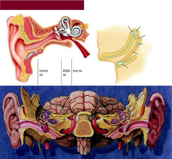
Fig. 1.1.1 (a) The ear is divided into three compartments: external, middle, and inner. The pinna is composed of cartilage covered by skin. The shape of the cartilage is very important, since it gives the shape of the auricle. Any necrosis of the cartilage may lead to cosmetic deformity. The external auditory canal measures approximately 2.5 cm. The outer one-third is cartilaginous and the inner two-thirds is bony. There is a narrowing at the bone–cartilage junction which causes foreign bodies to get stuck in this area. The skin of the bony part is very thin lying on the periosteum and does not contain glands, hair follicles, and any adnexal structures. There are two or three fissures in the cartilaginous external auditory canal which are called “Santorini fissures.” These fissures provide a potential route for the spread of infection from the external ear to the parotid area or infratemporal fossa and also of tumors from the parotid area to the external ear. The eustachian tube connects the middle ear to the nasopharynx. The posterior one-third of the adult eustachian tube is bony and lies within the petrous portion of the temporal bone. The anterior two-thirds is cartilaginous. In adults the tube lies at an angle of 45° in relation to the horizontal plane, whereas this inclination is only 10° in infants. The tube is longer in the adult than in the infant and young child. (b) Illustration showing the organs of hearing and the cerebellum. Sound
waves are channeled by the pinna (visible part of the ear) into the auditory canal (pink) toward the eardrum. The eardrum transmits the vibrations to three tiny bones – the malleus, incus, and stapes – in the middle ear. The stapes passes the vibrations to the inner ear structures (purple), the semicircular canals and the cochlea (spiral). Auditory sensations are picked up by the cochlear nerve (yellow) and transmitted to the medulla (brainstem), the thalamus, and ultimately the cerebral cortex (visual photos). (c) The external and middle ear develop from the branchial apparatus. The middle ear cavity is derived from the endodermal first branchial cleft. The inner ear develops from the otic placode. The first arch, or Meckel’s cartilage, contributes to the malleus and the incus. The tensor tympani muscle derives from the first branchial arch and is innervated by the nerve of the first branchial arch, which is mandibular branch of the trigeminal nerve. The second branchial arch, or Reichert’s cartilage, contributes to the suprastructure of the stapes. The stapes muscle is innervated by a facial nerve, which is the nerve of the second branchial arch. The chorda tympani nerve, a branch of the facial nerve (second arch nerve), joins the first arch nerve to the mandibular lingual nerve. The footplate of the stapes is derived from the otic capsule. Thus, a congenital abnormality can occur in one part while the other parts may develop normally

1.1 Ear Anatomy
a
Fig. 1.1.2 The innervation of the auricle is by the greater auricular nerve (C3), the lesser occipital nerve (C2, 3), the auriculotemporal nerve (V3), and sensorial branches of the VII and X cranial nerves
Fig. 1.1.3 The external auditory canal is not straight. To see the tympanic membrane, the ear canal should be straightened by pulling the auricle posteriorly and superiorly in adults (but inferiorly in infants)
b
Light cone
Fig. 1.1.4 (a, b) The tympanic membrane is elliptical and slightly conical in shape. The apex of this cone, the umbo, marks the inferior part of the manubrium. The diameter of the tympanic membrane measures approximately 9 mm (9–10 vertical; 8–9 horizontal). The surface area is 85–90 mm2. The tympanic membrane is composed of three layers: an outer epidermal layer, an inner mucosal layer, and a middle fibrous layer. The area above the short process of the malleus is known as pars flaccida and the area below as pars tensa. The pars flaccida does not have a middle fibrous layer, therefore it is flaccid. The pars tensa thickens peripherally forming the tympanic annulus. The tympanic annulus does not exist superiorly around the pars flaccida. There is a light triangle in the anterior–inferior quadrant of the tympanic membrane. The position of this triangle changes superiorly and becomes shorter when the tympanic membrane is retracted
3
A E R
O N
E S
H T
O R
AT
K C E N D N A

4 |
Chapter 1 Ear |
Fig. 1.1.5 False-color scanning electron micrograph (SEM) of the three smallest bones in the human body responsible for conduction of sound waves in the middle ear. At the top left is the malleus (hammer), which strikes the incus (anvil – right of malleus); the incus is joined to the stapes (stirrups), which conducts sound toward the inner ear. Sound waves enter the ear through the external auditory meatus and cause the eardrum to vibrate. Vibrations from the eardrum are passed to the malleus and then the stapes via the incus. The stapes transmits the vibrations to the fluid-filled cochlea of the inner ear where the vibrations are converted to nerve impulses. Lever effect: the manubrium mallei is 1.3 times longer than the long process of the incus. This difference in the lengths of the manubrium mallei and long process of the incus contributes a lever factor of 1.3 to increase the intensity of the sound (visual photos)
Fig. 1.1.6 Due to the differences in the physical properties of air and water, sound vibrations in the air are largely reflected away from the surface of water (99.9% of the energy of air-borne sound is reflected away), with only 0.1% entering the water. Although the surface area of the tympanic membrane is 85–90 mm2, the effective vibrating area of the tympanic membrane is 55 mm2. The surface area of the footplate is 3.2 mm2. The ratio of the surface areas of the tympanic membrane and the footplate is 55/3.2 = 17.1. This represents the hydraulic ratio of the tympanic membrane and stapes footplate, producing an increase force at the oval window of 17 times for the human ear, since the sound pressure level is equal to the force divided by the surface area (P = F/a). The final transformer ratio of the human tympanic membrane and ossicular chain is the product of the lever ratio of 1.3 times the hydraulic ratio of 17, which equals 22. This gain compensates the loss due to the air–bone difference
1.1 Ear Anatomy
a |
b |
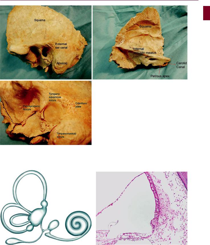
Fig. 1.1.7 (a–c) The temporal bone contains the ear. It has five parts: |
sure, petrosquamous suture, tympanosquamous suture, tympano- |
the bony external ear canal, the styloid process, the squamous por- |
mastoid suture etc. The mastoid process is not present at birth, which |
tion, the petrous portion, and the mastoid process. There are suture |
makes the facial nerve very superficial |
lines between these various portions such as the petrotympanic fis- |
|
5
A E R
O N
E S
H T
O R
AT
K C E N D N A
Fig. 1.1.8 The inner ear comprises the cochlea and the labyrinth. The labyrinth consists of three semicircular canals (superior, posterior, and lateral) and two otolithic organs (utricle and saccule). The utricular duct and the saccular duct join to form the endolymphatic duct
Fig. 1.1.9 The cochlea has three fluid-filled compartments: the scala tympani, the scala vestibuli, and the scala media, which contains the organ of Corti (courtesy of Paparella, Paparella otopathology lab director)