

Учебники / Diagnosis in Otorhinolaryngology Onerci 2009
.pdf2.9 Septum
2.9
Septum
a
b |
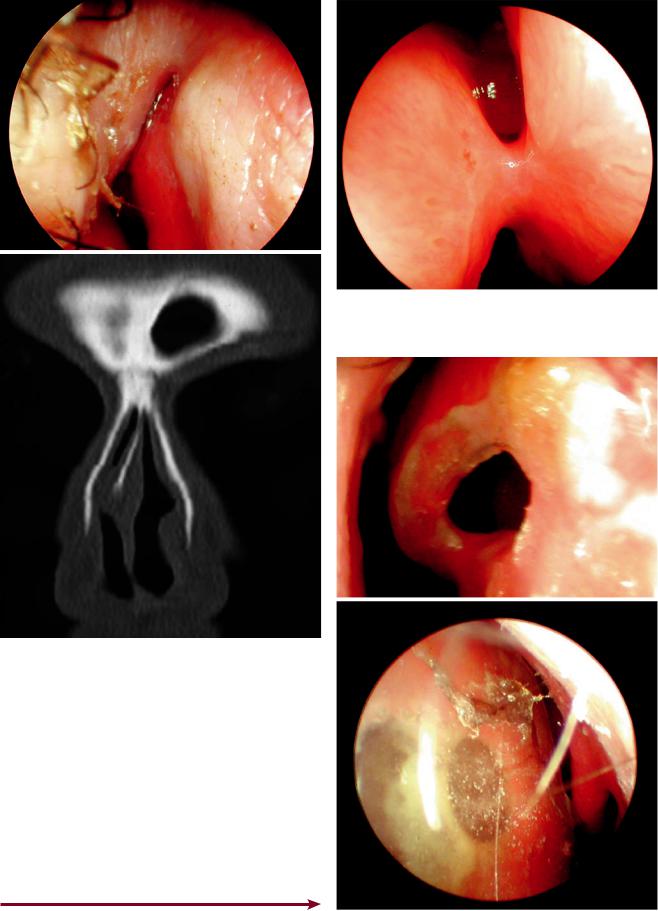
Fig. 2.9.2 Septal spur at the anterior septal area completely obstruct- |
ing the airway |
Fig. 2.9.1 (a) Septal deviation to the left side. (b) Compensatory Fig. 2.9.3 Severe caudal dislocation of the septum to the right inferior turbinate hypertrophy on the opposite side
97
A E R
O N
E S
H T
O R
AT
K C E N D N A
98 |
Chapter 2 Nose |
a
b
Fig. 2.9.4 (a) Septal deviation to the right, which narrows nasal valve area. (b) Coronal CT shows narrowing of the nasal valve area
Fig. 2.9.6 Septal perforation. There are many causes of septal perforation. The most common cause is previous septal surgery. Other causes include chronic trauma such as nose-picking, use of cocaine, septal hematomas, and infections like tuberculosis. The majority of perforations are located in the anterior cartilaginous portion of the septum. However, syphilitic infection involves the posterior bony septum. In small perforations a whistling sound may be heard during inspiration. Crusting may cause obstruction. As the crusts break off, bleeding occurs. Steam inhalations, nasal douching, and softening ointments may decrease the crusting. (a) Septal perforation after submucous resection. (b) Septal button to close the perforation temporarily. Small and medium-sized perforations can be closed by surgery. For bigger perforations, a silastic nasal septal button can be used for occlusion
Fig. 2.9.5 Synechia between septum and inferior turbinate
a
b
2.9 Septum
a |
b |
Fig. 2.9.7 (a, b) Saddle nose deformity. This was due to dorsal collapse of the nose as a result of cartilage destruction in a patient with tuberculosis
a |
b |
c
99
A E R
O N
E S
H T
O R
AT
K C E N D N A
Fig. 2.9.8 External rhinoplasty. (a) A transverse incision across the columella, (b) elevation of the nasal skin superiorly. (c) Suturing lower lateral cartilages

100 Chapter 2 Nose
Fig. 2.9.9 (a) Cartilage and |
a |
c |
|
bony hump removal. (b) To |
|||
|
|
||
rotate the nasal tip superiorly, |
|
|
|
the septal cartilage, lower and |
|
|
|
upper lateral cartilages may be |
|
|
|
shortened. (c) Medial and lateral |
|
|
|
osteotomies to narrow the nose |
|
|
b
Fig. 2.9.10 Infected cartilage graft at the nasal dorsum after rhinoplasty
2.10
Epistaxis
Although the majority of cases of epistaxis are self-limiting, it may sometimes be very severe and very difficult to manage. In Table 2.10.1, local and systemic factors that may cause epistaxis are presented.
Epistaxis may be from the anterior or posterior septum or nasal cavity. Posterior epistaxis is usually from the sphenopalatine artery and sometimes may be difficult to control and manage. Bleeding from Little’s area may be cauterized by silver nitrate.
The patient sits in the upright position. The head should be kept straight and should not be extended in order to keep the intracranial pressure low. The nose should be cleared of clots by blowing. Cotton wool pledgets soaked in appropriate solution are placed in the nasal cavity. In hypertensive patients, adrenaline should not be used. These pledgets show whether the bleeding is from the anterior or posterior side. If the bleeding is from Little’s area, it can be cauterized. Anterior packing should be placed in layers. If the epistaxis is from the posterior septal area, then posterior packing may be necessary. Systemic diseases should be treated.
Table 2.10.1 Etiology of epistaxis
Local
Trauma
Inflammation
Postoperative
Foreign body
Nasal and paranasal sinus tumors
Hereditary hemorrhagic telangiectasia
Atrophic rhinitis
Systemic
Hypertension
High venous pressure (mitral stenosis)
Blood dyscrasias (leukemia, hemophilia, vitamin K deficiency)
Anticoagulant drugs
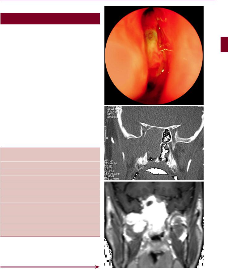
Fig. 2.10.1 Unilateral bleeding and nasal obstruction in a young male patient should raise the suspicion of juvenile nasopharyngeal angiofibroma (JNA). (a) Endoscopic view of the JNA in the nose.
(b)Coronal CT shows widening of the pterygomaxillary fissure.
(c)Hypervascularity of the tumor
2.10 Epistaxis
a
b
c
101
A E R
O N
E S
H T
O R
AT
K C E N D N A

102 Chapter 2 |
Nose |
a |
b |
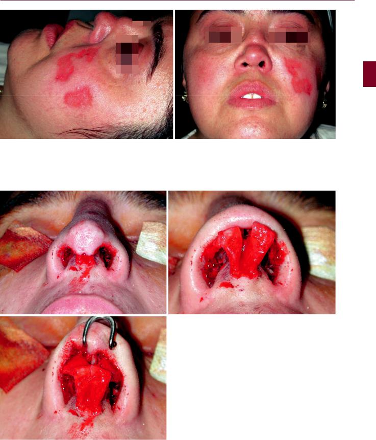
Fig. 2.10.2 (a) Hereditary hemorrhagic telangiectasia (Osler-Weber- Rendu disease) is characterized by small abnormal capillaries in the nasal mucosa. It may cause serious nasal bleeding, and hemoglobin levels may drop to very low levels, necessitating blood transfusion. In
Fig. 2.10.3 Kiesselbach’s plexus (Little’s area). It is localized at the anterior portion of the septum. Approximately 90% of bleeding occurs from this area. The external and carotid artery systems anastomose in this area. The anterior and posterior ethmoid arteries from the internal carotid artery system and the superior labial artery, greater palatine artery, and sphenopalatine artery from the external carotid artery system form a vascular plexus
the nasal mucosa, numerous leashes of bleeding vessels can be seen. Laser coagulation may be effective in the early stages. In severe cases, skin grafting may be necessary. (b) Angiography shows hypervascularization
Fig. 2.10.4 Bleeding vessels in Kiesselbach’s plexus (Little’s area)
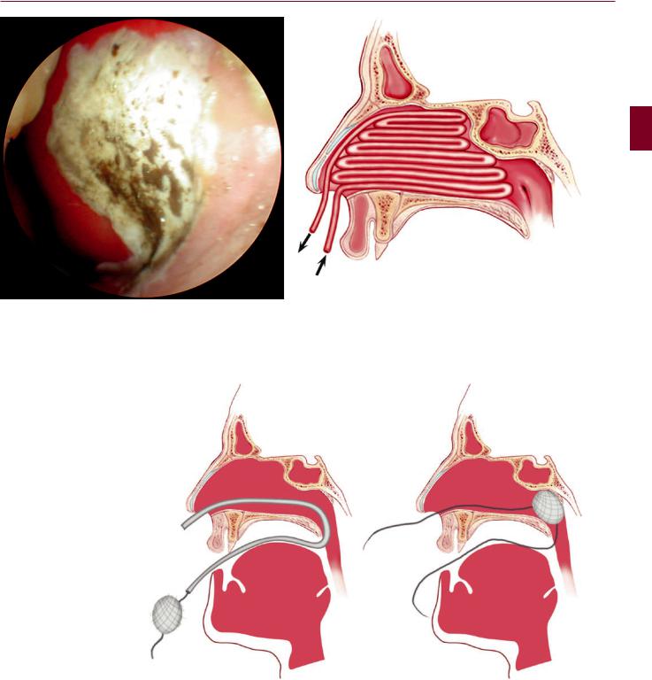
Fig. 2.10.5 Cauterization of Little’s area with silver nitrate sticks
Fig. 2.10.7 Posterior nasal packing. A postnasal pack is prepared from a tightly compressed ball of gauze according to the size of the postnasal space and tied with 0-silk ties. A soft rubber catheter is passed along the floor of the nose to the pharynx and taken out through the mouth. One end of the silk tie is attached to the tip of the catheter and drawn back through the nose. The pack is pushed behind the soft palate in the nasopharynx by exerting moderate pressure. The ends of the silk ties are attached near the nose and mouth corner. All patients with posterior nasal packing should be hospitalized for close observation
2.10 Epistaxis
Fig. 2.10.6 Anterior nasal packing with Vaseline-impregnated gauze. After cleaning the clots, the packing is done in layers
103
A E R
O N
E S
H T
O R
AT
K C E N D N A

104 Chapter 2 Nose
2.11
Traumas
Fig. 2.11.1 Nasal fracture. A blow coming from the lateral side caused the nose to be displaced to the other side. There is ecchymosis in the infraorbital area
Fig. 2.11.3 Coronal CT scan showing multiple fractures of the nasal bones
Fig. 2.11.4 Lateral X-ray showing nasal bone fracture. Depression of the nasal bones requires the elevation of the depressed bony fragments
Fig. 2.11.2 Lateral X-ray. Nasal fracture shows displacement of nasal bones
Fig. 2.11.5 Raccoon eyes or periorbital ecchymosis is a sign of basal skull fracture. It results from blood from the skull fracture tracking down into the soft tissue around the eyes (courtesy of Dr. Kıratlı)

2.11 Traumas
a |
b |
Fig. 2.11.6 Cerebrospinal fluid rhinorrhea after trauma. (a) Coronal CT showing herniation through the lamina cribrosa at the anterior ethmoid area. (b) Photograph of the herniated tissue
a |
b |
Fig. 2.11.7 (a, b) Air in the intracranial cavity after traumatic fracture in the
anterior and posterior frontal tables and anterior skull base
105
A E R
O N
E S
H T
O R
AT
K C E N D N A

106 Chapter 2 |
Nose |
a |
b |
Fig. 2.11.8 (a, b) Le Fort III maxillary fracture. Extensive facial fractures may require urgent intervention to prevent respiratory obstruction. Maxillary fractures were first classified by Dr. Le Fort and this classification system was called the“Le Fort classification.”In the Le Fort classification system there are three types of fractures: Le Fort I: The fracture line passes along the pyriform aperture and inferior wall of the antrum. It is a low maxillary fracture that separates the maxilla at the level of
the nasal floor. Le Fort II (pyramidal fracture): The fracture line passes across the nasal bone, the frontal processes of the maxilla, and the zygomatic-maxillary junction. It separates the central (middle) third of face from the base of the skull. Le Fort III (craniofacial dysjunction): The facial bones separate completely from the skull base. The fracture line extends along the nasofrontal, maxillofrontal, and zygomaticofrontal sutures. These fractures are reduced with miniplates
a |
b |
c |
d |
e
Fig. 2.11.9 Left-sided orbital blowout fracture. The soft tissue contents of the orbit have extruded into the maxillary antrum. Orbital blowout fracture is usually caused by blunt trauma to the eye globe. Increased pressure in the orbit causes fracture in the weakest part of the orbital cavity. The orbital contents extrude into the maxillary antrum. Entrapment of the inferior rectus muscle causes limitation of
eye movement. Orbital herniation leads to enophthalmos on longterm follow-up. (a–g) After a severe maxillofacial trauma, mistreated blowout fracture resulted in enophthalmos and restriction in the mobility of the left eye. The patient recovered from diplopia after surgery (courtesy of Dr. Şener)