

Учебники / Diagnosis in Otorhinolaryngology Onerci 2009
.pdf26 |
Chapter 1 Ear |
Fig. 1.5.8 Left ear. Adhesive otitis media. The tympanic membrane is thinned and lies on the promontorium. The short process of the malleus and malleus handle is more prominent due to retraction of the membrane. Fibrous annulus is clearly identified
Fig. 1.5.9 Right ear. Otitis media with effusion due to nasopharyngeal carcinoma. Amber color and retraction of the tympanic membrane. A unilateral otitis media with effusion in adults always necessitates a detailed search for nasopharyngeal carcinoma
Fig. 1.5.10 Mucoid material in the external ear canal after myringotomy in otitis media with effusion
Fig. 1.5.11 Thick and sticky mucoid material obtained from the middle ear in a patient with otitis media with effusion

1.5 Otitis Media with Effusion
Fig. 1.5.12 Right ear. Sheppard grommet ventilation tube, which |
Fig. 1.5.14 Sheppard grommet ventilation tube extrusion. A tube in |
was inserted due to otitis media with effusion 6 months earlier. The |
the external ear canal with wax around it, 8 months after insertion. |
tympanic membrane looks normal. The ventilation tubes ventilate |
After extrusion, tympanosclerosis or membrane scarring may occur. |
the middle ear. The appearance of the drum returns to normal after a |
A small tube causes less trauma but is extruded more rapidly |
while. The tubes should stay in place for at least 6 months or more to |
|
have a normal middle ear physiology |
|
Fig. 1.5.13 Left ear. A 1.25-mm Paparella drain tube in the posterior inferior quadrant of the tympanic membrane. The tympanic membrane appears normal after 3 months of insertion of the ventilation tubes. Behind the transparent tympanic membrane the inner flanges of the tube can be seen. A myringotomy incision is generally made in the posterior inferior quadrant of the tympanic membrane. Some surgeons prefer to insert it in the anterosuperior quadrant to reduce the risk of early extrusion. Insertion of the ventilation tubes in the posterosuperior quadrant of the tympanic membrane should be avoided since it may damage the incudostapedial joint or long process of the incus
27
A E R
O N
E S
H T
O R
AT
K C E N D N A

28 |
Chapter 1 Ear |
1.6
Acute Otitis Media
a |
b |
Fig. 1.6.1 (a) Normal tympanic membrane. The short process of the malleus and malleus handle is seen. The tympanic membrane is transparent, sometimes allowing the long process of the incus and
Fig. 1.6.2 Otitic barotrauma (hemotympanum). The tympanic membrane appears blue due to hemorrhagic fluid collected in the middle ear. It is due to an inability to ventilate the middle ear following an abnormal function of the eustachian tube. Otitic barotrauma is usually seen during descent in flight or during scuba diving. No treatment is needed. If there is associated upper respiratory tract infection or allergy, topical and systemic oral decongestants with antihistamines may help recovery. To prevent further episodes the patients are advised not to go scuba diving when their nose is obstructed because of the difficulty of inflating the eustachian tube. Frequent flyers and regular sufferers are advised to use prophylactic measures to prevent eustachian tube problems, such as topical nasal decongestants and chewing gum etc.
promontorium to be seen. Note the light reflex in the anteroinferior quadrant of the membrane. (b) Eustachian tube dysfunction. Note the vascularization along the manubrium mallei

a |
a |
b
b
c
Fig. 1.6.3 Bullous myringitis in (a) the right ear and (b) the left ear. The malleus handle is hardly visible. Bullous myringitis is due to a viral or Mycoplasma pneumoniae infection of the tympanic membrane. There is severe ear pain, but no hearing loss. Draining the blebs may provide immediate relief from pain. Only the outer epithelial layer should be punctured. Complete puncturing of the tympanic membrane may result in perforation
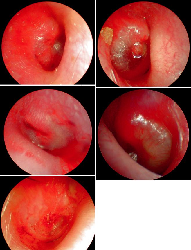
Fig. 1.6.4 Acute otitis media, hyperemia stage. (a) Hyperemia in the attic region of the left ear; the patient complains of ear pain only for the last one hour (b) Hyperemia in the attic region and the posterosuperior part of the tympanic membrane of the left ear; the patient complains of ear pain only for the last three hours (c) Hyperemia in the attic region and the posterosuperior part of the tympanic membrane of the right ear; slight bulging of the tympanic membrane has started
1.6 Acute Otitis Media 29
A E R
O N
E S
H T
O R
AT
K C E N D N A
30 |
Chapter 1 |
Ear |
|
a |
d |
b |
e |
c
Fig. 1.6.5 Acute otitis media. Different phases of the exudative stage.
(a) Bulging in the posterior half of the tympanic membrane (right ear). (b) Slight bulging of the tympanic membrane (left ear). (c) Due
to bulging the malleus handle cannot be differentiated (right ear). (d) More severe bulging (right ear). (e) More severe bulging and opaque tympanic membrane (left ear). There is conductive-type hearing loss

a
b
1.6 Acute Otitis Media |
31 |
AT O R H T E S O N R A E
Fig. 1.6.6 Acute hemorrhagic otitis media in the right ear. Bulging of the tympanic membrane due to hemorrhagic purulent material in the middle ear
Fig. 1.6.7 Acute hemorrhagic otitis media in the right ear. There is severe bulging associated with severe ear pain. Due to extensive bulging, white-colored epithelium is seen on the tympanic membrane. The malleus cannot be identified
Fig. 1.6.8 Suppurative stage in acute otitis media (right ear). (a) Puru lent material filling the external ear canal and preventing the drum from being seen. (b)The small perforations in the tympanic membrane are seen after cleaning the purulent material in the external ear
K C E N D N A

32 |
Chapter 1 Ear |
Fig. 1.6.9 Suppurative stage in acute otitis media (left ear). (a) Purulent material filling the external ear canal and preventing the drum from being seen. (b) The small perforations in the tympanic membrane are
seen after cleaning the purulent material in the external ear. Note the perforation is located at the anterior superior quadrant. If drainage is not adequate, myringotomy at the lower quadrants may be necessary
a |
c |
b
Fig. 1.6.10 Resolution stage. During healing small perforations are seen in different parts of the tympanic membrane. All these perforations close by themselves without any additional treatment. (a) Left ear; (b) right ear; (c) left ear, stapes muscle tendon is seen through the perforation; (d) left ear
1.6 Acute Otitis Media
Fig. 1.6.11 The perforations of the tympanic membrane close by the epithelium coming from the edges of the perforation. Sometimes minor interventions may be needed if the perforation is big
Fig. 1.6.12 White areas of tympanosclerosis. They do not cause any symptoms and any hearing loss. There is no need for treatment. Tympanosclerosis in the tympanic membrane generally follows the insertion of ventilation tubes. Previous otitis media may also cause tympanosclerosis in the tympanic membrane and ossicles. If tympanosclerosis reduces the mobility of the ossicles, conductive-type hearing loss may appear which is not so easy to treat
Fig. 1.6.13 The perforation closed by pseudomembrane formation
Table 1.6.1 Contributing factors for recurrent acute otitis media
Day care center
Smoking at home
Bottle feeding
No mother milk nutrition
Immune insufficiency
Cleft palate
Table 1.6.2 Pathogenesis of acute otitis media
Upper respiratory tract infection
Edema of the eustachian tube Nasal obstruction
Positive pressure in the nasopharynx Adenoid vegetation
Eustachian tube obstruction
Inhabit the pathogens
Table 1.6.3 Stages of acute otitis media
Hyperemia
Exudation
Suppuration
Coalescence
Complication
Resolution
33
A E R
O N
E S
H T
O R
AT
K C E N D N A

34 |
Chapter 1 Ear |
1.7
Chronic Otitis Media
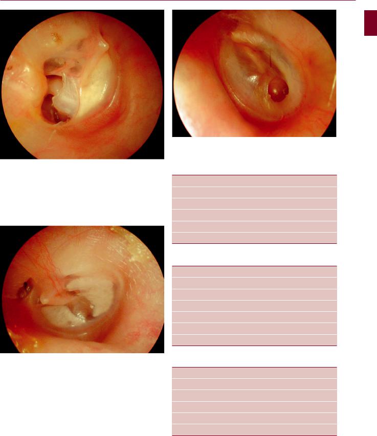
Fig. 1.7.1 Left ear. In the posteroinferior part of the tympanic membrane there is a central perforation. The epithelium goes into the middle ear from the anterior and inferior edges of the perforation. Anterior to the manubrium mallei the tympanic membrane is calcified. In the posterosuperior quadrant the tympanic membrane lies on the incus and stapes
Fig. 1.7.2 Left ear. A central perforation in the anterosuperior quadrant of the tympanic membrane. Through the perforation the eustachian tube orifice and tympanosclerotic middle ear mucosa are seen. The tympanic membrane appears to be tympanosclerotic
Fig. 1.7.3 Right ear. Central perforation in the anteroinferior quadrant of the tympanic membrane. Pseudomembranous tympanic membrane in the anterosuperior quadrant. The middle ear mucosa and the tympanic membrane are tympanosclerotic. Just posteroinferior to the umbo, the tympanic membrane is thickened by fibrous tissue and embedded with calcium. Tympanosclerosis is the collection of collagen and calcium in the submucosal layer in the tympanic membrane or middle ear mucosa. It is a healing process of the body. Especially after ventilation tube insertion, white calcium deposits may be seen due to submucosal bleeding. Such tympanosclerotic plaques may be large enough to interfere with normal tympanic membrane function. In the middle ear mucosa it may cause fixation of the ossicles and may lead to conductive-type hearing loss
Fig. 1.7.4 Left ear. Anteroinferior central perforation. At the posteroinferior location of the perforation, polypoid granulation tissue is present; the middle ear mucosa is hyperemic and edematous. There are calcified plaques in the tympanic membrane
1.7 Chronic Otitis Media
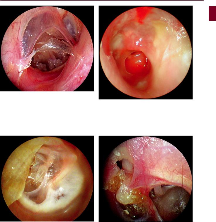
Fig. 1.7.5 Right ear. Central perforation with tympanosclerosis in the middle ear. The tympanic membrane tries to close the perforation and a pseudomembrane formation is seen
Fig. 1.7.7 Left ear. Acute infection in a patient with chronic otitis media. Purulent drainage in the external ear canal. There is a 3-mm central perforation in the tympanic membrane. Anterosuperior to the perforation there is polypoid tissue formation. The middle ear mucosa is edematous and hyperemic
35
A E R
O N
E S
H T
O R
AT
K C E N D N A
Fig. 1.7.6 Right ear. The anterior part is tympanosclerotic. The filter paper used to close the perforation is transported out onto the external ear canal and a replacement membrane closing the perforation is seen; however, there is a very small perforation still remaining between the filter paper and the pseudomembrane
Fig. 1.7.8 Right ear. Short process of the malleus and malleus handle can be seen. Just above the short process, attic perforation is seen. Posterior to the malleus handle, the tympanic membrane is severely retracted and lies on the promontorium and incudostapedial joint. The long process of the incus, lenticular process, incudostapedial joint, and stapes tendon are seen behind the adhesive and thinned tympanic membrane