

Учебники / Diagnosis in Otorhinolaryngology Onerci 2009
.pdf46 |
Chapter 1 Ear |
a
Fig. 1.10.5 Glomus jugulare. A pink–bluish silhouette of a glomus tumor behind the tympanic membrane
Fig. 1.10.6 Glomus jugulare tumor extending out to the external auditory canal
b
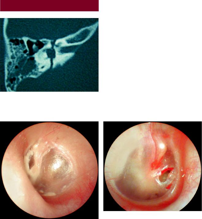
Fig. 1.10.7 (a) Temporal MR image. Glomus jugulare tumor on the right side extending to the hypotympanum. (b) Angiography shows hypervascularized glomus tumor
Fig. 1.10.8 Right ear. Through the tympanic membrane perforation, the glomus tumor is seen to be localized in the hypotympanum. It is in contact with the incudostapedial joint

Table 1.10.1 Etiology of hearing losses |
|
|
Cause |
Conductive hearing loss |
Sensorineural hearing |
|
|
loss |
Congenital |
Ear atresia |
Prenatal |
|
Ossicular pathologies |
Genetic |
|
|
Pregnancy |
|
|
Rubella |
Acquired |
External ear canal |
Birth (hypoxia jaundice) |
|
Wax |
Trauma (iatrogenic, acous- |
|
Foreign body |
tic trauma, head trauma) |
|
|
|
|
Middle ear |
Inflammatory (chronic otitis |
|
Otitis media with |
media, mumps, meningitis) |
|
|
|
|
effusion |
|
|
Chronic otitis media |
Degenerative presbyacusis |
|
Otosclerosis |
Ototoxicity |
|
Traumatic drum |
Neoplastic acoustic |
|
|
neurinoma |
|
Perforations |
Idiopathic Meniere disease |
|
|
Sudden hearing loss |
|
|
|
Table 1.10.2 Ototoxic drugs
Salicylic acid
Aminoglycosides
Streptomycin
Dihydrostreptomycin
Neomycin
Gentamycin
Kanamysin
Tobramycin Diuretics
Furosemide
Etacrynic acid Chemotherapeutic agents
Cisplatin/carboplatin
Nitrogen mustard
6-Amino nicotinamide
Vincristine/vinblastine
Misonidazole
Dichloro-methotroxate
Lonidamine
Paclitaxel
Others
Vancomycin
Polymyxin B
Iodoform
Tetanus antitoxin
Interferon alpha 2a
1.10 Hearing Loss 47
A E R
O N
E S
H T
O R
AT
K C E N D N A

48 |
Chapter 1 Ear |
1.11
Otalgia
Fig. 1.11.1 Nonotological causes of earache. The most common cause of otalgia is dental problems in adults
Fig. 1.11.2 Aphthous stomatitis behind the molar teeth in the gingivobuccal sulcus may cause severe ear pain
Fig. 1.11.3 Squamous cell carcinoma of the tongue which has caused ear pain

Fig. 1.11.4 Earache may also be seen in laryngeal carcinomas
1.11 Otalgia 49
A E R
O N
E S
H T
O R
AT
K C E N D N A
50 |
Chapter 1 Ear |
1.12
Temporal Bone Fractures
Fig. 1.12.1 Axial CT scan. Transverse temporal bone fracture on the left side. High-resolution CT scans of the temporal bone provide necessary information about the fracture. Audiometric examination helps in the diagnosis
Fig. 1.12.2 In longitudinal fractures, a fracture line at the posterosuperior part of the external ear canal may be seen
Fig. 1.12.3 Traumatic tympanic membrane perforation. These perforations are sometimes seen after a blow to the ear. To be sure that the perforation is really traumatic, the physician should check that the edges of the perforation are irregular and hemorrhagic

1.12 Temporal Bone Fractures
Fig. 1.12.4 Schematic representation of longitudinal and transverse fractures.Temporal bone fractures are classified into two main groups: longitudinal and transverse fractures. Longitudinal fractures are much more frequent, with the incidence of longitudinal fractures being four times greater than transverse fractures. Generally, temporal and parietal blows are associated with longitudinal fractures. Since the areas of the foramina are relatively weaker parts of the skull base, fractures tend to occur in their vicinity. Longitudinal fractures start in the squamous portion and go to the middle ear through the posterior and superior walls of the external ear canal and then to the petrous apex. Generally, conductive hearing loss is accompanied by longitudinal temporal bone fractures. Facial nerve injury may occur at the geniculate ganglion area and is only seen in 15% of longitudinal fractures. Tympanic membrane perforation or bleeding into the middle ear may also be seen. Transverse fractures generally occur due to frontal or occipital trauma. Since the blow comes from anterior-pos- terior or posterior-anterior direction, the fracture line occurs at a right angle to the axis of the petrous bone and the fracture line starts from the foramen magnum or jugular foramen and extends to the middle ear.Itfrequentlyaffectsthefacialnerveandinnerear.Hemotympanum may be associated with transverse fractures, but tympanic membrane perforation is not seen. Temporal bone fractures do not always follow these general guidelines, and some fractures are mixed. These fractures are evaluated according to the type of lesion
How to Make the Diagnosis of CSF Draining from the External Ear Canal
If the fluid is collected on a filtered paper or on a gauze, it forms a halo around the circle of blood. If the fluid can be collected in a tube, beta-2 transferrin positivity indicates CSF leak.
Table 1.12.1 How to treat CSF fistula
Head is elevated 30°
Gaita softeners are prescribed
Diuretics such as diazomide are given to decrease the CSF pressure
Wide-spectrum antibiotics are started as prophylaxis against meningitis
No packing is done to the external ear canal
Table 1.12.2 Differential diagnosis between longitudinal and transverse temporal fractures
|
Longitudinal |
Transverse |
Frequency (approximate %) |
80 |
20 |
Hearing loss |
Conductive |
Sensorineural |
Vertigo |
Rare |
Frequent |
Facial nerve paralysis |
Rare (10–15%) |
Frequent (50%) |
|
|
|
51
A E R
O N
E S
H T
O R
AT
K C E N D N A

52 |
Chapter 1 Ear |
1.13
Tinnitus
Fig. 1.13.3 Hydropic Reissner’s membrane in all turns of the cochlea (courtesy of Paparella, Paparella otopathology lab director). Endolymphatic hydrops causes roaring-type tinnitus
Fig. 1.13.1 Wax filling the ear canal may cause tinnitus
Fig. 1.13.4 Acoustic neuroma on the left side. Unilateral tinnitus, if associated with high-frequency hearing loss and low scores of speech discrimination, should be investigated for acoustic neurinoma
Fig. 1.13.2 Glomus jugulare tumor causing objective tinnitus

1.13 Tinnitus
Tinnitus is any abnormal noise in the ear. It may be objective or subjective. All pathologies in the external, middle, and inner ear may cause tinnitus. Unilateral tinnitus, if associated with high-frequency hearing loss, should be investigated and acoustic neurinoma eliminated.
Objective tinnitus can be heard by the examiner as well. Objectivetinnitusisrareandthecommonestformisvascular pathology such as glomus jugulare tumor, high jugular bulbus, arteriovenous malformations, and carotid body tumors. The normal pulsatile noise of blood passing through the internal carotid artery may also cause tinnitus. If you close the ear it becomes more apparent. Temporomandibular joint pathologies, insects in the external ear canal, and palatal myoclonus are other pathological conditions causing objectivetinnitusbesidesvascularcauses.Mostformsofobjective tinnitus are readily identifiable and curable.
Table 1.13.2 Ear diseases associated with objective tinnitus
Vascular pathologies
Arteriovenous malformation
Aneurysms
Carotid body tumor
Glomus jugulare
Palatal myoclonus
TMJ pathologies
Insects in the external ear canal
Table 1.13.1 Ear diseases associated with subjective tinnitus
External ear |
Wax |
|
Foreign body |
Middle ear |
Chronic otitis media |
|
Otosclerosis |
|
Otitis media with effusion |
|
Ossicular chain pathologies |
Inner ear |
Presbyacusis |
|
Acoustic trauma |
|
Ototoxic drugs |
|
Meniere’s disease |
|
Acoustic neuroma |
|
|
Table 1.13.3 Drugs that can cause or exacerbate tinnitus
Aspirin
Aminoglycosides
Loop diuretics
Quinine
Indomethacin
Alcohol
53
A E R
O N
E S
H T
O R
AT
K C E N D N A

54 |
Chapter 1 Ear |
1.14
Vertigo
Fig. 1.14.1 Normal appearance of Reissner’s membrane and the tectorial membrane (courtesy of Paparella, Paparella otopathology lab director)
Fig. 1.14.2 Hydropic Reissner’s membrane in one turn of the cochlea (courtesy of Paparella, Paparella otopathology lab director)
Table 1.14.1 Anamnesis in vertigo patient
Onset of vertigo
Character of vertigo, real vertigo, or dizziness
Duration
Relationship to the movements of the head
Other associated symptoms, tinnitus, hearing loss etc.
Fig. 1.14.3 Profound endolymphatic hydrops in all turns of the cochlea (courtesy of Paparella, Paparella otopathology lab director)
Fig. 1.14.4 Intracanalicular acoustic neuroma and serous labyrinthitis in the vestibule and cochlea (courtesy of Paparella, Paparella otopathology lab director)
Table 1.14.2 Differential diagnosis in vertigo according to duration
Duration |
No hearing loss |
With hearing loss |
Seconds |
Benign paroxysmal |
|
|
positional vertigo |
|
Minutes |
Vertebrobasilar |
|
|
insufficiency |
|
Hours |
|
Endolymphatic hydrops |
|
|
(Meniere disease) |
Days |
Vestibular neuritis |
Labyrinthitis |
Weeks |
Intracranial pathologies |
Acoustic neurinoma, |
|
Multiple sclerosis |
psychogenic |
|
|
|

1.14 Vertigo |
55 |
a
Fig. 1.14.5 Temporal bone section. Otosclerosis causing obstruction in the vestibular aqueduct and endolymphatic hydrops in all turns of the cochlea (courtesy of Paparella, Paparella otopathology lab director)
Table 1.14.3 Differential diagnosis in central and peripheral vertigo
|
Peripheral |
Central |
|
Unsteadiness |
Slight, moderate |
Severe |
|
Nausea, vomiting |
Severe |
Slight |
b |
Hearing symptoms |
Frequent |
Rare |
|
Neurologic symptoms |
Rare |
Frequent |
|
Compensation |
Fast |
Slow |
|
|
|
|
|
Table1.14.4 Differentialdiagnosisincentralandperipheralpositional vertigo
|
Peripheral |
Central |
Latent period |
+ |
– |
Adaptation |
+ |
– |
Fatigue |
+ |
– |
|
|
|
Fig. 1.14.6 (a) Balancing stone from inner ear. Color scanning electron micrograph of crystals of calcium carbonate on the surface of an otolith. An otolith or otoconium is a calcified stone that is found in the otolith organs of the inner ear. They are attached to sensory hairs, and, when the head tilts, the movement of the stones causes nerve impulses that form the basis of the sense of balance. In humans, otoconia can range in size from 3 to 30 µm (millionths of a meter) across (visual photos). (b) Dix Hallpike maneuver. The patient is brought to the supine position from the sitting position with the head turned to one side. The maneuver is repeated on the opposite side. The presence of nystagmus or any feeling of movement is recorded
K C E N D N A AT O R H T E S O N R A E