

Учебники / Diagnosis in Otorhinolaryngology Onerci 2009
.pdff
g
2.11 Traumas 107
A E R
O N
E S
H T
O R
AT
K C E N D N A
Fig. 2.11.9 (Continued)

108 Chapter 2 |
Nose |
a |
d |
b
e
c
Fig. 2.11.10 (a–e) After a severe traffic accident, fracture in the medial and inferior orbital walls resulted in enophthalmos and restriction in the mobility of the right eye. Gaze to the medial and inferior sides is limited. Note the herniation of the globe to the ethmoids (courtesy of Dr. Şener)

a |
b |
c
2.11 Traumas 109
A E R
O N
E S
H T
O R
AT
K C E N D N A
d |
e |
Fig. 2.11.11 Penetrating trauma to the right orbit and the intracranial cavity. The pencil was taken out, all foreign material cleaned, and CSF fistula repaired. (a, b) Pencil penetrating the skin just inferior and
medial to the orbit. (c–e) Coronal and axial CT scans showing trauma to the eye and the intracranial cavity (courtesy of Tesav)
110 Chapter 2 Nose
2.12
Nasolacrimal obstructions
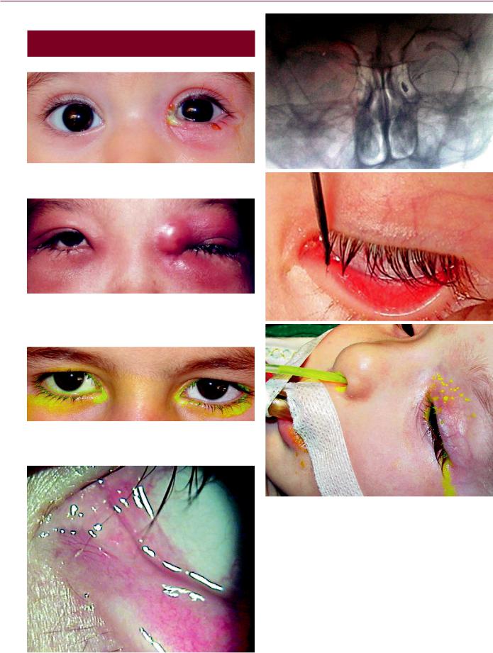
Fig. 2.12.1 Acute dacryocystitis on the left side
Fig. 2.12.2 Acute dacryocystitis with severe infection around the eye. Note the fluctuating mass in the left internal canthal area with hyperemia and edema around the left eye
Fig. 2.12.3 Congenital dacryostenosis with lengthened fluorescein drainage time on the right side
a
b
c
Fig. 2.12.5 Congenital dacryostenosis on the left side. (a) Macro dacryocystography. Opaque material on the left side was not drained to the nose. Opaque material on the right side has already been drained.
(b) Probing of the left nasolacrimal canal. (c) Fluorescein suctioned from the inferior meatus after injection to the nasolacrimal canal
Fig. 2.12.4 Punctum agenesis on the left side

a |
b |
c |
d |
2.12 Nasolacrimal obstructions 111
A E R
O N
E S
H T
O R
AT
K C E N D N A
Fig. 2.12.6 Endoscopic intubation of a patient with congenital dacryostenosis on the left side. (a) Inferior turbinate. (b) Medialization of the inferior turbinate. (c) The tip of the probe in the inferior meatus.
(d) Silicon tubes tied in the inferior meatus after they have been passed through the nasolacrimal canal under endoscopic control
Fig. 2.12.7 The loop of the silicon tube should not be very tight in order to avoid trauma to the canaliculi

112 Chapter 2 Nose
2.13
Tumors
a |
c |
|
b |
d |
|
Fig. 2.13.1 Inverted papillomas generally arise from the lateral nasal wall. They are unilateral masses. In 15% of cases there may be malignant transformation. The tumor should be excised completely and sent for histological examination to check for malignant transforma-
tion. Endoscopic removal is the treatment of choice. (a) Right-sided mass. (b) On sagittal MR image, the mass completely fills the nasal cavity. (c) Removal of the mass. (d) Histological picture of the inverted papilloma. Epithelium shows invaginations into the tissue

Fig. 2.13.2 Inverted papilloma that fills the nostril and is clearly visible
a
2.13 Tumors
b
113
A E R
O N
E S
H T
O R
AT
K C E N D N A
Fig. 2.13.3 Left-sided inverted papilloma. (a) Endoscopic appearance. (b) Coronal CT shows extension to the upper wall of the maxillary antrum

114 Chapter 2 Nose
a |
b |
|
d
c
e
f
Fig. 2.13.4 Juvenile nasopharyngeal angiofibroma. It usually arises superior and posterior to the sphenopalatine foramen. It only occurs in male adolescents. These patients present with the complaint of nasal obstruction and bleeding. (a) Endoscopic view. (b) On coronal CT scan. widening of pterygomaxillary fissure is diagnostic. (c) Axial CT
shows extension to the infratemporal fossa. (d) Angiography shows the hypervascularity of the tumor. (e) Embolization helps the surgeon to operate in a less bloody field. No vascularity after embolization is seen. (f) Endoscopic surgery is the treatment of choice. Specimen after total removal of the tumor

a |
b |
c
2.13 Tumors 115
A E R
O N
E S
H T
O R
AT
K C E N D N A
Fig. 2.13.5 Fibrous dysplasia is a fibro-osseous disease of the facial skeleton. It usually appears during the first two decades of life. On CT scan, ground glass presentation due to a mixture of fibrous and osseous components is characteristic of this tumor. If it does not
cause any deformity or functional disorder, no surgery is required.
(a) Fibrous dysplasia filling the left nostril completely. (b) Coronal CT showing fibrous dysplasia in the sphenoid bone. (c) Coronal CT showing fibrous dysplasia in the frontal bone
a |
b |
Fig. 2.13.6 Osteomas are common benign tumors of the sinuses. They occur most frequently in the frontoethmoid region. (a) Coronal CT showing osteoma in the right ethmoid area. (b) The specimen after removal

116 Chapter 2 Nose
a
c
b
d
Fig. 2.13.7 Malignant melanoma in the nasal cavity. (a) Mass in the right nasal cavity. (b) Pigmented mass after cleaning of purulent material. (c) Coronal MR image shows mass filling the ethmoids and
Fig. 2.13.8 Specimen of mucosal malignant melanoma in the nose after excision with safe margins. Malignant melanoma of the sinonasal tract comprises 1–2% of all melanomas. A pigmented polyp may be a malignant melanoma
maxillary sinus and extending to the palate. (d) Mass in the hard palate. Mass in the nasal cavity has eroded the palatal bone
Fig. 2.13.9 A unilateral mass in the nose that bleeds spontaneously should arouse the suspicion of malignancy. Esthesioneuroblastoma is an uncommon tumor that arises in the olfactory epithelium. These tumors very often invade the cranium. To show the intracranial extension, MR imaging is necessary