

Учебники / Diagnosis in Otorhinolaryngology Onerci 2009
.pdf36 |
Chapter 1 Ear |
Fig. 1.7.9 Correct instillation of ear drops. First, any debris or discharge in the external ear canal is cleaned. The auricle is pulled upward and backward. Subsequently, five to six drops (or more if needed) are introduced into the ear canal (help may be needed). Tragal massage may help the drops go into the middle ear. The patient is kept in that position for 2–3 min. Cotton wool is placed in the ear canal and can be removed 10 min later
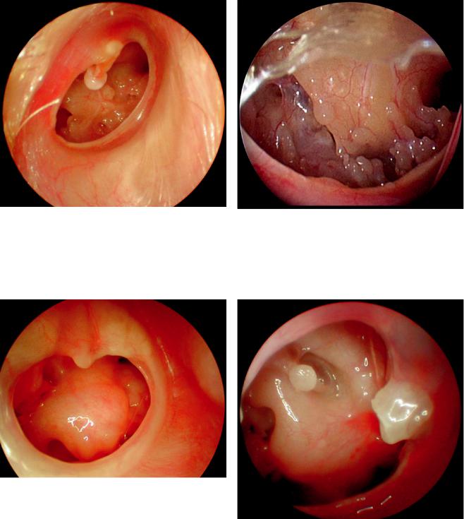
Fig. 1.7.11 Right ear. The tympanic membrane in the posterosuperior quadrant behind the malleus handle is retracted. The retraction pocket has cerumen and keratin debris and the apex of the retraction pocket cannot be identified
Fig. 1.7.10 Right ear. Attic perforation above the short process of the malleus after cleaning the epithelial debris
Fig. 1.7.12 Right ear. The tympanic membrane is retracted posterosuperiorly toward the antrum area. The bone over the retraction pocket is destroyed. The incus is eroded. The epithelium at the apex of the retraction pocket is seen. The chorda tympani nerve is visible behind the thinned retracted tympanic membrane

a
Fig. 1.7.13 Right ear. Behind the handle of the malleus, cerumen |
b |
|
|
and keratin debris in the retraction pocket can be seen. The rest of |
|
the tympanic membrane is opaque and vascularized |
|
1.7 Chronic Otitis Media 37
A E R
O N
E S
H T
O R
AT
K C E N D N A
Fig. 1.7.15 (a) Right ear. Attic perforation and attic cholesteatoma above the short process of the malleus. (b) Histologic section of the temporal bone shows cholesteatoma in the attic (courtesy of Paparella, Paparella otopathology lab director)
Fig. 1.7.14 Right ear. Cholesteatoma defect in the attic and antrum area. The cholesteatoma mass destroyed the bone at the attic region. The chorda tympani nerve is crossing the middle ear
38 |
Chapter 1 Ear |
Fig. 1.7.16 Right ear. Subtotal perforation of the tympanic membrane. Short process of the malleus, malleus handle, umbo, and fibrous annulus are seen. In the anterosuperior part of the middle ear is the eustachian tube orifice and just above it facial nerve protuberance; in the posteroinferior part of the middle ear, round window niche is visible
Fig. 1.7.18 Right ear. Nearly total tympanic membrane perforation. The middle ear mucosa is tympanosclerotic. In the anterosuperior part of the middle ear the eustachian tube orifice is seen
Fig. 1.7.17 Right ear. Subtotal perforation of the tympanic membrane. Fibrous annulus is still intact, which makes the perforation a central one. Spontaneous closure of the perforation is nearly impossible. Promontory; eustachian tube orifice in the superior–anterior part of the promontory. Round window niche posteroinferior to the promontory is visible
Fig. 1.7.19 Right ear. Nearly total tympanic membrane perforation. Through the perforation promontory, the round window niche can be seen; posterosuperior to the promontory stapes, the head of the stapes, footplate, and above the stapes the facial nerve canal are clearly visible

1.7 Chronic Otitis Media
Fig. 1.7.20 Right ear. Nearly total tympanic membrane perforation. Short process of the malleus, malleus handle, long process of the incus, incudostapedial joint, stapes, and posterior crus of the stapes are seen. The mucopurulent material is seen around the footplate and in the round window niche
Fig. 1.7.22 Left ear. Marginal perforation of the tympanic membrane in the posterior part. The remnant of the tympanic membrane is opaque and thickened. Ivory-colored cholesteatoma mass is filling the whole middle ear cavity
39
A E R
O N
E S
H T
O R
AT
K C E N D N A
Fig. 1.7.21 Left ear. Nearly total tympanic membrane perforation. Posterior to the promontory, round window niche; posterosuperior to the promontory, stapes, footplate, and stapes tendon; above the stapes, facial nerve canal; and in the anterosuperior part of the middle ear, tensor tympani muscle are seen
Fig. 1.7.23 A cholesteatoma mass removed from the ear, measuring approximately 4 cm in diameter

40 |
Chapter 1 Ear |
Fig. 1.7.24 Left ear. Atticoantrotomy cavity after keratin debris has been cleaned. The head of the malleus and the short process of the incus are seen in the cavity. There is a central perforation of 2 mm in the anteroinferior part of the tympanic membrane
Fig. 1.7.26 Left ear. After myringoplasty the grafted tympanic membrane is very well vascularized. In the anterior part the fibrous annulus and membrane remnant are seen
Fig. 1.7.25 Right ear. View after Bondy mastoidectomy. The tympanic membrane is slightly vascularized and retracted. The cholesteatoma is marsupialized into the external ear canal by the atticoantrotomy operation. Bondy mastoidectomy is performed in cases of cholesteatoma that develop from the attic region and extend to the antrum cavity. Since the cholesteatoma mass does not extend into the middle ear cavity, there is no need to open the middle ear cavity. The cholesteatoma is followed and marsupialized into the external ear canal

1.8
Facial Nerve Paralysis |
a |
|
|
|
|
Fig. 1.8.1 Facial nerve paralysis due to facial nerve absence at birth (Courtesy of TESAV)
b
1.8 Facial Nerve Paralysis 41
A E R
O N
E S
H T
O R
AT
K C E N D N A
Fig. 1.8.3 Ramsey Hunt syndrome. (a) Vesicles in the right auricle.
(b) Right-sided facial nerve paralysis; temporal MR image shows enhancement of the right facial nerve denoting viral infection of the facial nerve (courtesy of Dr. Sarac)
Fig. 1.8.2 Bell’s palsy. When the patient closes his eyes, the eye on the paralyzed side rolls up (Courtesy of TESAV)

42 |
Chapter 1 Ear |
|
|
|
|
Table 1.8.1 Branches of the facial nerve |
|
|
|
|
Origin |
Nerve |
Function |
Type |
|
Geniculate ganglion |
N. petrosus superficialis major |
Lacrimation |
Parasympathetic |
|
Pyramidal segment |
N. stapedius |
Contraction of stapes muscle |
Motor |
|
Vertical segment |
N. chorda tympani |
Taste for the anterior two-thirds of the |
Sensorial parasympathetic |
|
|
|
tongue salivation for sublingual and |
|
|
|
|
submandibular salivary glands |
|
|
|
|
|
|
Table 1.8.2 Staging of nerve injury |
|
|
|
Severity |
Pathology |
Recovery |
Sequela |
Neuropraxia |
Only edema, temporary block in axon flow |
Complete |
None |
Axonotmesis |
Myelin sheath degeneration, block in axonal flow |
Nearly complete |
None |
Neurotmesis |
Damage to the endo-, peri-, or epineurium |
Not always, incomplete |
Recovery (if it occurs) with sequela |
|
|
|
|
Table 1.8.3 Causes for intratemporal facial nerve paralysis
Congenital
Traumatic
Infectious
Acute
Chronic
Herpes zoster Neoplasms
Glomus jugulare
Acoustic neurinoma
Bell paralysis (Bell’s palsy)
Other
Sarcoidosis
Polyneuropathy
Table 1.8.4 Topographic tests
Schirmer |
Lacrimation |
Stapes reflex |
Contraction of stapes muscle |
Taste |
Taste for anterior two-thirds of the tongue |
Salivation |
The amount of salivation of submandibular gland |
|
|

1.9 Complications of Otitis Media
1.9
Complications of Otitis Media
The aditus ad antrum connects the attic and middle ear to the mastoid cavity. Since it is a narrow passage, any inflammation may cause mucosal edema and thickening which in turn blocks the aditus ad antrum. Purulent material collects in the mastoid cells. This purulent material applies pressure on the mucosalveinsandcausesanoxiaandacidosis.Acidosisleads todecalcification.Osteoclastscomeintothefieldandremove decalcified bony lamella. All mastoid cells coalesce and make one mastoid cavity. This stage is called the coalescence stage and is the first danger sign of important complications.
Fig. 1.9.2 Acute mastoiditis during acute otitis media. Note the pinna was pushed anteriorly and laterally. The postauricular sulcus disappeared
Fig. 1.9.1 Sagging in the posterosuperior wall of the external ear |
Fig. 1.9.3 Right peripheral facial nerve paresis following acute |
canal in an acute mastoiditis patient |
mastoiditis |
43
A E R
O N
E S
H T
O R
AT
K C E N D N A

44 |
Chapter 1 Ear |
a
b
Fig. 1.9.4 Right temporal lobe abscess in a chronic otitis media patient with cholesteatoma. (a) Coronal view, (b) axial view
Table 1.9.1 Complications of middle ear infections
Extracranial
Acute mastoiditis
Facial nerve paralysis
Acute suppurative labyrinthitis
Petrositis
Intracranial
Meningitis
Intracranial abscess
Extradural abscess
Subdural abscess
Brain abscess
Lateral sinus thrombophlebitis
Otitic hydrocephalus
Table 1.9.2 Main symptoms in otogenic complications
Disease |
Fever |
Neurologic |
CSF findings |
|
|
findings |
|
Mastoiditis |
+++ |
– |
– |
Petrositis |
++ |
+ |
– |
Lateral sinus |
+++ |
– |
– |
thrombophlebitisa |
|
|
|
Otitic hydrocephalus |
– |
± |
Pressure over 300 |
Bacterial meningitis |
+++ |
+ |
+ |
Extradural abscess |
± |
– |
– |
Subdural abscess |
+ |
± |
– |
Brain abscess |
+ |
+ |
+ |
|
|
|
|
a Chills are a highly diagnostic symptom associated with lateral sinus thrombophlebitis

1.10 Hearing Loss
1.10
Hearing Loss
Fig. 1.10.1 Inflammation in the labyrinth of a patient with total hearing loss following suppurative labyrinthitis (courtesy of Paparella, Paparella otopathology lab director)
Fig. 1.10.3 In stapes surgery, the stapes suprastructure is removed and a hole is created in the footplate for a Teflon piston. The Teflon piston is placed in the footplate and hung on the long process of the incus which creates a bridge instead of the stapes (courtesy of Sennaroğlu)
Fig. 1.10.2 Temporal bone section. Otosclerosis causing obstruction |
|
in the vestibular aqueduct and endolymphatic hydrops in all turns of |
|
the cochlea (courtesy of Paparella, Paparella otopathology lab |
Fig. 1.10.4 Extruded metal piston in the external auditory canal in a |
director) |
patient who had undergone otosclerosis surgery in the past |
45
A E R
O N
E S
H T
O R
AT
K C E N D N A