

Учебники / Diagnosis in Otorhinolaryngology Onerci 2009
.pdf2.13 Tumors
|
Fig. 2.13.12 Lateral rhinotomy approach had been used for removal |
Fig. 2.13.10 Neuroblastomas are sarcomas of nervous system origin |
of tumors of the nasal cavity and paranasal sinuses. In the lateral rhi- |
affecting mostly infants and children up to 10 years of age |
notomy approach, an incision is made along the nasofacial sulcus. |
|
This incision is extended into the nasal cavity along the nasolabial |
|
sulcus. The nasal flap is prepared and rotated upward and medially. |
|
Due to poor cosmetic results, this technique was replaced by other |
|
techniques |
b
Fig. 2.13.13 Epidermoid carcinoma in the medial canthus of the right eye (courtesy of Dr. Kıratlı)
117
A E R
O N
E S
H T
O R
AT
K C E N D N A
Fig. 2.13.11 (a) Retinoblastoma is the most frequent intraocular |
|
malignant tumor in childhood affecting mostly infants and children |
Fig. 2.13.14 Epidermoid carcinoma in the left nasal vestibule |
up to 3 years of age. (b) Axial MR image (courtesy of Dr. Kıratlı) |

118 Chapter 2 Nose
Fig. 2.13.15 Epidermoid carcinoma in the right nasal ala
Fig. 2.13.16 Epidermoid carcinoma invading the columella and extending into both nasal cavities
Fig. 2.13.17 Epidermoid carcinoma that invades the nasal tip and nasal cavities
Fig. 2.13.18 Epidermoid carcinoma filling the left gingivobuccal sulcus and invading the left maxillary sinus

2.13 Tumors
Fig. 2.13.19 Total nose excision due to squamous cell carcinoma of |
|
the nose |
Fig. 2.13.21 Excision of the tumor with exenteration of the eye |
Fig. 2.13.20 Epidermoid carcinoma causing destruction of the face
119
A E R
O N
E S
H T
O R
AT
K C E N D N A

122 Chapter 3 Throat & Neck
3.1
Acute Tonsillopharyngitis
The term sore throat is used to define all kinds of acute inflammatory symptoms in the throat. Sore throat is more common in children than in adults. Children may experience six to eight upper respiratory tract infections. Half of these infectionsareassociatedwithpharyngitis.Aviralpharyngitis is frequently accompanied by a runny nose and cough.
Table 3.1.1 Etiology of sore throats
Acute pharyngitis
Acute tonsillitis
Lingual tonsillitis
Peritonsillar abscess
Vincent’s angina
Diphtheria
Candidiasis
Infectious mononucleosis
Acute leukaemia
Innormalchildrenbothviral(15–40%)andbacterialinfec- tions (30–40%) are common.In adults infections are generally viral. Group A beta-hemolytic streptococcus (GABHS) isgenerallytheprimarycausativeorganismratherthanasecondary invader. GABHS infections are rarely seen in adults and in children younger than 2 years.
It is important to differentiate GABHS infections in children. The antibiotic therapy should be started within 9 days after the onset of infection to prevent potential cardiac and renal complications. It is not possible to make a diagnosis of GABHSinfectionbyclinicalexaminationalone.Throatswabs are necessary to identify Streptococcus (Fig. 3.1.1). Generally, a cherry red tongue and perioral pallor suggest GABHS infection. In infectious mononucleosis, the cervical lymph nodes are enlarged and petechial hemorrhages may be seen on the palate. In some patients, hepatosplenomegaly may be palpable. A positive Paul-Bunnell test result or identification of atypical lymphocytes in the peripheral blood is diagnostic forinfectiousmononucleosis.Administrationofampicillinin infectious mononucleosis cases causes skin rash. Biopsy of lymphnodesduringacuteinfectiousmononucleosismaylead to an erroneous diagnosis of lymphoma. In GABHS infections the primary antibiotic treatment is penicillin. Antibiotic treatment should be continued at least for 10 days. To date, there is no vaccine developed against GABHS.
In immunocompromised patients fungal infections such as candidiasis should be kept in mind.
Thefirstsignsofacuteleukemiamaybeorallesions.Enlarged tonsils with ulcerative lesions, petechial lesions and bleeding in the oral cavity, gingival ulcerations, a low-grade fever, and cervicallymphadenopathyshouldalertthephysiciantothepossibility of acute leukemia.
IspenicillinprophylaxisnecessaryinrecurrentGABHS infections?
Unlessthereisananamnesisofpreviousacuterheumatic fever, there is no evidence that penicillin prophylaxis prevents recurrent attacks of acute tonsillopharyngitis.
3.1 Acute Tonsillopharyngitis 123
a
b
Fig. 3.1.2 Acute pharyngitis. The pharyngeal mucosa is hyperemic and edematous. The patient shows all the symptoms and signs of infection and sore throat
a
c
b
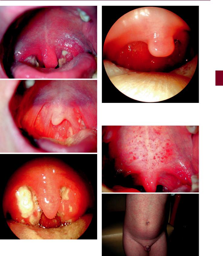
Fig. 3.1.1 (a) Acute tonsillitis due to group A beta-hemolytic streptococcal (GABHS) infection. (b) Acute tonsillitis and pharyngitis. Exudative lesions are seen both on the tonsils and pharyngeal mucosa.
(c) Acute tonsillitis in a patient with infectious mononucleosis
Fig. 3.1.3 (a) Petechial hemorrhages are seen on the soft palate in a patient with infectious mononucleosis. (b) Ampicillin rash. Ampicillin treatment in patients with infectious mononucleosis causes rash. The ampicillin rash resembles measles
K C DENN ATA O R H E S O N R A E

124 Chapter 3 Throat & Neck
a
Fig. 3.1.4 Generally a cherry red tongue and perioral pallor suggest GABHS infection
a
b
b
Fig. 3.1.6 (a, b) Chronic tonsillitis. There is no straightforward diagnosis for chronic tonsillitis. Deep tonsillar crypts, white debris in these crypts, and the vascularization of the anterior pillars are seen. This white debris consisting of stagnated food remnants in the crypts may cause halitosis
Fig. 3.1.5 Hypertrophic tonsils. (a) The right tonsil is more hypertrophic than the left one. (b) Kissing tonsils. The tonsils cause obstruction and sleep apnea
Fig. 3.1.7 Tonsilloliths. Stagnated food remnants may stay in the crypts for a long period, become hardened, and look like small stones called tonsilloliths

Fig. 3.1.8 Tonsils removed with their capsules after tonsillectomy
Table 3.1.2 Indications of tonsillectomy
Recurrent acute tonsillitis (more than five episodes in 1 year, five attacks per year for two consecutive years, or three attacks per year for three consecutive years)
Recurrent acute tonsillitis with recurrent febrile seizures or cardiac complications
Chronic tonsillitis
Peritonsillar abscess
Obstructive tonsillar hypertrophy causing disturbances with respiration and nutrition
Obstructive sleep apnea syndrome
Asymmetric growth or tonsillar lesion suggestive of neoplasm
3.1 Acute Tonsillopharyngitis |
125 |
|
Table 3.1.3 Contraindications to tonsillectomy |
A E |
|
Bleeding disorders |
||
Recent acute infection |
R |
|
Children under the age of 3 years |
|
|
|
||
Young children weighing less than 15 kg have a greater risk asso- |
N |
|
ciated with blood loss |
||
|
E S O |
|
|
||
|
|
|
|
|
|
|
ATA O R H |
|
|
|
|
|
DENN |
|
|
K C |
|

126 Chapter 3 Throat & Neck
3.2
Adenoids
Adenoids are an accumulation of lymphoid tissue located at the posterosuperior wall of the nasopharynx above the level of soft palate. Recurrent upper respiratory tract infections cause adenoids to enlarge. Adenoids reach their greatest size at the age of 6 years and then gradually regress. If adenoids obstruct the eustachian tube they may cause otitis media with effusion. Adenoids also serve as a reservoir for microorganisms and may be the cause of recurrent infections. Adenoids may cause nasal obstruction and sleep apnea. Adenoid hypertrophy alone or together with tonsillar hypertrophy with chronic mouth breathing may cause craniofacial growth abnormalities. The anterior facial height is increased, the midface is flat, and the palateishighlyarched.Thistypicalfacialappearanceiscalled adenoid face. If adenoid face develops it is difficult to correct this skeletal deformity. Adenoidectomy will not reverse dental changes that have already occurred. Orthodontic assistance is
important but it is not always helpful. Maxillofacial surgery may be needed for correction in some cases.
In adults, nasopharyngeal carcinoma should be suspected if nasal obstruction is associated with a unilateral serous otitis media.
Fiberopticorrigidendoscopesprovidesufficientvisualization of the nasopharynx. Lateral X-rays may show the size of the adenoids and the degree of obstruction. Correct positioning of the child is very important. A wrongly angled X-ray mayerroneouslypointtolargeadenoids.PlainX-raysshould beavoidedinchildrenduetotheharmfuleffectsofradiation. Endoscopy may help rule out other diseases such as Thornwald cysts or malignancy causing nasal obstruction.
Adenoidectomy should be undertaken in patients with symptoms.Submucouscleftpalateshouldalwaysbechecked forbeforetheoperationandadenoidectomyshouldbeavoided in the presence of a submucous cleft. A bifid uvula may be a sign of a possible submucous palatal cleft. The adenoids are curetted. Since surgery is blind and the adenoid tissue does not have a capsule as in the tonsils, complete removal of the adenoids is almost impossible.
Fig. 3.2.3 Adenoid face is a typical facial appearance with increased anterior facial height and flat midface
Fig. 3.2.1 Adenoids are an accumulation of lymphoid tissue located at the posterosuperior wall of the nasopharynx above the level of the soft palate
Fig. 3.2.2 Adenoids reach their greatest size at the age of 6 years and then gradually regress
Fig. 3.2.4 The palate is high arched in children with adenoidal hypertrophy

3.2 Adenoids 127
a |
b |
Fig. 3.2.5 Lateral X-ray of the postnasal space. (a) Adenoid tissue is not obstructing the airway; (b) adenoid tissue is almost completely obstructing the airway
Fig. 3.2.6 Orthodontic apparatus to correct hard palate deformity
due to adenoid hypertrophy
Fig. 3.2.8 Endoscopic photograph of adenoidal hypertrophy
K C DENN ATA O R H E S O N R A E
Fig. 3.2.7 Inferior turbinate hypertrophies may accompany adenoid |
|
hypertrophy. Sometimes they are the only cause of nasal obstruction |
Fig. 3.2.9 Endoscopic photograph of Thornwald cyst |

128 Chapter 3 Throat & Neck
Fig. 3.2.10 Adenoidectomy technique
Table 3.2.1 Indications for adenoidectomy
Adenoid hypertrophy with chronic mouth breathing
Adenoid hypertrophy with sleep apnea
Chronic adenoiditis with middle ear effusions
Suspicion of a nasopharyngeal malignancy (for biopsy purposes)
Fig. 3.2.11 Adenoid tissue after removal
Table 3.2.2 Contraindications for adenoidectomy
Bleeding disorders
Recent upper respiratory tract infection
Submucosal cleft palate