

created by aurellro

Color Atlas of Otoscopy
From Diagnosis to Surgery
Mario Sanna, MD
Professor of Otolaryngology
Department of Head and Neck Surgery
University of Chieti
Chieti, Italy
Istituto Scientifico Ospedale San Raffaele
Rome, Italy
Gruppo Otologico
Piacenza, Italy
Alessandra Russo, MD |
Giuseppe De Donato, MD |
Gruppo Otologico |
Gruppo Otologico |
Piacenza, Italy |
Piacenza, Italy |
with the collaboration of
Essam Saleh, Abdelkader Taibah, Maurizio Falcioni, Fernando Mancini
464 illustrations, most in color
Thieme
Stuttgart • New York 1999
IV
Library of Congress Cataloging-in-Publication Data
Sanna, M.
Color atlas of otoscopy: from diagnosis to surgery / Mario Sanna, Alessandra Russo, Giuseppe De Donato; with the collaboration of Essam Saleh...[et al.].
p. cm.
Includes bibliographical references and index. ISBN 3-13-111491-6 (hardcover)
1. Otoscopy-Atlases. 2. Ear-Diseases-Atlases. 3. Ear-Surgery- Atlases. I. Russo, Alessandra. II. Donato, Giuseppe De. III. Title.
[DNLM: 1. Ear Diseases-diagnosis atlases. 2. Otoscopes. 3. Ear Diseases-surgery atlases. WV 17S228c 1998]
RF 123. S26 1998 617.8'07545-dc21 DNLM/DLC
for Library of Congress
Mario Sanna, MD
Professor of Otolaryngology, Head and Neck Surgery
University of Chieti, Chieti, Italy
Gruppo Otologico
Piacenza, Italy
Alessandra Russo, MD
Abdelkader Taibah, MD
Giuseppe De Donato, MD
Maurizio Falcioni, MD
Fernando Mancini, MD
Gruppo Otologico
Piacenza, Italy
All rights reserved. This book, including all parts thereof, is legally protected by copyright. Any use, exploitation or commercialization outside the narrow limits set by copyright legislation, without the publisher's consent, is illegal and liable to prosecution. This applies in particular to photostat or mechanical reproduction, copying, or duplication of any kind, translating, preparation of microfilms, and electronic data processing and storage.
Cover design by Renate Stockinger, Stuttgart
© 1999 Georg Thieme Verlag, RiidigerstraBe 14, D-70469 Stuttgart, Germany
Thieme New York, 333 Seventh Avenue, New York, NY 10001 USA.
Typesetting and Photolitho: BEFORE S.r.l., Grottammare (AP),
Italy
Printed in Germany by Staudigl Druck, Donauworth
ISBN 3-13-111491-6 GTV |
|
ISBN 0-86577-721-7 TNY |
1 2 3 4 5 6 |
Essam Saleh, MD
Department of Otolaryngology, Head and Neck Surgery
University of Alexandria, Egypt
Important Note: Medicine is an ever-changing science. Research and clinical experience are continually expanding our knowledge, in particular our knowledge of proper treatment and drug therapy. Insofar as this book mentions any dosage or application, readers may rest assured that the authors, editors, and publishers have made every effort to ensure that such references are in accordance with the state of knowledge at the time of production of the book.
Nevertheless, this does not involve, imply, or express any guarantee or responsibility on the part of the publishers with respect to any dosage instructions and forms of application stated in the book. Every user is requested to examine carefully the manufacturer's leaflets accompanying each drug and to check, if necessary in consultation with a physician or specialist, whether the dosage schedules mentioned therein or the contraindications stated by the manufacturers differ from the statements made in the present book. Such examination is particularly important with drugs that are either rarely used or have been newly released on the market. Every dosage schedule or every form of application used is entirely at the user's risk and responsibility. The authors and publishers request every user to report to the publishers any discrepancies or inaccuracies noticed.
Any reference to or mention of manufacturers or specific brand names should not be interpreted as an endorsement or advertisement for any company or product.
Some of the product names, patents, and registered designs referred to in this book are in fact registered trademarks or proprietary names, even though specific reference to this fact is not always made in the text. Therefore, the appearance of a name without designation as proprietary is not to be construed as a representation by the publisher that it is in the public domain.
Foreword
The good fortune of otology resides in the fact that in most cases a diagnosis can be established through careful otoscopic examination: the tympanic membrane is the window to the middle ear.
Otoscopy constitutes the first phase in the examination of the patient. The initiation of the young otologist begins with this basic step. Colleagues of my generation will recall the long months of training which were necessary to understand and identify something in the depths of a narrow, tortuous, and sensitive external canal, often obstructed by physiologic or pathologic secretions. It was difficult to find good textbook illustrations. There were only drawings and lengthy pages of description not worthy of comparison with the unparalleled iconography of Politzer or Toynbee in the last century... Photographs were either absent; or when included, were of such mediocer quality, that they were of limited interest. We experienced a feeling of frustration in that era of the electron microscope and of space probes bringing back photos of the earth taken from the moon...
Modern optical systems, in particular the binocular microscope, have permitted an unfettered approach and the detailed observation of the tympanic membrane under optimal conditions of lighting and magnification. The addition of observer tubes and video cameras have helped to further familiarize ourselves with the various pathologic conditions. However, the tympanic membrane has long defended itself from photographic intrusion. Inclined in relation to the three spatial planes, and of a diameter of 1 cm (while the normal canal accepts only a 4 mm speculum), it is only through progressive scanning that we view the totality of the surface. Our brain reconstructs the virtual image. Thus, otoscopic photography faces a formidable challenge: to reproduce not what one sees but what one imagines. The solution came with the introduction of the Hopkins optical system, which provides wide angle capability through a narrow diameter endoscope, affording an enlarged field of vision and greater depth of field with increased light transmission. The principle is simple; however, utilization of the equipment necessitates a certain degree of experience to obtain quality pictures with regularity. Through my father, to whom I am indebted, I acquired a passion for photography, permitting me to acquire
the necessary experience and subsequently to share it. This is the reason for which I feel honored, as friend and colleague, to preface this remarkable volume.
Having perfectly mastered the technical problems, we note with real pleasure that Dr. Sanna and his collaborators offer us more than an "Atlas of Otoscopy", as the title of the volume modestly suggests. It is truly a "Manual of Otology" in that it covers all aspects of inflammatory, infectious, and tumor pathology of the ear, as seen through modifications of the otoscopic image.
The reader, initially attracted by a book of pictures, will be further captivated by a concise text, where, with style and precision, the principal pathologic conditions are described: definition, nature, pathogenesis, and classification accompanied by diagrams. The text indicates as well the complementary examinations indispensable for diagnosis and available therapeutic options. Thus, radiographic images (CT scan, MRI) are juxtaposed with the otoscopic view when deemed appropriate. All pertinent information conforms to the most recently available sources and reflects the consensus of the scientific community.
A particularly interesting and original aspect is represented by the last chapters which deal with the pathology of the skull base: cholesteatoma of the petrosa, glomus tumors, meningoencephalic herniations, areas in which Dr. Sanna has special experience which he shares with us.
The resident or practitioner desirous of an initiation into otology will find a presentation of auricular pathology which is both general and detailed. Such a structure is thoroughly complementary to the knowledge acquired during his or her medical training. The well-informed otorhinolaryngologist will find an update of the most recent clinical, radiologic, and therapeutic acquisitions in a field which is in constant evolution.
We thank and warmly congratulate the author and his collaborators for this exceptional work which reflects the level of their talent and experience. It clearly represents a significant advance in the field of Otology.
Dr. C. Deguine
Lille, France
VI
Preface
Despite advances in diagnostic techniques and imaging modalities, otoscopy remains the cornerstone in the diagnosis of otologic diseases. Every otolaryngologist, pediatrician, or even general practitioner dealing with ear diseases should have a good knowledge of otoscopy.
This atlas is based on 15 years of experience in the Gruppo Otologico in the treatment of otologic and neurotologic disorders. It presents a vast collection of otoscopic views of a variety of lesions that can affect the ear and temporal bone. Many examples are given for each disease so that the reader becomes acquainted with the variable presentations each pathology can have.
While otoscopy alone can establish the diagnosis in some cases, parameters such as history, or audiological and neuroradiological evaluation are required in others. An important aspect of this atlas is that it juxtaposes, when appropriate, the clinical picture, radiological diagnosis, and intraoperative findings with the otoscopic findings of the patient. Needless to say, every patient should be considered as a whole and in some particular cases, the otoscopic findings might only be the "tip of the iceberg." Otalgia, otorrhea, and granulations in the external auditory canal are manifestations of otitis externa, but when they persist, particularly in the elderly, they should arouse suspicion of malignancy. Otitis media with effusion can be a simple disease when seen in children, whereas unilateral persistent otitis media with effusion in an adult may be the only sign of a nasopharyngeal carcinoma. A small attic perforation in the presence of facial nerve paralysis and sensorineural hearing loss may be all that is
seen in a giant petrous bone cholesteatoma. The manifestation of an aural polyp can vary from a mucosal polyp associated with chronic suppurative otitis media to the much less common but more dangerous glomus jugulare tumor. A small retrotympanic mass may represent an anomalous anatomy such as a high jugular bulb or an aberrant carotid artery. It may also represent frank pathology such as facial nerve neuroma, congenital cholesteatoma, or even en-plaque meningioma.
In each chapter, a surgical summary that lists the different approaches for the management of the pathology dealt with is provided. Throughout the book, emphasis is on how the otoscopic view and the clinical picture may affect the choice of treatment and the surgical technique.
At the end of this atlas, a chapter on postsurgical conditions is presented. The presence of previous surgery poses special difficulties because of the distorted anatomy. Moreover, the otologist should be able to distinguish between what is considered to be normal postsurgical healing and complications that need further intervention.
The authors would like to thank Dr. Clifford Bergman, medical editor at Georg Thieme Verlag, for his excellent cooperation and help. Thanks also go to Paolo Piazza, neuroradiologist, for his continuous cooperation and to Maurizio Guida for the illustrations included in the book.
Mario Sanna, MD
Alessandra Russo, MD
Giuseppe De Donato, MD

Contents
1 Methods of Otoscopy. |
|
|
|
2 The Normal Tympanic Membrane |
|
|
4 |
Anatomy |
4 |
Histology |
5 |
3 Diseases Affecting the External Auditory Canal |
|
||
Exostosis and Osteoma |
7 |
Pathologies Extending to the External |
|
Furunculosis |
10 |
Auditory Canal |
17 |
Myringitis and Meatal Stenosis |
10 |
Carcinoid Tumors |
17 |
Otomycosis |
14 |
Histiocytosis X |
19 |
Eczema |
15 |
Other Pathologies |
20 |
Cholesteatoma of the External Auditory Canal |
15 |
Carcinoma of the External Auditory Canal. . . |
|
4 Secretory Otitis Media (Otitis Media with Effusion) |
26 |
||
5 Cholesterol Granuloma |
|
34 |
|
6 Atelectasis, Adhesive Otitis Media |
|
38 |
|
7 Non-Cholesteatomatous Chronic Otitis Media |
|
46 |
|
General Characteristics of Tympanic |
|
Perforations Complicated by or Associated |
|
Membrane Perforations |
46 |
with Other Pathologies |
54 |
Posterior Perforations |
47 |
Tympanosclerosis |
56 |
Anterior Perforations |
49 |
Tympanosclerosis Associated with Perforation. |
57 |
Subtotal and Total Perforations |
51 |
Tympanosclerosis with Intact Tympanic |
|
Posttraumatic Perforations |
53 |
Membrane |
|
8 Chronic Suppurative Otitis Media with Cholesteatoma |
59 |
||
Epitympanic Retraction Pocket |
60 |
Cholesteatoma Associated with Atelectasis... |
68 |
Epitympanic Cholesteatoma |
61 |
Cholesteatoma Associated with Complications |
70 |
Mesotympanic Cholesteatoma |
66 |
|
|
9 Congenital Cholesteatoma of the Middle Ear |
73 |
||
10 Petrous Bone Cholesteatoma |
|
75 |
|
11 Glomus Tumors (Chemodectomas) |
|
83 |
|
Differential Diagnosis with Other Retrotympanic |
|
|
|
Masses |
98 |
|
|

VIII
12 |
Meningoencephalic Herniation |
|
109 |
13 |
Postsurgical Conditions |
|
115 |
Myringotomy and Insertion of a Ventilation |
|
Myringoplasty |
|
Tube |
115 |
Tympanoplasty |
|
References |
|
142 |
|
Index |
|
145 |
|
1
1 Methods of Otoscopy
A preliminary examination is carried out using a head mirror or an otoscope.
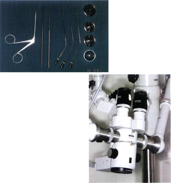
For proper otoscopy, the external auditory canal should be cleaned. Few instruments are used for this step, namely, aural speculi of different sizes, a Billeau
ear loop, Hartman auricular forceps, and suction tips (Fig. 1.1). In cases with a history of recurrent otitis, we prefer to clean the ear with the aid of a microscope (Fig. 1.2).
Fig. 1.1
Fig. 1.2
2 1 Methods of Otoscopy
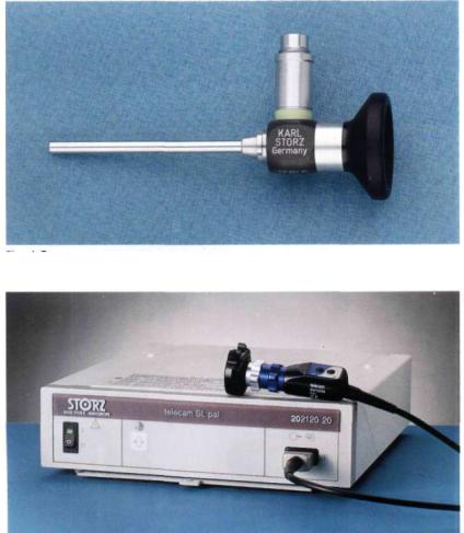
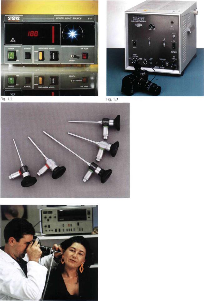
The use of a rigid 0° 6-cm endoscope (1215AAStorz, Fig. 1.3) connected to a video system enables the patient to see the pathology involving his/her ear (Figs. 1.4 and 1.5 show the Endovision Telecam SL 20212001 and the Xenon Light Source 615-Storz). With the help of a video printer connected to the monitor, instant photos of the pathology can be obtained. The rigid 30° endoscope allows evaluation of attic retraction pockets, the extent of which cannot always be determined using the microscope or the 0° endoscope (Fig. 1.6 shows a series of rigid endoscopes -Storz).
During the last few years, instant photography has also been used in the operating room. A copy of the important steps of the operation is given to the patient while another copy is kept in the patient's chart. The patient is also photographed during the follow-up visit. Thus, for each patient pre-, intra-, and postoperative photographic documentation is obtained.
All the photos in this book were obtained with an Olympus OM 40 camera mounted to the endoscope with a Storz 593-T2 objective. The focus is adjusted to infinity and the diaphragm to 140. We use the TTL- Computer-Flash-Unit Model 600 BA Storz (Fig. 1.7). The film used is a Kodak Ektachrome 64T Professional Film (Tungsten).
Methods of Otoscopy
Fig. 1.6
In all the cases, the examiner sits to the side of the patient whose head is slightly tilted towards the contralateral side. The examiner holds the camera attached to the endoscope with his right hand. With the ring and middle finger of the left hand, the examiner pulls the patient's auricle backwards and outwards to straighten the external auditory canal. The endoscope is advanced over the index finger of the examiner's left hand into the patient's external auditory canal. In this manner, any undue injury to the external auditory canal is prevented (Fig. 1.8).
Fig. 1.8