

38
6 Atelectasis, Adhesive Otitis Media
Adhesive otitis media is characterized by complete or partial adhesions between the thin retracted and atrophic pars tensa and the medial wall of the middle ear. Necrosis of the long process of the incus or the stapes' suprastructure can also occur with a resultant natural myringostapedopexy. It should be differentiated from atelectasis and from simple drum retraction in which the tympanic membrane is mobile with the Valsalva or Toynbee maneuvers.
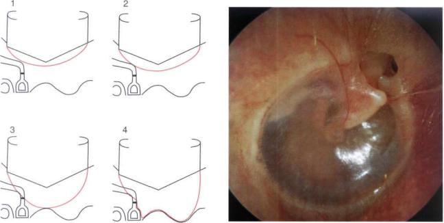
Sade (1979) distinguished five grades of atelectasis (Fig. 6.1): grade I is characterized by a mild retraction of the tympanic membrane; in grade II the retracted tympanic membrane comes in contact with the incus or the stapes; in grade III the tympanic membrane touches the promontory; grade IV is adhesive otitis media; and in grade V there is a spontaneous perforation of the atelectatic ear drum with otorrhea and polyp formation.
Nakano (1993) proposed two types of adhesive otitis: type A in which the retracted and atrophic tympanic membrane adheres completely to the promontory, and type B in which retraction and adhesion affect
mainly the posterior part of the tympanic membrane, usually without retraction of its anterior half.
Histologically, the tympanic membrane is atrophic due to thinning or even absence of the lamina propria. It can be hypothesized that the negative middle ear pressure caused by eustachian tube dysfunction or persistent secretory otitis media leads to atrophy of the elastic fibers of the pars tensa. An occasional episode of acute suppurative otitis media might form adhesions between the mucosa of the promontory and the retracted tympanic membrane.
Figure 6.1 Classification (modified) of atelectasis according to Sade (1979) (see text).
Figure 6.2 Right ear. Grade I atelectasis according to Sade (1979). The tympanic membrane is retracted but does not come into contact with the middle ear structures. A mild retraction of the pars flaccida, through which the head of the malleus is visible, is also noted. The base of the retraction pocket is under control with no sign of cholesteatoma. It is also possible in this case to assume that the drum is mobile on Valsalva or Toynbee maneuvers. This patient presented with very mild conductive hearing loss and a normal tympanogram (type A) (see Figs. 6.3 and 6.4).

Atelectasis, Adhesive Otitis Media |
39 |
120 |
|
|
|
|
|
|
|
|
|
|
|
125 |
250 500 |
1K |
2K |
4K |
8K |
16KHz |
-200 |
-100 |
0 |
+100 |
+200 |
Figure 6.3 |
Audiogram |
of the same case. |
Mild |
conductive |
Figure 6.4 |
Tympanogram of the same case. Normal or type A. |
|||||
hearing loss.
0
10
20
30
40
50
60
70
80
90
100
110
120
125 |
250 |
500 |
1K |
2K |
4K |
8K |
16KHz |
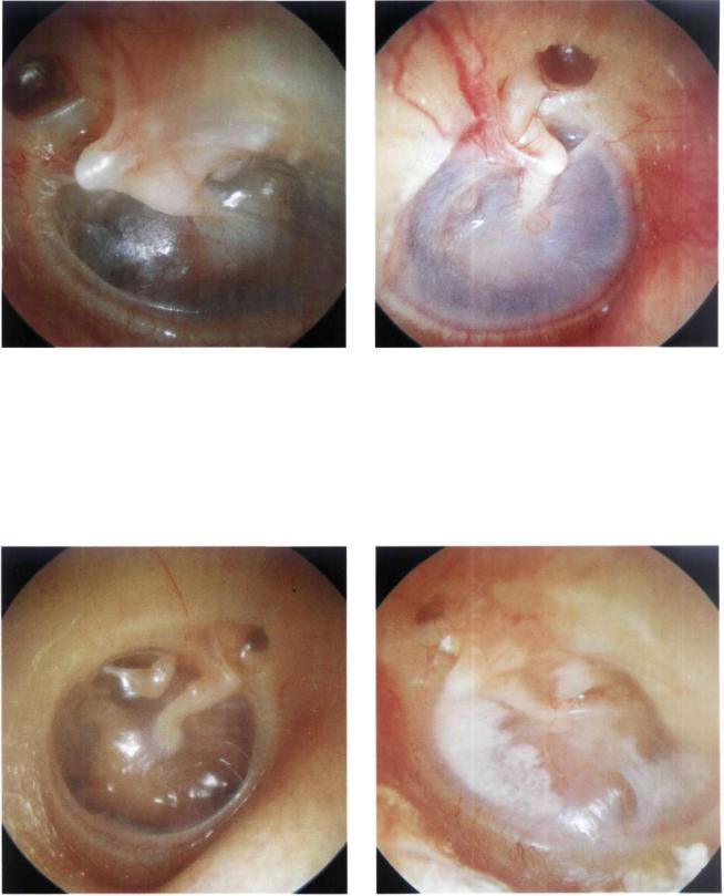
Figure 6.5 Right ear. Grade I atelectasis with the malleus slightly medialized. An epitympanic retraction pocket is also seen. Middle ear effusion with yellowish color can be appreciated. Pure tone audiogram revealed a 40-dB conductive hearing loss (Fig. 6.6), whereas the tympanogram was type B, i.e., typical of middle ear effusion (Fig. 6.7). In this case, the insertion of a ventilation tube is indicated to avoid further retraction of the tympanic membrane, to aerate the middle ear, and to improve hearing.
Figure 6.6 Audiogram of the same case showing a 40-dB conductive hearing loss.

40 6 Atelectasis, Adhesive Otitis Media
10
9
8
7
6
5
4
3
2
1
0
-200 |
-100 |
0 |
+100 |
+200 |
Figure 6.7 Tympanogram type B of the same case, typical of middle ear effusion.
Figure 6.8 Right ear. Grade I atelectasis. The tympanic membrane is markedly thinned due to partial resorption of the lamina propria. The incus is seen in transparency. Pure tone audiogram is normal (Fig. 6.9), whereas the tympanogram has a very high compliance (Fig. 6.10). As the tympanic membrane is mobile with the Valsalva maneuver, insertion of a ventilation tube is not indicated.
10 dBHL
10
20
30
40
50
60
70
80
90
100
110
120
125 |
250 |
500 |
1K |
2K |
4K |
8K |
16KHz |
Figure 6.9 Audiogram of the same case (see text).
10
9
8
7
6
5
4
3
2
1
0
-200 |
-100 |
0 |
+ 100 |
+200 |
Figure 6.10 Tympanogram of the same case, type AD according to the classification of Liden-Jerger, 1976 (see text).
Atelectasis, Adhesive Otitis Media |
41 |
Figure 6.11 Left ear. Grade II atelectasis with marked epitympanic retraction. The tympanic membrane touches the incus. The malleus is medialized. Air-fluid levels are seen in the anteroinferior quadrant. The insertion of a ventilation tube is necessary to restore normal conditions.
Figure 6.12 Right ear. Grade II atelectasis. A condition similar to the previous case but with the onset of thickening of the tympanic membrane.
Figure 6.13 Right ear. Grade II atelectasis. The tympanic membrane is very thin due to absence of the fibrous layer. The membrane adheres to the incudostapedial joint and the tensor tympani tendon. Insertion of a ventilation tube is indicated.
Figure 6.14 Left ear. Grade III atelectasis. The tympanic membrane touches the promontory and the incus. An air-fluid level and a tympanosclerotic plaque can be seen in the anterior quadrant.
42 6 Atelectasis, Adhesive Otitis Media
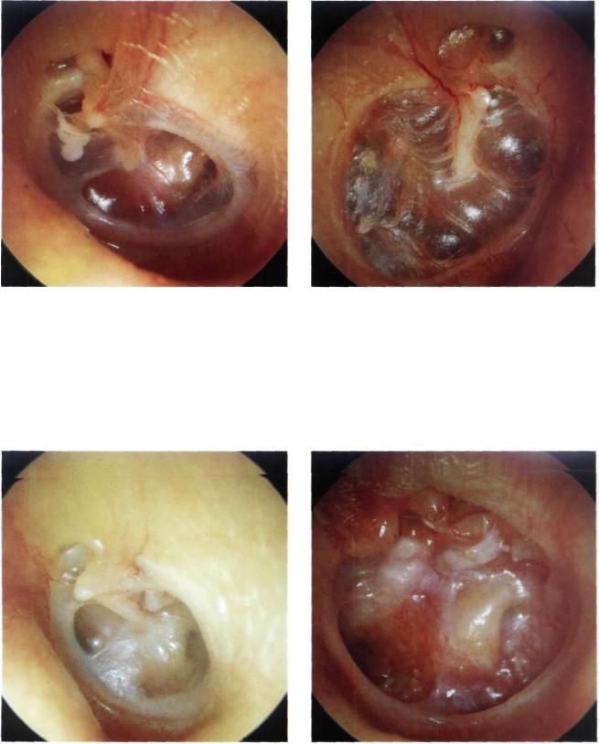
Figure 6.15 Left ear. Grade III atelectasis. The thin and atrophic tympanic membrane is in contact with the promontory. Middle ear effusion is seen. A tympanosclerotic plaque is present in the anterosuperior quadrant. The head of the malleus is visible through an epitympanic retraction pocket. The insertion of a ventilation tube is indicated.
Figure 6.16 Right ear. Adhesive otitis media or grade IV atelectasis associated with a mild epitympanic retraction pocket. The thin and atrophic tympanic membrane completely covers the promontory. The tympanic membrane retraction has caused erosion of the long process of the incus with a subsequent spontaneous myringostapedopexy As the patient does not complain of hearing loss, surgery is not indicated.
Figure 6.17 Left ear. Grade IV atelectasis. The malleus is medialized and adherent to the promontory. The tympanic membrane is atrophic. The epidermal layer of the membrane is adherent to the incudostapedial joint, the promontory, and the round window. A retraction pocket corresponding to the eustachian tube orifice is also seen. Middle ear effusion is present. Insertion of a ventilation tube is indicated.
Figure 6.18 Left ear. Adhesive otitis media. This case represents the long-term sequela of persistent secretory otitis
media with chronic eustachian tube dysfunction. The fibrous and mucosal layers of the tympanic membrane were resorbed, whereas the epidermal layer is completely adherent to the medial wall of the middle ear. The promontory, round and oval windows, as well as residues of the ossicular chain are all visible. The handle of the malleus is completely medialized and partially eroded. The long process of the incus is eroded, whereas the stapes suprastructure is completely absent. As the patient does not suffer from otorrhea, surgery is not advised.

Atelectasis, Adhesive Otitis Media |
43 |
Figure 6.19 Right ear. The thin and atrophic tympanic membrane adheres to the promontory, incudostapedial joint, pyramidal process, and stapedius tendon. The long process of the incus is partially eroded. Calcifications are present in the anterior quadrants. As hearing is normal, surgery is not indicated.
Figure 6.20 Right ear. Atelectasis associated with marked epitympanic erosion through which the head of the malleus and body of the incus are seen covered with epithelial squa-
Figure 6.21 Left ear. Posterior retraction pocket. The tympanic membrane remains adherent to the stapes' head even after Valsalva maneuver (myringostapedopexy). The remaining part of the tympanic membrane is thick and shows tympanosclerosis. Audiometry revealed normal hearing. Cases with myringostapedopexy generally have good hearing; therefore, surgery is not indicated except if conductive hearing loss develops and/or a posterior retraction pocket is associated with frequent otorrhea. Surgery varies from simple myringoplasty (when the tympanic membrane needs reinforcement) to tympanoplasty (in which the ossicular chain is eroded and needs ossiculoplasty).
mae. The tympanic membrane is thin and transparent due to absence of the fibrous layer. The handle of the malleus is amputated. The long process of the incus is eroded and a natural myringostapedopexy is noted. The promontory, round window, head of the stapes, and oval window can be seen through the thin tympanic membrane. Despite the attic epithelialization, a true cholesteatoma has not yet formed. Regular follow-up of such cases is fundamental. Should the disease progress with cholesteatoma formation, surgery in the form of an open tympanoplasty is indicated.
Figure 6.22 Right ear. The tympanic membrane, being adherent to the long process of the incus, caused erosion of the latter with subsequent conductive hearing loss (see Fig. 6.23). The second portion of the facial nerve is seen superior to the oval window. The head of the stapes and the stapedius tendon are also visible. Tympanoplastic surgery was performed on this patient. The tympanic membrane was reinforced and the incus interposed between the handle of the malleus and the stapes.
44 6 Atelectasis, Adhesive Otitis Media
0
10
20
30
40
50
60
70
80
90
100
110
120
125 |
250 |
500 |
1K |
2K |
4K |
8K |
16KHz |
Figure 6.23 Audiogram of the same case showing conductive hearing loss.
Figure 6.24 Left ear. Mesoand epitympanic retraction pockets that adhere to the head of the malleus, the partially eroded long process of the incus, and the incudostapedial joint. A ventilation tube has been inserted in the anterior quadrant to avoid further retraction that might lead to cholesteatoma.
Figure 6.25 Right ear. Grade IV atelectasis. All of the middle ear structures can be seen in transparency. Starting from the malleus and moving in a clockwise direction, we can distinguish the tubal opening, the hypotympanum, the promontory, the round window, the stapedius tendon, and the incudostapedial joint.
Figure 6.26 Right ear. Large mesotympanic retraction pocket that caused erosion of the incus and stapes suprastructure. The second portion of the facial nerve passing superior to the oval window, the promontory, and the round window can all be seen in transparency. In cases with good social hearing and no otorrhea, surgery is not indicated.
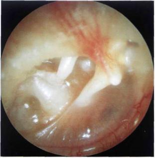
Figure 6.27 Right ear. Posterior retraction pocket. The tympanic membrane adheres to the promontory, the round window, the partially eroded long process of the incus, the head of the stapes, and the stapedius tendon. The processus cochleariformis is clearly visible between the malleus and the long process of the incus. Middle ear effusion can be observed anterior to the malleus and in the region of the oval window. In this case, ventilation tube insertion is indicated in an attempt to prevent further erosion of the ossicular chain and the formation of mesotympanic cholesteatoma.
Atelectasis, Adhesive Otitis Media |
45 |
Summary
In grade I, II, and III atelectasis, a long-term ventilation tube is usually inserted to prevent further retraction of the tympanic membrane. However, in cases with marked conductive hearing loss that denotes erosion of the incus or the superstructure of the stapes, ossiculoplasty is performed after extraction and sculpturing of the eroded incus or using a homologous incus. A large disk of tragal cartilage is used to reinforce the tympanic membrane.
Indications for surgery in adhesive otitis media include cases with tympanic membrane perforation (grade V according to Sade 1979), with or without polypi, granulation or otorrhea, those cases with a large infected retraction pocket causing frequent otorrhea, or those with conductive hearing loss due to ossicular chain erosion. In all these cases a tympanoplasty is performed using a postauricular incision. A disk of tragal cartilage is used with the perichondrium adherent to its lateral surface. If the handle of the malleus is present, it is incorporated into the cartilaginous disk after creating a triangular defect for its accommodation. This technique has the advantage of preventing retraction and adhesions between the tympanic membrane and the promontory. At the same time, it enables repair of the tympanic membrane perforation with the tragal perichondrium.
It can be concluded that there is no single treatment for the atelectatic ear. The milder the degree of atelectasis, the more conservative the treatment is. It should be noted, however, that in the long term conservative treatment (e.g., ventilation tube) was not found to modify further evolution of atelectasis. As atelectasis results from eustachian tube dysfunction, the ideal solution would be correction of this defect. At present, there is no acceptable "functional" surgery for the eustachian tube. Individual treatment should be administered according to the consequences of this dysfunction in each case. Such a strategy, however, requires a high mental elasticity and versatile surgical techniques.