

115
13 Postsurgical Conditions
As seen in the previous chapters, some otoscopic views may be difficult to interpret. This difficulty increases in cases involving previous surgery because of the distortion of the normal anatomy. The examiner should be competent and experienced enough to distinguish between cases with normal postoperative healing and those with recurring pathology and/or immediate and late postoperative complications.
In this chapter, postoperative otoscopic views with and without complications and/or recurrence are presented.
•Myringotomy and Insertion of a Ventilation Tube
The indications of myringotomy and ventilation tube insertion have been discussed previously. Myringotomy is usually performed in the anteroinferior quadrant of the tympanic membrane in the region of the cone of light. The incision is made in a radial direction using a myringotomy knife. In cases with a hump of the anterior wall of the external auditory canal, myringotomy can be performed immediately inferior to the umbo in the posteroinferior quadrant. The incision should never be made in the posterosuperior quadrant to avoid injury to the ossicular chain. The operation is performed under general anesthesia in children. In adults, however, local anesthesia is sufficient. After making a radial incision of the tympanic membrane, the middle ear effusion is aspirated and the ventilation tube is inserted. In the majority of cases, hearing improves immediately.
The patient is instructed to avoid water entering the ear by blocking it with cotton anointed with petrolatum when taking a shower or with rubber earplugs when swimming. Infection could occur if water were to enter the middle ear through the ventilation tube. Should this occur, ear lavage with a disinfectant solution consisting of 2% boric acid in 70% alcohol is indicated. When the tube is obstructed by cerumen or crusts, the administration of hydrogen peroxide drops is usually sufficient to restore its patency.
There are many types of commercially available ventilation tubes, but they can be generally grouped into shortand long-term tubes. Tubes with a larger inner flange usually remain in place longer. Once extruded, the myringotomy site closes spontaneously in about 98% of cases.
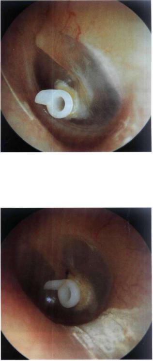
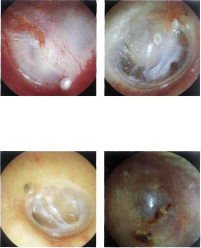
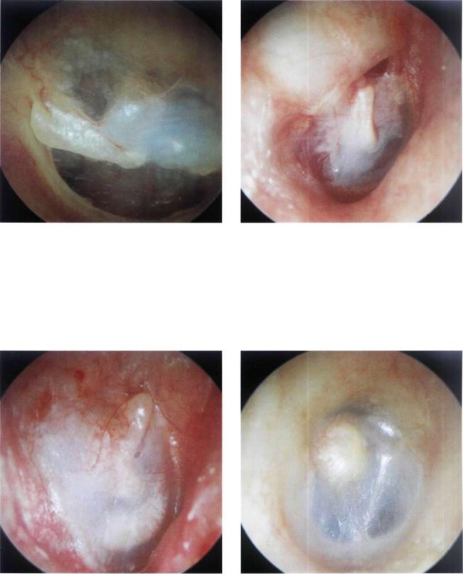
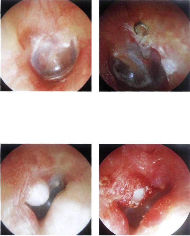
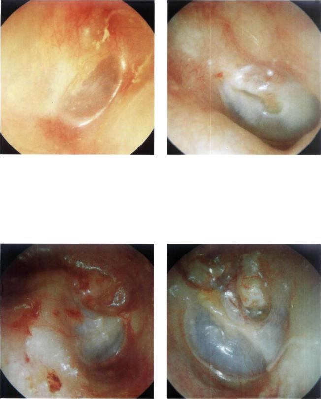
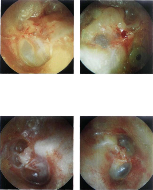
Figure 13.1 Left ear. The Sultan ventilation tube. This type has two small wings: an outer one with which the tube can be held using the ear forceps and an inner one, viewed through the tympanic membrane, which facilitates tube insertion and prevents rapid extrusion. If properly inserted, the Sultan ventilation tube can remain for about 6 to 18 months before extrusion.
Figure 13.2 Left ear. In this case, the tube has been placed inferior to the umbo due to the presence of an anterior hump in the anterior canal wall.
116 13 Postsurgical Conditions
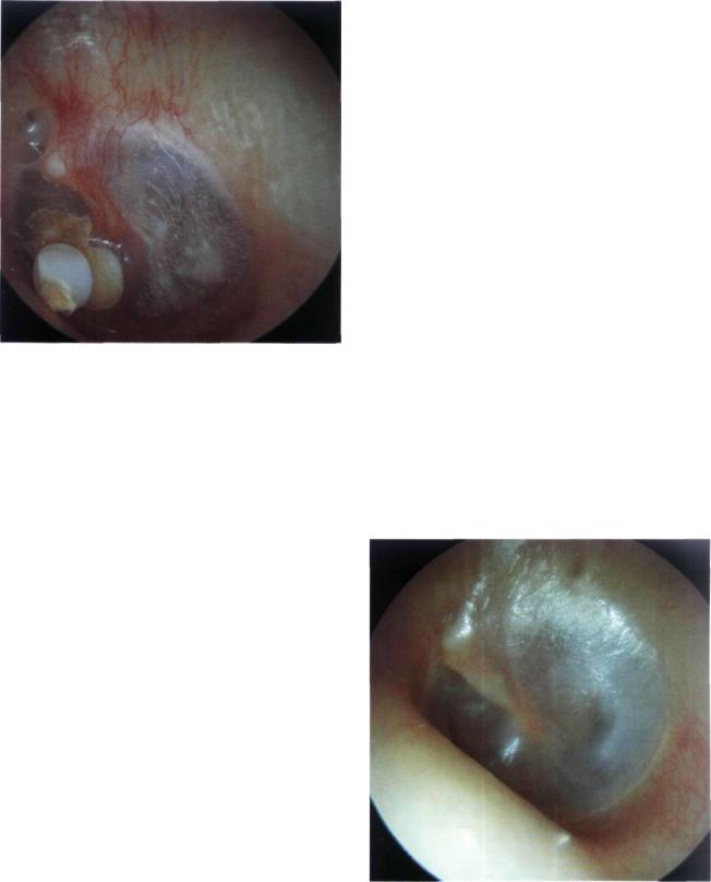
Figure 13.3 Right ear. The consequences of a misplaced ventilation tube is shown. A healed myringotomy is seen in the posterosuperior quadrant (at 9 o'clock). Two months later the tube was extruded. During tube insertion, however, dislocation of the incus occurred. The dislocated incus fell to the hypotympanum where its body and short process can be clearly seen. In the anteroinferior quadrant, immediately under the umbo, another healed myringotomy site (this time correctly placed) is visible. In the latter, tube extrusion occurred 1 year later.
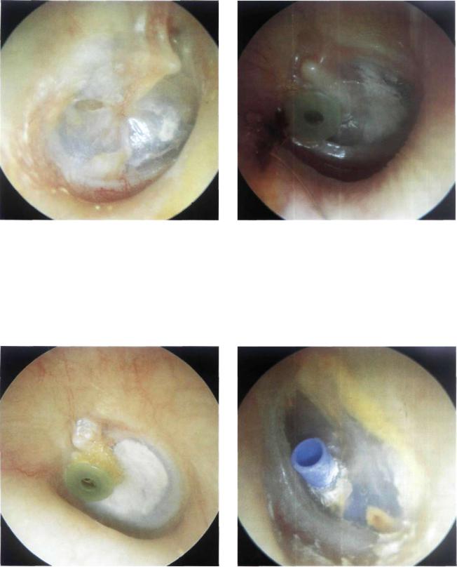
Figure 13.4 Left ear. A long-term ventilation tube inserted 6 months after tympanoplasty because of an observed tendency for graft retraction. The graft is seen in an optimal condition with no evidence of retraction, indicating patency of the ventilation tube. This tube has been in situ for more than 10 years.
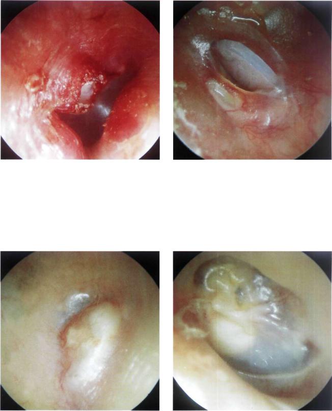
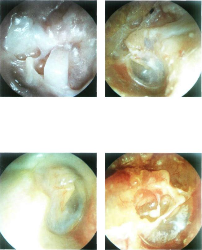
Figure 13.5 Left ear. Long-term ventilation tube. A large tympanosclerotic plaque that formed 1 year after the tube insertion can be clearly seen. Such plaques result from hemorrhagic infiltrate between the epidermal and fibrous layers of the tympanic membrane secondary to the myringotomy and are asymptomatic.
Figure 13.6 Left ear. Example of a long-term "T" tube inserted in the anteroinferior quadrant of the tympanic membrane. After its insertion the two wings of the tube open by virtue of their retained "memory," thereby preventing tube extrusion.
Myringoplasty 117
quadrant where the graft is detached from the anterior residues of the tympanic membrane and falls into the middle ear. When an overlay technique is utilized, blunting of the anterior angle can occur with resultant conductive hearing loss. Lateralization, in which the graft is detached from the handle of the malleus, is another possible complication that leads to conductive hearing loss. It occurs mostly when the graft is placed lateral rather than medial to the handle of the malleus. Stenosis of the external auditory canal, due either to inflammatory reaction or as a result of bad repositioning of the meatal flaps, can also occur.
Figure 13.7 Left ear. A ventilation tube in the process of extrusion. It is preferable not to take out the tube but rather wait for self-extrusion to occur. Closure of the myringotomy site occurs in about 98% of cases.
•Myringoplasty
The aim of reconstructing a tympanic membrane perforation is twofold: first, to allow the patient to have a normal social life with no restrictions, even regarding water entry into the ear, and second, to correct the hearing loss resulting from the perforation.
There are essentially two techniques for myringoplasty. The underlay technique is utilized in the presence of an anterior residue (at least the annulus) of the tympanic membrane, under which the graft can be placed. In the absence of any anterior residue of the membrane, the overlay technique is used. In such cases, the graft is positioned against the anterior wall of the external auditory canal.
Normally, the tympanic membrane forms an acute angle with the anterior wall of the external auditory canal. While performing myringoplasty, it is generally possible to respect this angulation when the annulus is present anteriorly.
The myringoplasty operation is considered a success when the reconstructed tympanic membrane is intact, well epithelialized, and has normal angulation with the external auditory canal. These characteristics allow the patient to have a normal social life (hearing improvement and possibility of water entry into the ear). Reperforation is a frequent complication of myringoplasty that occurs in about 5 to 10% of cases in the best series. Reperforation occurs more commonly in the underlay technique, particularly in the anterior
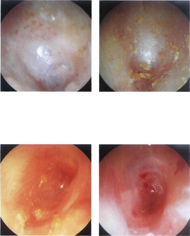
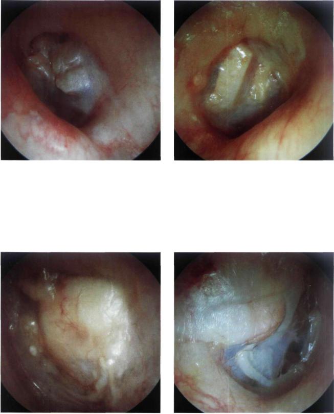
Figure 13.8 Left ear. Normal aspect of the reconstructed tympanic membrane. The posterior quadrant is slightly elevated. In this case, a posterior perforation was grafted with temporalis fascia using an underlay technique.
118 13 Postsurgical Conditions
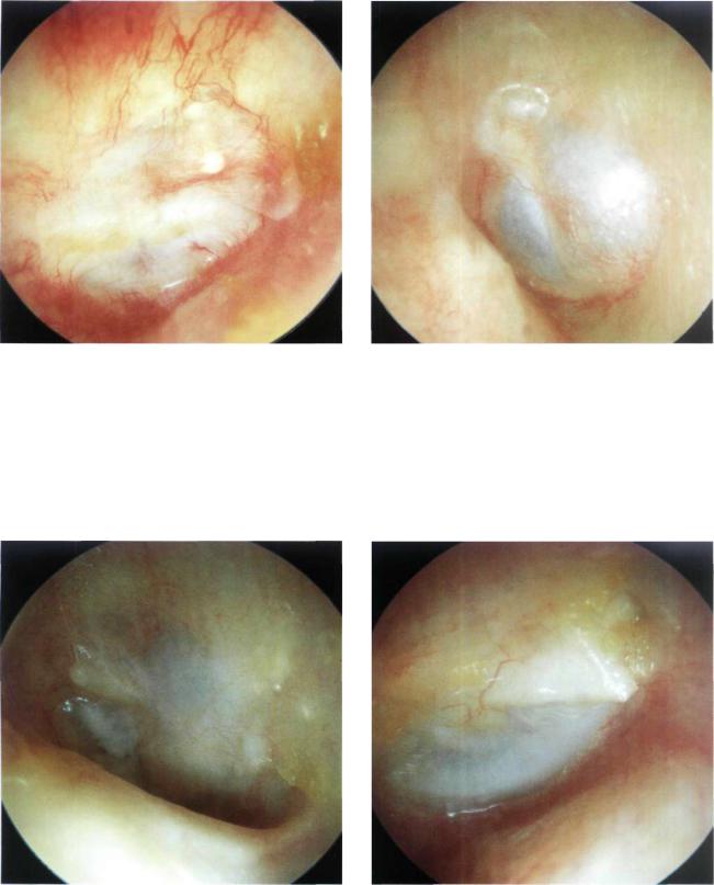
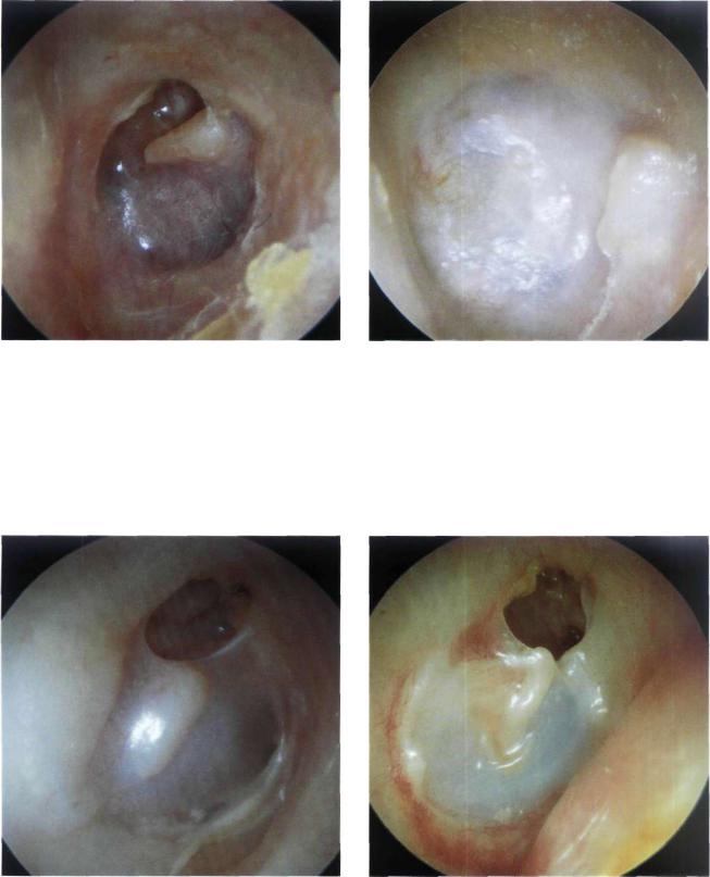
Figure 13.9 Right ear. Myringoplasty with an underlay technique. The reconstructed tympanic membrane is thicker than normal. The anterior angle is maintained. The handle of the malleus is clearly visible except for the umbo, which is detached from the membrane. Tympanosclerotic plaques are also visible.
Figure 13.10 Left ear. Another example of a tympanic membrane perforation that was repaired using an underlay technique with preservation of the anterior residue. The posterior quadrants are slightly lateralized, making it difficult to see the handle of the malleus.
Figure 13.11 Left ear. Similar case. The repaired tympanic membrane is well attached to the malleus except for the area of the umbo due to lateralization of the posteroinferior quadrant.
Figure 13.12 Right ear. Underlay myringoplasty. The malleus is slightly medialized. The repaired tympanic membrane is whitish in its anterior quadrants and vascularized in the posterior ones. The anterior angle is normal.
Myringoplasty 119
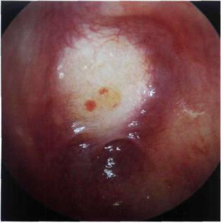
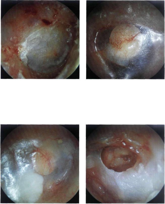
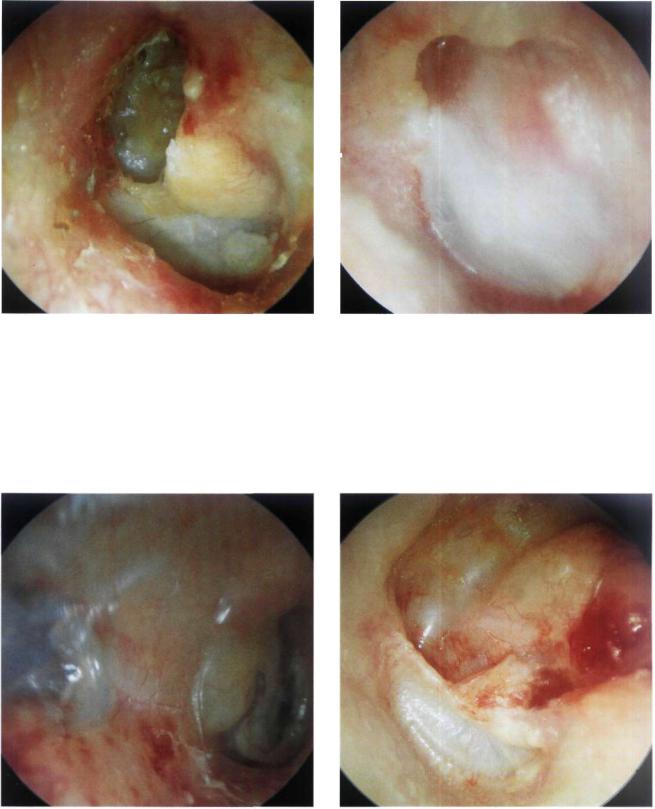
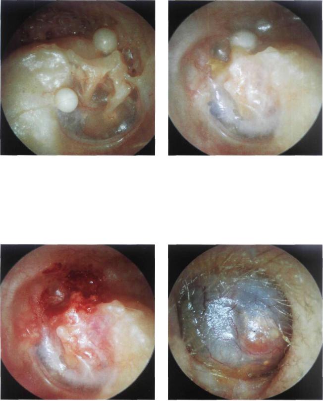
Figure 13.13 Left ear. The repaired tympanic membrane retains a normal anterior angle and is well vascularized, though thicker than normal. A small cholesteatomatous pearl is observed. This pearl can be easily removed in the outpatient clinic under the microscope.
Figure 13.14 Right ear. The repaired tympanic membrane has normal thickness. The short process of the malleus can be observed, although the handle is not visible due to lateralization.
Figure 13.15 Left ear. Another example of a graft that is detached from the handle of the malleus using an underlay technique.
Figure 13.16 A lateralized reconstructed tympanic membrane with blunting of the anterior angle following an overlay technique. Both complications lead to altered mobility of the tympanic membrane with consequent conductive hearing loss.
120 13 Postsurgical Conditions
Figure 13.17 The external auditory canal is wide but the repaired tympanic membrane is lateralized and shows blunting.
Figure 13.18 Similar case. The reconstructed tympanic membrane is lateralized with marked blunting of the anterior angle.
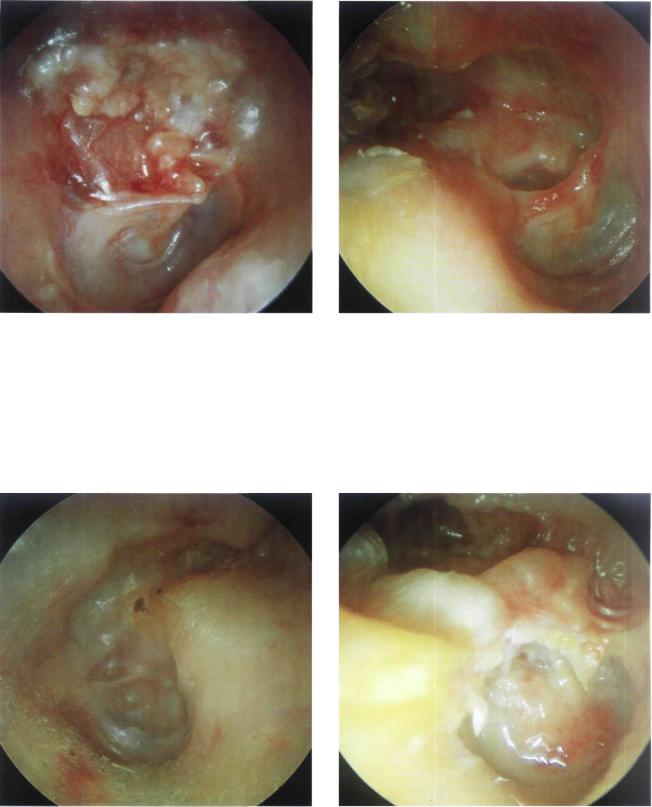
Figure 13.19 Postoperative myringitis. The tympanic membrane is hyperemic, thickened, and lateralized following a tympanoplasty. The epidermal layer is substituted by granulation tissue. Myringitis is a rare complication that usually resolves with local steroid applications. In very rare cases, reoperation is necessary. The pathological tympanic membrane is removed followed by grafting.
Figure 13.20 A patient who has undergone quadruple myringoplasty. In these cases, myringitis and canal stenosis are frequent; therefore, it is necessary to remove the pathological tissues, perform canalplasty, and use free skin flaps.
Myringoplasty 121
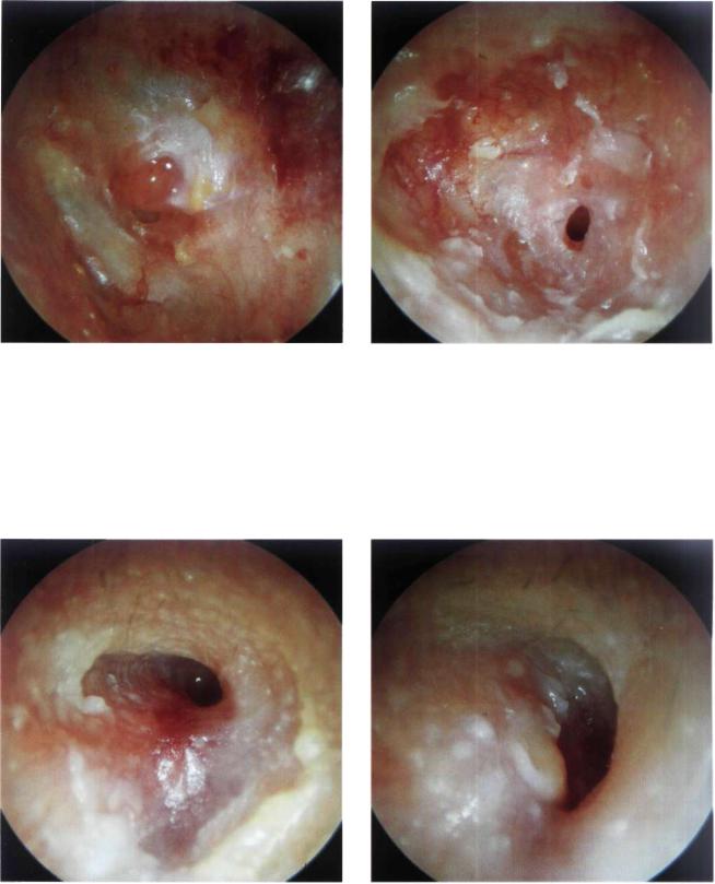
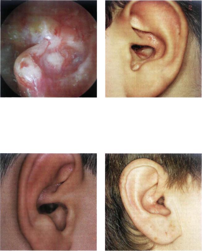
Figure 13.21 Left ear. Reperforation of the tympanic membrane with granulations near the perforation. In such cases, curettage of the granulation and freshening of the edges under the microscope may lead to spontaneous closure of the perforation.
Figure 13.22 Reperforation of the tympanic membrane. Myringitis with otorrhea can be appreciated. Lavage and freshening of the perforation edges as well as insertion of Gelfoam (in the middle ear) can favor spontaneous closure of the perforation.
Figure 13.23 Left ear. Stenosis of the external auditory |
Figure 13.24 Right ear. Partial stenosis of the external |
canal following myringoplasty. |
auditory canal following myringoplasty. For the management |
|
of this complication, it is usually sufficient to incise the skin of |
|
the canal and insert a plastic sheet for about 20 days, while |
|
using local medication of steroid lotion. |
122 13 Postsurgical Conditions
Figure 13.25 Retrotympanic cholesteatoma following myringoplasty. This iatrogenic cholesteatoma can be explained by the entrapment of epidermal residues in the middle ear or malpositioning of the meatal flap at the level of the anterior angle. It can be managed by incision of the cholesteatoma sac, aspiration of its contents, and insertion of a plastic sheet in the external auditory canal for about 20 days to favor healing.
•Tympanoplasty
Tympanoplasty operations can be classified into those without mastoidectomy, performed with chronic otitis media in which the tympanic membrane perforation is associated with necrosis of the ossicular chain, and those with mastoidectomy, performed in chronic suppurative otitis media with cholesteatoma. As mentioned previously, tympanoplasty with mastoidectomy can be either closed or open.
In closed tympanoplasty, the posterior wall of the external auditory canal is kept intact. This technique is employed in children and in patients with very pneumatized mastoids to avoid having a large cavity. Regular otoscopic follow-up is essential to identify the formation of a retraction pocket or a recurrent cholesteatoma. Should these occur, there should be no hesitation in switching to an open technique.
In open tympanoplasty, the posterior wall of the external auditory canal is removed. The indications of this technique in the treatment of cholesteatoma include: a wide erosion of the posterosuperior wall, cholesteatoma in the only hearing ear, bilateral cholesteatoma, cholesteatoma in patients with Down's syndrome, the presence of a contracted mastoid, a large labyrinthine fistula, and recurrent cholesteatoma following a closed tympanoplasty. Because the posterior canal wall is removed, the mastoid cavity is exteriorized and on otoscopy the external auditory canal and the mastoid appear as one communicating cavity. If properly performed, the cavity appears rounded in shape, dry, and well epithelialized. On the other hand, a badly performed cavity may appear wet, irregular, and be lined with granulation tissue in addition to accumulated debris. There may also be the possibility of a residual cholesteatoma.
In cases of tympanoplasty, it is usually possible to see the reconstructed ossicular chain through the tympanic membrane. We generally prefer to utilize an autologous or homologous incus for reconstruction. In our experience (more than 1000 tympanoplasties) we never encountered any case of extrusion when the incus was used. In contrast, variable rates of extrusion were noticed when biological materials (e.g., plastipore, ceramics, hydroxyapatite) were utilized. Although the use of homologous ossicles has never been proven to transmit slow viruses (e.g., Creutzfeldt-Jakob disease), the theoretical risk makes it more prudent to use predominantly autologous tissue or biomaterial of better characteristics that might appear in the future.
Later on in this chapter, some otoscopic views of cases managed by the modified Bondy technique are shown. This is an open technique indicated in epitympanic cholesteatoma with a good preoperative hearing in which the tympanic membrane and the ossicular chain are intact. Some cases of radical mastoidectomy are also shown. This technique is used mainly in elderly patients with sensorineural hearing loss in which the only goal of surgery is to have a dry and safe ear.
Tympanoplasty 123
Figure 13.26 Left ear. The sculptured incus is visible under the handle of the malleus. The reconstructed tympanic membrane appears very thin but intact. The anterior angle is perfect. A piece of cartilage placed over the incus is clearly visible.
Figure 13.27 Right ear. Staged closed tympanoplasty. The tympanic membrane has a normal angle and is well attached to the handle of the malleus. The cartilage used for reconstructing the attic is visible. In this region, a small self-cleaning retraction pocket can be seen.
Figure 13.28 Right ear. Staged closed tympanoplasty performed 10 years previously for the management of a cholesteatoma. The tympanic membrane is whitish, slightly thicker than normal, but retains a good anterior angle. The annulus is well seen anteriorly. The handle of the malleus is in a good position. There are no signs of resorption of the posterior canal wall.
Figure 13.29 Right ear with a previous tympanoplasty. The tympanic membrane is thin with mild blunting. The sculptured incus is visible.
124 13 Postsurgical Conditions
Figure 13.30 Right ear. Otoscopic view after a secondstage tympanoplasty in which the incus was used for ossiculoplasty. The tympanic membrane and the handle of the malleus are excellently positioned.
Figure 13.31 Left ear. Perfect reconstructed tympanic membrane with optimal thickness and no blunting. The sculptured incus is in contact with the handle of the malleus: It is slightly elevated with respect to the level of the tympanic membrane.
Figure 13.32 Left ear. Another example of the incus positioned under the handle of the malleus.
Figure 13.33 Left ear. Ossiculoplasty. The tympanic membrane is retracted and the malleus is medialized. The sculptured incus is displaced posteriorly and is adherent to the posterior mesotympanum. Two tympanosclerotic plaques are noted anteriorly and interiorly.
Tympanoplasty 125
Figure 13.34 Left ear. Posteriorly displaced incus that was used for ossiculoplasty. The trough created on the incus to fit the handle of the malleus is clearly seen. Revision surgery is necessary to reposition the displaced incus and improve the patient's hearing.
Figure 13.35 Right ear. Slightly retracted reconstructed tympanic membrane. A T-shaped collumela from homologous cartilage is visible. The collumela has been placed between the tympanic membrane and the footplate of the stapes.
Figure 13.36 Left ear. Ossiculoplasty. A piece of cartilage that was interposed between the reconstructed ossicular chain and the tympanic membrane can be visualized. It appears as a whitish thick mass that causes elevation of the posterior quadrants of the tympanic membrane.
Figure 13.37 Right ear. Closed tympanoplasty. Sculptured incus in a perfect position under the reconstructed tympanic membrane. The cartilage used to reconstruct the posterosuperior wall of the external auditory canal is also visible.
126 13 Postsurgical Conditions
Figure 13.38 Right ear. Post tympanoplasty. Good position of the tympanic membrane. In this case, it is difficult to identify the type of ossicular chain reconstruction due to the thickness of the tympanic membrane, particularly noted at its posterior quadrants.
Figure 13.39 Left ear. In the posterosuperior quadrant a TORP (total ossicular replacement prosthesis) with its circular head is noted. The overlying cartilage is partially resorbed. There are no signs of extrusion.
Figure 13.40 Right ear. Another example of a TORP that is visible through the tympanic membrane. The overlying cartilage, which is whitish in color, has been displaced into the posteroinferior quadrant. There are no signs of extrusion.
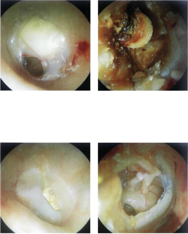
Figure 13.41 Left ear. Posterosuperior perforation of the reconstructed tympanic membrane with extrusion of the TORP. The shaft of the prosthesis has caused an erosion of the footplate of the stapes (which appears through the perforation as a rounded dark area).
Tympanoplasty 127
Figure 13.42 Left ear. Anteroinferior reperforation due to an acute otitis media, occurring 3 years after a staged closed tympanoplasty. A rectangular cartilage used for ossiculoplasty is visible. The cartilage is well integrated in the tympanic membrane residue.
Figure 13.43 Right ear. An example of TORP extrusion that occurred 1 year after a second-stage tympanoplasty. The head of the prosthesis can be seen despite the surrounding wax. The tympanic membrane residue is atelectatic.
Figure 13.44 Left ear. Gold prosthesis in the process of extrusion in a staged closed tympanoplasty.
Figure 13.45 Right ear. Post tympanoplasty. Large reperforation. In the posterosuperior quadrant a Teflon prosthesis is interposed between the medialized malleus and the footplate of the stapes. The round window is visible in the posteroinferior quadrant. The anterior residue of the tympanic membrane is tympanosclerotic.
128 13 Postsurgical Conditions
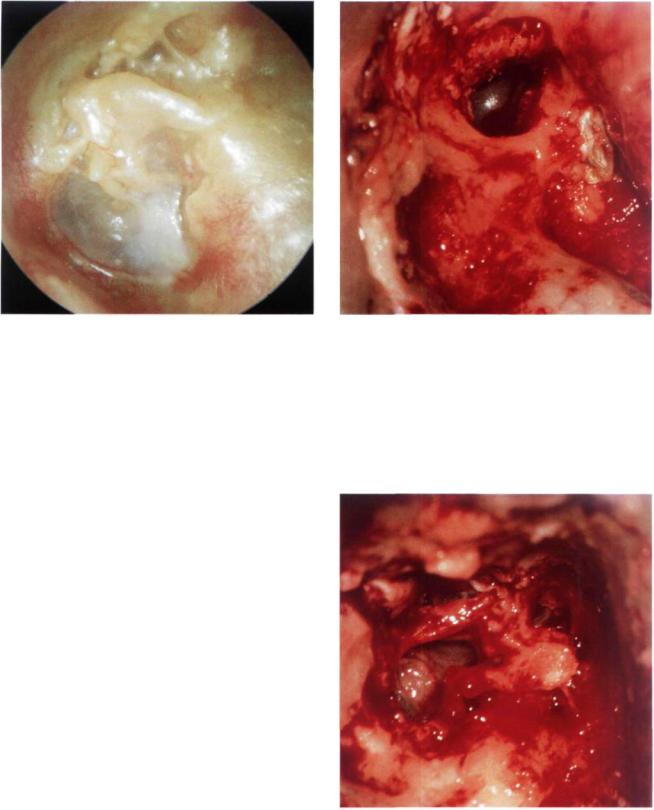
Figure 13.46 Right ear. Post stapedectomy. The atticotomy is seen in the posterosuperior quadrant. The preserved chorda tympani is well appreciated.
Figure 13.47 Left ear. A rare case of extrusion of a stapes prosthesis. The metallic ring is seen extruding through a microperforation covered with epidermal squames. The Teflon shaft of the prosthesis can be visualized through the tympanic membrane.
Figure 13.48 Right ear. A cholesteatomatous pearl in the |
Figure 13.49 Incision of the skin over the cyst. |
external auditory canal in a patient who had previously under- |
|
gone a stapedectomy. The tympanomeatal flap was not cor- |
|
rectly repositioned. This skin was thus folded in on itself and |
|
the entrapped epithelium gave rise to this pearl. This compli- |
|
cation was easily resolved in the outpatient clinic by incising |
|
the skin (see Fig. 13.49) and removing the cholesteatomatous |
|
cyst (see Fig. 13.50). |
|
Tympanoplasty 129
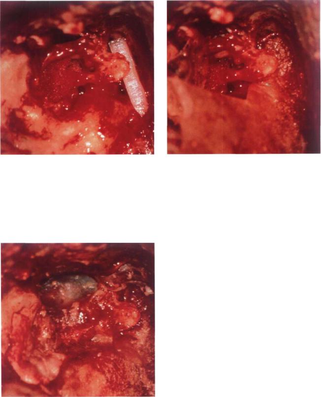
Figure 13.50 Removal of the cholesteatomatous cyst.
Figure 13.51 Left ear. Silastic sheet in extrusion through a posterosuperior perforation. The handle of the malleus is clearly visible anteriorly. In general, Silastic is inserted in firststage tympanoplasty. This material is usually placed in the middle ear to favor the restoration of the normal mucosal lining of the middle ear and to avoid the formation of adhesions in the meantime. It is removed during the second-stage tympanoplasty, except in cases showing a tendency towards atelectasis.
Figure 13.52 Right ear. Post tympanoplasty. A white retrotympanic mass (cholesteatoma of the anterior angle) is noted causing bulging of the tympanic membrane. The cholesteatoma is probably the result of inadequate removal of the epithelium in an overlay technique. The entrapped skin led to the formation of the cholesteatoma.
Figure 13.53 Left ear. Good anterior angle of the reconstructed tympanic membrane. An anteromalleolar cholesteatomatous cyst is seen. An epitympanic retraction pocket that is adherent to the head of the malleus and body of the incus is also observed.
130 13 Postsurgical Conditions
Figure 13.54 Left ear. In the posterosuperior quadrant, the sculptured incus with the short process pointing anteriorly is seen through the retracted tympanic membrane. In cases with hearing loss, repeat surgery is indicated to reinforce the tympanic membrane and improve the hearing. Surgery entails dissection of the retraction pocket from the incus, and the placement of cartilage between the sculptured incus and the tympanic membrane. This cartilage prevents (or delays) the reformation of a retraction pocket and corrects the hearing deficit.
Figure 13.55 Left ear. Another example of retraction of the tympanic membrane leading to inclination of the sculptured incus. The dislocated incus becomes fixed to the posterior mesotympanum, resulting in hearing loss.
Figure 13.56 Right ear. Sculptured incus seen through the tympanic membrane in a case of closed tympanoplasty. An epitympanic retraction pocket is seen. This pocket should be followed up regularly to guard against the formation of a recurrent cholesteatoma. Should this occur, the closed technique must be transformed into an open one to avoid further recurrence of the cholesteatoma.
Figure 13.57 Right ear. Recurrent epitympanic cholesteatoma following closed tympanoplasty. The reconstructed tympanic membrane (pars tensa) shows an optimal anterior angle and is perfectly attached to the handle of the malleus. In this case, transformation to an open technique is indicated while conserving the tympanic membrane and ossicular chain if there is no hearing loss.
Tympanoplasty 131
:
Figure 13.58 Left ear. Another example of a staged closed tympanoplasty 6 years after the second stage. A large resorption of the posterosuperior wall of the external auditory canal associated with recurrent cholesteatoma is observed. In the posterosuperior quadrant, the cartilage used for ossiculoplasty is seen. Revision surgery was performed with transformation into an open technique.
Figure 13.59 Left ear. A small epitympanic retraction pocket is observed. Though smail and shallow, this pocket should be followed up regularly as it may become deeper with time, leading to the formation of a recurrent cholesteatoma.
Figure 13.60 Right ear. Partial resorption of the posterior wall of the external auditory canal about 7 to 8 mm from the annulus following a closed tympanoplasty. The atrophic area appears bluish due to lack of underlying bone. No cutaneous retraction is seen. However, due to the lack of bone, the skin can invaginate into the mastoid cavity giving rise to recurrent cholesteatoma. In such cases, regular long-term follow-up is indicated.
Figure 13.61 Left ear. Total resorption of the posterior wall of the external auditory canal 3 years after a closed tympanoplasty. The otoscopic view is similar to that observed after an open tympanoplasty. Repeat surgery was necessary. The facial ridge was lowered and all bony irregularities were smoothed to avoid the retention of squamous debris with subsequent otorrhea. An adequate meatoplasty was also performed.
132 13 Postsurgical Conditions
Figure 13.62 Right ear. An example of a successful closed tympanoplasty 11 years postoperatively. The tympanic membrane is in perfect position and angulation. The posterior canal wall is intact and there is no evidence of recurrent cholesteatoma (see previous cases in Figs. 13.60 and 13.61).
Figure 13.63 Left ear. Another example of a closed tympanoplasty 2 years postoperatively. The attic was reconstructed using cartilage and bone pate and shows no signs of erosion. A small cholesteatomatous pearl is seen in the posterosuperior quadrant. It can be easily removed in the outpatient clinic under microscopic control.
Figure 13.64 Right ear. A well performed open tympanoplasty. The cavity is epithelialized and the facial ridge is adequately lowered. In the attic region, the material used for obliteration can be noted.
Figure 13.65 Left ear. Open tympanoplasty. Attic obliteration with autologous bone.
Tympanoplasty 133
Figure 13.66 Right ear. Open tympanoplasty. Partial obliteration of the attic with bone pate.
Figure 13.67 Right ear. A patient with bilateral cholesteatoma. An open tympanoplasty with obliteration was performed. The material used for obliteration of the attic (cartilage and bone pate) has nearly totally resorbed. The cavity is humid, granulating, and wet. Hearing is poorer than that of the other side in which an open technique without obliteration was performed (see next figure).
Figure 13.68 Same patient, left ear. The cavity is dry, |
Figure 13.69 Right ear. A badly performed open tym- |
smooth, well epithelialized, and the facial ridge is low. |
panoplasty. The cavity is irregular, with undermined borders, |
|
and a very high facial ridge. Purulent secretion is present in |
|
the middle ear and the rest of the cavity. |
134 13 Postsurgical Conditions
Figure 13.70 Right ear. Another example of a badly performed open tympanoplasty. Purulent secretions and a high facial ridge are observed.
Figure 13.71 Left ear. Open tympanoplasty. A large perforation of the reconstructed tympanic membrane is seen. Cholesteatomatous pearls are observed in the attic.'
Figure 13.72 Right ear. Open tympanoplasty. The facial ridge has not been sufficiently lowered in this case. This leads to accumulation of cerumen and cellular debris in the cavity with subsequent infection, secretion, and maceration of the skin lining the cavity.
Figure 13.73 Left ear. TORP in extrusion following a sec- ond-stage open tympanoplasty. In the first stage, a cholesteatoma involving the attic and mesotympanum and causing erosion of the ossicular chain was removed. In the second stage, a TORP was used for reconstruction. It was placed between the footplate of the stapes and the tympanic membrane. One year postoperatively, early extrusion of the prosthesis is observed. To avoid this complication, a tragal cartilage has to be placed between the prosthesis and the tympanic membrane.

Tympanoplasty 135
Figure 13.74 Left ear. An example of a correctly performed open tympanoplasty. The cavity shows perfect epithelialization. The facial ridge is low. The tympanic membrane is well positioned with excellent contact with the handle of the malleus.
Figure 13.75 Left ear. Open tympanoplasty with a well epithelialized cavity. The tympanic membrane shows a tympanosclerotic plaque anteriorly; posteriorly, the ossicular chain reconstruction is observed.
Figure 13.76 Right ear. Open tympanoplasty. The cartilage used for obliteration of the attic is seen in the superior part.
Figure 13.77 Right ear. Open tympanoplasty. The tympanic bone was drilled in this case because it was involved with the cholesteatoma. The inferior annulus is visible. Superiorly, the chorda tympani is observed close to the incus used for reconstruction of the ossicular chain.
136 13 Postsurgical Conditions
Figure 13.78 Left ear. Example of a modified Bondy technique. In this case, the preoperative pure tone average was 20 dB. The patient conserved his preoperative hearing. The modified Bondy technique is indicated in epitympanic cholesteatoma with an intact tympanic membrane and ossicular chain. It is an open technique in which the attic and the mastoid are exteriorized (Fig. 13.79) and the facial ridge is lowered until the level of the annulus. The ossicular chain and the tympanic membrane are left in situ (Fig. 13.80). If necessary, the attic is obliterated with a piece of cartilage; this procedure helps to reduce the risk of retractions around the ossicles (Fig. 13.81). Fascia is then inserted with two anterior tongues; one is positioned under the incus body, the other between the handle of the malleus and the long process of the incus (Figs. 13.82, 13.83). A meatoplasty according to the size of the cavity is performed at the end of the procedure.
Figure 13.79 Same case, intraoperative view. Exteriorization of the attic and the mastoid, lowering the facial ridge until the level of the annulus.
Figure 13.80 The ossicular chain and the tympanic membrane are left in situ.
Tympanoplasty 137
Figure 13.81 The attic is obliterated with a piece of carti- |
Figure 13.82 Fascia is inserted with two anterior tongues: |
lage. |
one is positioned under the incus body, another between the |
|
handle of the malleus and the long process of the incus. |
Figure 13.83 At the end of the procedure the skin flaps are repositioned over the fascia.
138 13 Postsurgical Conditions
Figure 13.84 Left ear. Another case of the modified Bondy technique. Note the intact ossicular chain.
Figure 13.85 Right ear. The modified Bondy technique. A ventilation tube was inserted because of the presence of middle ear effusion that did not respond to medical treatment.
Figure 13.86 Left ear. The modified Bondy technique. Although an attic retraction is noted recurrent cholesteatoma is uncommon with this technique. The tympanic membrane is retracted and middle ear effusion is noted. In this case, the insertion of a ventilation tube is indicated.
Figure 13.87 Right ear. The modified Bondy technique. The attic is obliterated with cartilage.
Tympanoplasty 139
Figure 13.88 Left ear. Open tympanoplasty. The ossicular chain was reconstructed using an autologous cartilage that was not extruded despite the presence of atelectasis of the tympanic membrane.
Figure 13.89 Left ear. Another case of the modified Bondy technique. As the incus was slightly eroded, a piece of cartilage was placed between it and the malleus. The attic was obliterated with cartilage.
Figure 13.90 Right ear. The modified Bondy technique. The tympanic membrane is normal and the cavity is dry and perfectly epithelialized.
Figure 13.91 Right ear. In this case of a modified Bondy technique, incus erosion occurred 3 years postoperatively due to the presence of a significant retraction pocket. The middle ear shows a catarrhal effusion.
140 13 Postsurgical Conditions
Figure 13.92 Right ear. A modified Bondy technique. Two cholesteatomatous pearls are present in the cavity. They are easily removed in the outpatient clinic. The attic, antrum, and mastoid were exteriorized. The ossicular chain was left in situ.
Figure 13.93 Left ear. A cholesteatomatous pearl seen in the attic following a modified Bondy technique.
Figure 13.94 Same patient after removal of the pearl in the outpatient clinic.
Figure 13.95 Radical mastoidectomy. A mucosal cyst causes complete obstruction of the external auditory canal.
Tympanoplasty 141
Figure 13.96 Right ear. Radical mastoidectomy. The second portion of the facial nerve is uncovered. Scars around the nerve produced an H.B. grade III palsy.
Figure 13.97 Example of a well performed meatoplasty in an open tympanoplasty. The performance of an adequate meatoplasty that suits the dimension of the cavity is fundamental to assure proper aeration and prevent accumulation of epithelial debris and cerumen in the cavity.
Figure 13.98 Another example of a meatoplasty performed |
Figure 13.99 Example of a meatoplasty that shows mild |
in a 10-year-old boy who underwent surgery for bilateral epi- |
stenosis. |
tympanic cholesteatoma using a modified Bondy technique. |
|