


73
9 Congenital Cholesteatoma of the Middle Ear
Congenital cholesteatoma is defined as an epidermoid cyst that develops behind an intact tympanic membrane in a patient with no history of otorrhea, trauma, or previous ear surgery. Michaels studied fetal temporal bones and demonstrated the presence of an epidermoid structure between 10 and 33 weeks of gestation. This structure tends to involute spontaneously until it completely disappears. Michaels hypothesized that the persistence of this structure could act as an anlage and lead to congenital cholesteatoma. The fact that the most classic location of congenital cholesteatoma, namely in the anterosuperior part of the tympanum, corresponds to the site of the fetal Michaels structure supports this theory. In our cases, however, and contrary to the few studies reported in the literature (Derlacki and Clemis 1965, Friedberg 1994, Levenson et al. 1989, Cohen 1987), the most common site of congenital cholesteatoma was the posterior mesotympanum (see Table 9.1).
As no existing theory can truly explain the origin of congenital cholesteatoma in the posterior location, a strong conjecture can be made that these lesions might represent a different entity from those of the anterior location and may originate from epithelial cell rests that are trapped in the posterior mesotympanum during the development of the temporal bone. Diagnosis is either occasional in the asymptomatic patient, or the patient may complain of hearing loss due to erosion of the ossicular chain or of recurrent attacks of secretory otitis media due to occlusion of the tubal orifice by the cholesteatomatous mass. A high degree of suspicion and thorough examination are essential in detecting the presence of these lesions.
Table 9.1 Classification of congenital cholesteatoma of the middle ear
Type |
Location |
Number |
Percent |
Type A |
Mesotympanic |
23 |
52.27 |
Type A1 |
Premalleolar |
2 |
4.54 |
Type A2 |
Retromalleolar |
21 |
46.72 |
Type B |
Epitympanic |
3 |
6.81 |
Type A/B |
Mixed |
18 |
40.90 |
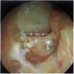
Figure 9.1 Right ear. Congenital cholesteatoma seen as a white retrotympanic mass causing bulging of the posterior guadrants of the tympanic membrane. Neither drum perforation nor bony erosion are detected.
Figure 9.2 Right ear. A small whitish retrotympanic mass is clearly seen. The mass lies posterior to the malleus (type A2). By definition, a cholesteatoma is considered congenital when the tympanic membrane is intact and there is no history of otorrhea or previous ear operations (including myringotomy or ventilation tube insertion).
74 9 Congenital Cholesteatoma of the Middle Ear
Figure 9.3 Left ear. A case similar to that in Figure 9.2. The cholesteatoma caused erosion of the long process of the incus with resultant conductive hearing loss.
Figure 9.4 Right ear, intraoperative view. A small premalleolar congenital cholesteatoma (type A1) can be seen behind the intact tympanic membrane.
Summary
Congenital cholesteatoma of the middle ear is an infrequent pathology during infancy and childhood. It presents behind an intact tympanic membrane, either anterior or posterior to the handle of the malleus.
Anterosuperior cholesteatoma can be removed through an extended tympanotomy that permits the preservation of the tympanic membrane and ossicular chain integrity. Posterior cholesteatoma, however, requires a staged closed tympanoplasty. The second stage serves to check for any residual cholesteatoma. The ossicular chain, which is generally eroded in the posterior type, can be reconstructed at this stage.
Figure 9.5 Same case, intraoperatively, after elevation of the tympanomeatal flap.

75
10 Petrous Bone Cholesteatoma
Unlike middle ear cholesteatoma, petrous bone cholesteatoma represents an epidermoid cyst that involves the petrous part of the temporal bone. This type of cholesteatoma involves and/or is related to very important structures (namely, the facial nerve, posterior labyrinth, cochlea, internal carotid artery, internal auditory canal, and posterior and middle fossa dura); therefore, the management of such lesions should be performed in centers specialized in otoneurology and skull base surgery. The main presenting symptom is fetid otorrhea, which frequently reoccurs in an open mastoid cavity. The second most common symptom is progressive facial nerve palsy, that occurs in more than 50% of cases. Hearing loss can be conductive, sensorineural, or mixed. About 50% of cases complain of vertigo, but it is rarely the motive for the patient's visit to the doctor. Otoscopy may be irrelevant or only demonstrates pars flaccida perforation or an open mastoid cavity with evidence of suppurative discharge. A computed tomography (CT) scan and magnetic resonance imaging (MRI) are fundamental to evaluate the extension of the lesion and to determine the surgical approach.
Petrous bone cholesteatoma is defined as congenital when it develops from epithelial cell rests entrapped in the petrous bone during embryological development. In such cases, the first symptoms are facial nerve paralysis, vertigo, and deaf ear due to invasion of the facial nerve and labyrinth. This type represents about 3% of all cases of cholesteatoma and is localized at the level of the petrous apex.
Petrous bone cholesteatoma is defined as acquired when a middle ear cholesteatoma follows the cell tracts of the temporal bone in a lateral to medial direction and invades the underlying structures. The most frequent symptoms in such cases are fetid otorrhea followed by hearing loss (conductive, perceptive, or mixed), facial nerve paralysis, and vertigo.
The iatrogenic form also develops in an old radical cavity or as a late occurrence following tympanoplasty. The most common symptoms in such cases are also fetid otorrhea, facial nerve paralysis, hearing loss, and vertigo.
We classify petrous bone cholesteatoma into five types according to its localization and extension: supralabyrinthine, infralabyrinthine, massive labyrinthine, infralabyrinthine apical, and apical.
Figure 10.1 The supralabyrinthine type of petrous bone cholesteatoma is centered on the region of the geniculate ganglion. Most frequently, it extends anteriorly towards the basal turn of the cochlea and the internal carotid artery. Less commonly, it grows towards the retrolabyrinthine air cells. This localization is typical of congenital cholesteatoma of the petrous bone. It may also arise due to a deep growth of an epitympanic cholesteatoma.
TS: |
Transverse sinus |
pc: |
Posterior clinoid |
Lv: |
Labbe's vein |
V2: |
Trigeminal 2 |
SS: |
Sigmoid sinus |
V3: |
Trigeminal 3 |
ev: |
Emissary vein |
C1: |
First cervical vertebra |
JB: |
Jugular bulb |
VII: |
Facial nerve |
JV: |
Jugular vein |
IX: |
Glossopharyngeal nerve |
ICA: |
Internal carotid artery |
X: |
Vagus nerve |
pp: |
Pterygoid processes |
XI: |
Spinal accessory nerve |
za: |
Zygomatic arc |
XII: |
Hypoglossal nerve |
et: |
Eustachian tube |
|
|
Figure 10.2 The infralabyrinthine type of petrous bone cholesteatoma is usually encountered in an old radical mastoid cavity. It is localized in the region of the hypotympanum and the infralabyrinthine air cells. It may extend posteriorly towards the posterior cranial fossa or anteriorly towards the internal carotid artery, petrous apex, and clivus.

76 |
10 Petrous B o n e C h o l e s t e a t o m a |
ICA
CLIVUS
JVICA
Figure 10.3 The massive labyrinthine type of petrous bone cholesteatoma largely involves the posterior labyrinth and the cochlea. It may extend anteriorly towards the internal carotid artery, medially towards the internal auditory canal, posteriorly towards the posterior fossa, or interiorly towards the infralabyrinthine compartment. Abbreviations are given in Figure 10.1.
ICA
CLIVUS
JV ICA
Figure 10.5 The apical type of petrous bone cholesteatoma is a rare congenital lesion. It may solely involve the apical compartment, causing erosion of it. It may involve the trigeminal nerve or more posteriorly the posterior cranial fossa. It may also engulf the horizontal portion of the internal carotid artery.
ICA
CLIVUS
JV ICA
Figure 10.4 The infralabyrinthine apical type of petrous bone cholesteatoma originates from the infralabyrinthine or apical compartments. When it originates from the former, it extends into the petrous apex. In some cases it may grow towards the sphenoid sinus or the horizontal portion of the internal carotid artery.

Petrous Bone Cholesteatoma |
77 |
Figure 10.6 Left acquired or iatrogenic supralabyrinthine petrous bone cholesteatoma in a radical cavity. A whitish retrotympanic mass is seen at the level of the second portion of the facial nerve. The patient presented with progressive facial nerve paralysis and total hearing loss. A correct diagnosis depends not only on otoscopy but also on the symptomatology (facial paralysis, anacusis) and a high-resolution CT scan.
Figure 10.7 CT scan of the case presented in Figure 10.6, axial section. Involvement of the lateral semicircular canal and the vestibule is well visualized. The cholesteatoma invades the cochlea anteriorly, while medially it reaches the fundus of the internal auditory canal. The posterior semicircular canal is not invaded.
Figure 10.8 CT scan of the case presented in Figure 10.6, coronal section. The medial extension of the cholesteatoma can be appreciated.
Figure 10.9 Postoperative CT scan. A transcochlear approach was performed and the operative cavity was obliterated with abdominal fat.

10 Petrous Bone Cholesteatoma
Figure 10.10 Right acquired supralabyrinthine petrous bone cholesteatoma. A whitish mass is present in the mastoid cavity of an open tympanoplasty. The mass occupies the whole epitympanum and extends interiorly behind the tympanic membrane. The patient presented with ipsilateral facial paralysis and conductive hearing loss.
Figure 10.11 CT scan of the case presented in Figure 10.10. The cholesteatoma invades the cochlea. Total removal of the pathology was accomplished using a transcochlear approach with obliteration of the operative defect using abdominal fat. The external auditory canal was closed as cul- de-sac. The facial nerve was infiltrated at the level of the geniculate ganglion and was repaired using a sural nerve graft.
Figure 10.12 Another example of right acquired supralabyrinthine petrous bone cholesteatoma. The patient presented with right facial nerve paralysis. Otoscopy reveals a right epitympanic erosion.
Figure 10.13 CT scan of the case presented in Figure 10.12, coronal view. Typical location and erosion of acquired small supralabyrinthine petrous bone cholesteatoma.

Petrous Bone Cholesteatoma |
79 |
Figure 10.14 Left congenital supralabyrinthine petrous bone cholesteatoma with extension towards the apex. Otoscopy is negative. The patient complained of progressive facial nerve paralysis of 5 years' duration as well as conductive hearing loss.
Figure 10.15 CT scan of the case presented in Figure 10.14. Coronal view showing extension of the cholesteatoma into the internal auditory canal.
Figure 10.16 CT scan of the case presented in Figure 10.14. Axial view showing cholesteatoma extending into the petrous apex.
Figure 10.17 Right congenital infralabyrinthine apical petrous bone cholesteatoma in a 30-year-old female patient. In the posterosuperior quadrant a white retrotympanic view is observed. The patient had complained of right anacusis since childhood and instability of 1 year duration. The facial nerve was normal.

80 |
10 Petrous Bone Cholesteatoma |
Figure 10.18 CT scan of the case presented in Figure 10.17. Coronal view demonstrating the involvement of the infralabyrinthine apical compartment by the cholesteatoma.
Figure 10.19 CT scan of the case presented in Figure 10.17. A more anterior coronal view at the level of the cochlea.
Figure 10.20 Postoperative CT scan showing total removal of the cholesteatoma through the transcochlear approach and obliteration of the operative cavity using abdominal fat.
Figure 10.21 Polyp in the external auditory canal in a patient who had undergone a tympanoplasty (see CT scan, Fig. 10.22). The patient presented with otorrhea and hearing loss.

Petrous Bone Cholesteatoma |
81 |
Figure 10.22 CT scan of the case presented in Figure |
Figure 10.23 Postoperative CT scan showing total removal |
10.21. A large infralabyrinthine apical petrous bone |
|
cholesteatoma extending to the cavernous sinus and to the |
|
sphenoid sinus can be seen. Total removal was achieved using |
|
an infratemporal fossa approach type B. |
|
Figure 10.24 Left acquired petrous bone cholesteatoma of the massive type. The patient had complained of fetid otorrhea and hearing loss since early childhood. Six months before presentation, he started to experience facial nerve paralysis. A radical mastoidectomy was performed in another center with partial removal of the pathology. The second and third portions of the facial nerve can be observed in the mastoid cavity. The patient underwent surgery using a transcochlear approach to obliterate of the cavity with abdominal fat.
Figure 10.25 CT scan of the case presented in Figure 10.24 demonstrating cholesteatoma invading the labyrinth.
82 10 Petrous Bone Cholesteatoma
Figure 10.26 Left radical mastoid cavity. This patient was operated on using a combined middle cranial fossa and transmastoid approach for the removal of a petrous bone cholesteatoma. The facial nerve was left as a bridge in the middle of the cavity. On follow-up the patient complained of recurrent episodes of facial nerve paralysis due to accumulation of cerumen and debris in the cavity. Therefore, the patient underwent a second operation for obliteration of the cavity with abdominal fat and closure of the external auditory canal as cul-de-sac.
Summary
When a patient presents with hearing loss (sensorineural or mixed) and/or facial nerve paralysis with or without a retrotympanic mass, the probability of petrous bone cholesteatoma should be considered. In such cases, it is necessary to perform a high - resolution CT scan of the temporal bone.
The ideal treatment for petrous bone cholesteatoma is radical surgical removal, although destruction of the labyrinth and rerouting of the facial nerve may be required. The status of the contralateral ear must also be considered.
The modified transcochlear approach is the most appropriate for the removal of petrous bone cholesteatoma. This approach offers direct lateral access to the petrous bone and allows the removal of all types of petrous bone cholesteatoma with their possible extension into the clivus or sphenoid sinus. In addition, it has the advantage of minimizing the occurrence of cerebral spinal fluid (CSF) leak and allows control of the different vital structures, including the internal carotid artery. Closure of the external auditory canal as cul-de-sac and obliteration of the operative cavity with abdominal fat avoids the risk of infection and the need for frequent toilet of a very deep cavity.
The middle cranial fossa approach and the radical mastoidectomy can be used in cases with noncom-
promised inner ear function. The former is utilized in small supralabyrinthine cholesteatoma, while the latter is utilized in small infralabyrinthine cholesteatoma with no involvement of the internal carotid artery.