

46
7 Non-Cholesteatomatous Chronic Otitis Media
The difference between acute and chronic otitis media is not the duration of the disease but rather the anatomopathological characteristics. Untreated acute otitis media persisting for months is still a process that tends essentially to return to normality. On the other hand, a chronic otitis, even if the ear stops discharging, has anatomopathological sequelae of clinical importance.
The most commonly encountered forms are active chronic suppurative otitis media characterized by otorrhea and inactive chronic suppurative otitis media in which the ear is dry. Naturally, the active form may become quiescent either spontaneously or following treatment. The ear becomes dry and the condition is designated inactive. A dry perforation, however, may be infected, leading to ear discharge. In this latter case, the mucosa may be hyperplastic and thick due to interstitial edema, fibrosis, or cellular infiltration. In some cases, polypi are formed which may be large enough to occupy the external auditory canal. In other cases, persistence of suppuration can lead to ulceration of the mucosa, formation of granulation tissue, and even bone resorption. The anatomical sequelae of chronic otitis media vary. They may be in the form of a simple central tympanic membrane perforation, erosion of the ossicular chain, or formation of tympanosclerosis. Both the active and inactive forms cause functional alterations such as conductive or mixed hearing loss (very rarely sensorineural). The absence of squamous epithelium in the middle ear has led to the designation of this form as a "safe type" of otitis media. This is to distinguish it from cholesteatoma, which is considered "unsafe" due to the potential complications that may arise from the presence of keratinized squamous epithelium in the middle ear.
•General Characteristics of Tympanic Membrane Perforations
Tympanic membrane perforations are usually present at the pars tensa. Pars flaccida perforations are generally associated with epitympanic cholesteatoma.
If a tympanic membrane perforation does not heal spontaneously, the epithelial and mucosal layers creep and meet along the borders of the perforation. This pathological communication between the middle and external ear can be considered a true "air fistula." In the presence of a tympanic membrane perforation, the patient is subject to recurrent infections and ear discharge.
Whenever tympanic membrane perforations are diagnosed, the following three considerations must be fulfilled: 1) At the level of the perforation the site, size, and state of the remainder of the tympanic membrane
around the perforation should be determined. 2) At the level of the middle ear, the state of the mucosa, the condition of the ossicular chain (if possible), and the presence or absence of epithelialization should be evaluated. 3) The otoscopic examination has to be complemented with the pure tone audiometry to have a better understanding of the ossicular chain (possible erosion of the incus, fixity of the chain).
Pars tensa perforations can be either central or marginal. Marginal perforations lie at the periphery of the tympanic membrane with absence of the fibrous annulus. Marginal perforations are considered "unsafe" because the skin of the external auditory canal, in the absence of the annulus, can easily advance towards the middle ear, giving rise to cholesteatoma.
Otoscopic examination can often define the junction between the skin and mucosa at the borders of the tympanic membrane perforation. At this junction the squamous epithelium has a "velvety" appearance. The presence of a red de-epithelialized ring along the perforation rim indicates the evagination of the mucosa towards the external surface of the tympanic membrane residue. However, invagination of the skin towards the inner surface of the tympanic membrane residue is more difficult to diagnose. This inward skin migration is favored by the atrophy of the mucosa which occurs as a result of the perforation. At the time of myringoplasty, freshening of the edge of the perforation not only favors the attachment of the graft but also greatly reduces the risk of leaving entrapped skin on the undersurface of the drum, which may lead to iatrogenic cholesteatoma.
Conductive hearing loss caused by tympanic membrane perforation has two main causes: 1) Reduction of the tympanic membrane surface area on which the acoustic pressure exerts its action. 2) Reduction of the vibratory movements of the cochlear fluids because sound reaches both windows at nearly the same time without the dampening and phase-changing effect of the intact tympanic membrane.
The site of the perforation cannot be correlated to a particular audiometric pattern. However, it is generally observed that hearing loss occurs more in the low frequencies and that for perforations of the same size, hearing loss occurs more in posterior perforations than in anterior ones.
The majority of posttraumatic and postotitic perforations heal spontaneously. When large portions of the tympanic membrane are lost or when chronic or recurrent infections occur, the perforation may become permanent. In these cases, the tympanic membrane must be repaired (myringoplasty) to restore the normal physiology of the ear.

Posterior Perforations |
47 |
Posterior Perforations
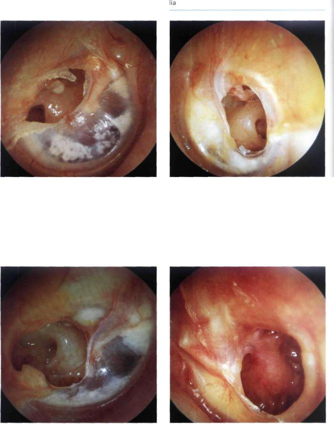
Figure 7.1 Left ear. The tympanic membrane is very thin due to atrophy of the fibrous layer. A posterosuperior marginal perforation is seen. This perforation is risky because the skin of the external auditory canal can easily advance into the middle ear, forming a cholesteatoma. In this case, a myringoplasty using an endomeatal approach is indicated.
Figure 7.2 Right ear. Marginal posterosuperior perforation through which the intact incudostapedial joint, the stapedius tendon, and the pyramidal process can be seen.
Figure 7.3 Left ear. Perforation of the posterosuperior quadrant of the tympanic membrane. Visualized through the perforation are the incudostapedial joint, the stapes, the stapedius tendon, the pyramidal process, the promontory, and the round window. The residue of the tympanic membrane is very thin due to absence of the fibrous layer. Tympanosclerosis can be seen in the marginal part of the drum residue. From the surgical point of view, posterior perforations are the easiest to repair especially when partial reconstruction of the tympanic membrane is all that is required. When the residue of the tympanic membrane is transformed into a rigid tympanosclerotic
plaque, it is advisable to remove it, conserving the epidermal layer to be laid over the graft.
Figure 7.4 Right ear. Large perforation of the posterior quadrants. Normal middle ear mucosa. The incudostapedial joint is intact. The oval window with the annular ligament surrounding the footplate can be seen. The pyramidal eminence, the stapedius tendon, the round window, and Jacobson's nerve running on the promontory are also visible. The remaining anterior quadrants of the tympanic membrane are tympanosclerotic and rigid, blocking the mobility of the malleus.
48 7 Non-Cholesteatomatous Chronic Otitis
Figure 7.5 Right ear. Presence of chronic otitis media. Dry perforation of the posterior quadrants of the tympanic membrane through which the head of the stapes and the round window are visible. The long process of the incus is necrosed. The middle ear mucosa is normal. The tympanic membrane residue shows tympanosclerosis with alternating areas of calcification and areas of thinned membrane due to atrophy of the fibrous layer. The operation, performed through a postauricular incision, will also include the reconstruction of the ossicular chain using the autologous incus.
Figure 7.6 Left ear. Posterior nonmarginal perforation. The incudostapedial joint, the promontory, and the round window are all discernible.
Figure 7.7 Right ear. Presence of simple chronic otitis media; a posteroinferior drum perforation. The middle ear mucosa is normal. The round window and Jacobson's nerve running on the promontory are seen. The incus can also be appreciated posterior to a retromalleolar tympanosderotic plaque. The tympanic membrane residue shows areas of atrophy alternating with areas of tympanosclerosis.
Figure 7.8 Left ear. Perforation of the posterior quadrants of the tympanic membrane. The skin advances along the posterosuperior border of the perforation towards the incudostapedial joint. The middle ear mucosa appears hypertrophic. Mucoid discharge is also present. A tympanosderotic plaque can be seen in the residue of the tympanic membrane.

Anterior Perforations |
49 |
Figure 7.9 Right ear. Marked posterior marginal perforation through which the skin penetrates into the middle ear. The ossicular chain is not identifiable.
Anterior Perforations
Figure 7.10 Left ear. Anterior perforation of the tympanic membrane through which the tubal orifice is visible. A white mass is present behind the anterosuperior margin of the perforation. This mass can be either a cholesteatoma or a tympanosclerotic plaque. The consistency of the mass can be tested using an instrument under the microscope; the cholesteatoma is soft and will break, whereas tympanosclerosis is generally hard.
Figure 7.11 Right ear. Anterior perforation in a patient with anterior and posterior humps of the external auditory canal as well as exostosis of the superior canal wall. In this case, canalplasty should be performed at the same time as myringoplasty.

50 7 Non-Cholesteatomatous Chronic Otitis Media
Figure 7.12 Left ear. Dry anteroinferior perforation. The middle ear mucosa is normal. The tympanic membrane residue shows tympanosclerosis, giving it a white aspect. The tubal orifice can be seen from the anterior margin of the perforation.
Figure 7.13 Right ear. Anteroinferior perforation. The posterior and anterior residues of the tympanic membrane show tympanosclerosis. The anteroinferior residue of the drum is de-epithelialized. The tubal orifice is also visible.
Figure 7.14 Right ear. Anteroinferior perforation. Two tympanosclerotic plaques are appreciated: one anteromalleolar and the other retromalleolar. The middle ear mucosa is normal. The hypotympanic air cells are seen through the perforation.

Subtotal and Total Perforations |
51 |
• Subtotal and Total Perforations
Figure 7.15 Right ear. Large tympanic membrane perforation. The tubal orifice, the hypotympanic air cells, the promontory, the round and oval windows, and the intact stapes can be viewed. An onset of necrosis of the incus can be distinguished.
Figure 7.16 Right ear. Perforation of the inferior quadrants of the tympanic membrane. All the tympanic membrane residue shows dense tympanosclerosis. Removal of these sclerotic plaques during myringoplasty assures an adequate vascularity to the graft, and thus a high success rate for the operation.
Figure 7.17 Right ear. Similar case. The promontory and the round window are visible. A tympanosclerotic plaque that engulfs the ossicular chain is seen at the level of the posterosuperior border of the perforation.
Figure 7.18 Left ear. Subtotal perforation. The annulus as well as a fibrous rim are visualized along the inferior border of the perforation. The handle of the malleus is medialized. The tubal orifice, the hypotympanic air cells covered with mucosa, Jacobson's nerve on the promontory, and the long process of the incus are visible. The residue of the tympanic membrane is thickened. In cases in which only a small anterior residue of the tympanic membrane is found, an overlay technique in which the graft is put over the annulus is used, thus preventing detachment of the anterior part of the graft leading to reperf oration.

52 7 Non-Cholesteatomatous Chronic Otitis
Figure 7.19 Left ear. Total perforation of the tympanic membrane through which evolving tympanosclerotic plaques are visible. The stapes and the stapedius tendon are visible. The long process of the incus is partially eroded. The handle of the malleus is medialized and adherent to the promontory. The tubal orifice and the hypotympanic air cells are also noted.
Figure 7.20 Left ear. Subtotal perforation of the tympanic membrane. The middle ear mucosa is normal. The tympanic membrane residue is de-epithelialized. The incudostapedial joint, the medialized handle of the malleus, and the hypotympanic air cells are visible.

Posttraumatic Perforations |
53 |
• Posttraumatic Perforations
Figure 7.21 Left ear. Posttraumatic perforation of the tympanic membrane in the region of the cone of light. The blood clot over the perforation has not been removed. This clot helps to guide spontaneous healing of the drum.
Summary
The presence of a tympanic membrane perforation that does not heal spontaneously as in chronic otitis media represents an anatomical and functional defect that needs surgical correction in the majority of cases.
Myringoplasty is indicated in cases with and without otorrhea, with a small or a large air-bone gap, and with no age limit. It is contraindicated when the tympanic membrane perforation is present in the only hearing ear.
Myringoplasty is generally performed using a postauricular incision under local anesthesia-except for children where general anesthesia is used. The tympanic membrane is repaired by an autologous temporalis fascia graft. We prefer the underlay technique in the majority of cases because it gives better results both anatomically and functionally. The overlay technique is used in selected cases when the anterior residue of the tympanic membrane is pathologic or absent. When properly performed, the overlay technique gives optimal results in these cases. Canalplasty is done whenever bony humps of the external canal are present that limit control of the perforation borders. If reperforation occurs after myringoplasty (in about 5% of cases), a revision operation is indicated after a few months. The results of the first and second operations in terms of graft take and reperforation are generally comparable.
Figure 7.22 Left ear. Posttraumatic perforation in the posterosuperior quadrant. The characteristic radial tear, running in the same direction as the fibers of the tympanic membrane, is apparent. Hemorrhagic points separating the epidermal layer from the fibrous layer are visible. These tiny hemorrhages are typical of posttraumatic perforations. This type of tympanic membrane perforation has a very high incidence of spontaneous healing.
547 Non-Cholesteatomatous Chronic Otitis
•Perforations Complicated or Associated with Other Pathologies
Figure 7.23 Right ear. Total perforation. Epidermization is present in the regions of the mesotympanum and the ossicular chain. The round window, hypotympanic air cells with thickened mucosa, Jacobson's nerve running on the promontory, and the tubal orifice are well visualized. This case is an example of chronic otitis media complicated by the presence of skin in the middle ear. Tympanoplasty should be staged. In the first stage, the skin is removed without traumatizing the ossicular chain, and the tympanic membrane is reconstructed.
In the second stage, the middle ear is checked for any residual skin, and the ossicular chain is reconstructed.
Figure 7.24 Left ear. Large perforation with diffuse epidermization of the middle ear associated with purulent otorrhea. In these cases, even if the ossicular chain proves intact, mastoid exploration should be done. A second stage is performed 1 year after the first operation to check for any skin residues.
Figure 7.25 Right ear. Perforation of the inferior quadrants of the tympanic membrane, the residues of which show tympanosclerosis. Epidermization is evident over the promontory. Since epidermization is limited in this case, a single-stage tympanoplasty can be performed.
Figure 7.26 Right ear. Another example of chronic otitis media complicated with diffuse epidermization of the middle ear. Surgery follows the same rules as for cholesteatoma.

Perforations Complicated or Associated with Other Pathologies |
55 |
Figure 7.27 Right ear. Large tympanic membrane perforation. The anterior drum residue shows tympanosclerosis. The ossicular chain is difficult to identify because of the presence of epidermization at this level. The round window is visible. A staged tympanoplasty is also indicated in this case.
Figure 7.28 Right ear. Granulomatous otitis media. A roundish mass fills the middle ear. Serous otorrhea is present.
Figure 7.29 Right ear. Small perforation of the inferior quadrants of the tympanic membrane with eversion of the mucosa onto the outer layer of the membrane. Tympanosclerosis, both anteroand posteromalleolar, can be noted.
Figure 7.30 Right ear. Case similar to that in Figure 7.29. The mucosa has replaced the epithelial layer. Ear discharge is also present. During myringoplasty, curettage of the everted mucosa is necessary until the fibrous layer of the tympanic membrane is reached.

56 7 Non-Cholesteatomatous Chronic Otitis Media
Figure 7.31 Left ear. Perforation of the anterior quadrants. Skin envelopes the handle of the malleus. During myringoplasty, curettage of the skin is necessary before reconstruction.
Figure 7.32 Right ear. Posterior perforation. The residues of the tympanic membrane appear whitish and bulging. During surgery, the middle ear was occupied by granulomatous tissue that proved to be tuberculosis (TB) on histopathological examination. This patient had a past history of pulmonary TB. Tuberculous otitis media should be suspected in cases of pulmonary TB presenting with otorrhea.
•Tympanosclerosis
Tympanosclerosis is characterized by fibroblastic invasion of the submucosal spaces of the middle ear followed by thickening, hyalinization, and fusion of collagen fibers into a homogenous mass with calcium deposits and phosphate crystals. Though the pathogenesis is not yet clear, it seems that chronic otitis media is a predisposing factor.
Two distinct forms are recognized:
Tympanosclerosis with Intact tympanic membrane.
This is characterized by calcareous plaques (chalk patches) in the fibrous layer of the tympanic membrane. The anteroand posteromalleolar regions are usually involved. The periannular region of the inferior quadrants is also affected, forming a horseshoe pattern. The pars tensa is rigid, thick, and loses its elasticity, assuming a whitish aspect. Atrophic and thinned areas can also occur. Infrequently, in very advanced cases, the tympanosclerotic plaques occupy all the middle ear spaces, attic, and aditus and completely block the ossicular chain. The tympanic membrane in these cases is very thick or even replaced by the plaques.
Tympanosclerosis associated with tympanic membrane perforation. The perforation is frequently central or subtotal and the annulus, infiltrated by calcium deposits, is well visualized. Frequently, submucous nodular deposits are encountered in the middle ear. Ossicular fixation or erosion due to devitalization as a
result of loss of blood supply can also occur. The middle ear mucosa is very thin with reduced vascularity. In some cases, tympanosclerotic plaques are seen extruding from the mucosa to present as white middle ear masses.

Tympanosclerosis Associated with Perforation |
57 |
•Tympanosclerosis Associated with Perforation
Figure 7.33 Right ear. Tympanosclerosis associated with perforation. The tympanic membrane residues and the middle ear (promontory and hypotympanum) show the characteristic plaques. The malleus is blocked by tympanosclerosis.
Figure 7.34 Right ear. Tympanosclerosis with perforation. A large tympanosclerotic plaque is noted in the anterior residue of the tympanic membrane. The middle ear is also involved. The promontory, oval window, stapes footplate, and round window can be appreciated.
Figure 7.35 Right ear. Perforations of the inferior quadrants with tympanosclerosis involving the residues of the tympanic membrane and the middle ear.
Figure 7.36 Right ear. Tympanosclerosis with perforation. The tympanosclerotic process involves the anterior residues of the tympanic membrane and the mucosa of the promontory reaching to the posterior mesotympanum. At this level, ossification of the stapedius tendon is seen. The tympanic segment of the fallopian canal is covered by a sclerotic plaque. The long process of the incus is eroded.

58 7 Non-Cholesteatomatous Chronic Otitis Media
Tympanosclerosis with Intact Tympanic Membrane
Figure 7.37 Left ear. Tympanosclerosis and intact drum. The majority of the tympanic membrane is thinned due to atrophy of the fibrous layer. Two tympanosclerotic plaques are present near the anterior and posterior margins.
Figure 7.39 Left ear. Tympanosclerosis with intact drum. A large plaque is visible in the posterior quadrants of the tympanic membrane. The anterior quadrants are thinned and atrophic, allowing visualization of the tubal orifice.
Figure 7.38 Left ear. The intact tympanic membrane shows tympanosclerotic plaques lying both anterior and posterior to the malleus that alternate with areas of atrophy (in the inferior quadrants).
Summary
Chronic otitis media associated with tympanosclerosis represents a more complex anatomopathological entity. In cases with intact tympanic membrane, surgery is indicated in the presence of a significant air-bone gap, signifying ossicular chain affection. Should erosion or fixation of the ossicles be found, ossiculoplasty is performed. Fixation of the stapes is an indication for stapedotomy.
In cases associated with tympanic membrane perforation, it is often possible to perform a single-stage reconstruction in which myringoplasty is performed with or without ossiculoplasty. A fixed stapes, however, is an indication for staging where myringoplasty is performed first, followed by a second-stage stapedotomy after a few months. In all suspected cases, the patient should be informed preoperatively of the possibility of staging surgery.
In a small percentage of cases of chronic otitis media with tympanosclerosis, a good postoperative functional level can deteriorate with time due to refixation of the ossicular chain with consequent air-bone gap. In such cases, after achieving closure of the tympanic membrane, a hearing aid is recommended.