

Учебники / Diagnosis in Otorhinolaryngology Onerci 2009
.pdf3.10 Neck Masses 159
3.10
Neck Masses
Thyroglossal Duct Cyst
The most common congenital neck mass in children is a thyroglossalductcyst.Thyroglossalductcystsaremostcommonly located below the hyoid bone; however, they may be found anywhere between the base of the tongue and the superior border of the thyroid gland. They move on swallowing and on tongue protrusion. Unless infected they are asymptomatic. The patient only complains about a lump in the neck. Treatment is complete excision of both the cyst and the entire thyroglossal duct up to the foramen cecum at the base of the tongue. Therefore, it is necessary to remove the central portion of the hyoid bone.
Dermoid Cyst
Dermoid cysts are usually located in the submental region in themidline.Theyareepithelialremnantsoccurringalongthe lines of fusion in the embryo. Dermoids are lined by epidermis and may contain hair follicles, epidermis, and sebaceous glands. Dermoid cysts should be excised.
Branchial Arch Anomalies
Branchialarchanomalies(sinuses,fistulas,cysts)resultfrom abnormalities in the normal development of the branchial apparatus. They are present at birth but usually present in the second or third decade of life.
anomaliesareduplicationsofboththemembranousandbony external canal. Their course may pass above or below the facial nerve.
Second Branchial Cleft Anomalies
Secondbranchialcleftanomaliesarethemostcommonbranchial defects. They have an opening in the lower half of the neck and their course is along the anterior border of the sternocleidomastoidmuscle.Theyopeninthetonsillarfossa. They are lateral to the structures in the neck.
Laryngoceles
Laryngoceles are dilations of the saccule, the lateral end of theventricle,andarecausedbyincreasedintralaryngealpressures. They may be classified as external or internal according to their relationship to the laryngeal cartilages. Internal laryngocelesarewithintheendolarynx,andtheyliemedialto the laryngeal cartilage. External laryngoceles extend outside thelarynxthroughthethyrohyoidmembrane,generallyatthe point where the superior laryngeal nerve passes. The treatment is surgical excision if they are symptomatic.
Cystic Hygromas
Cystic hygromas are anomalies of the lymph channels. Fifty percent present by 1 year of age, and 90% by age 2. They are soft, irregular swellings. They may be located in the floor of mouth or in the lateral neck area. Treatment is surgical excision. Surgery may be postponed until the child is 3–4 years of age, because of the possibility of involution and the relative technical easiness of the surgery at an older age if there is no risk of airway compromise or rapid growth.
First Branchial Cleft Anomalies
First branchial arch anomalies are not so common. These anomalies are commonly found at the angle of the mandible, and a fistula or sinus tract opens into the external auditory canal at the bony cartilaginous junction. First branchial cleft
Hemangiomas
Hemangiomas are benign tumors that are seen in the neonatal period. Spontaneous regression may occur as the child grows.
K C DENN ATA O R H E S O N R A E

160 Chapter 3 |
Throat & Neck |
a |
b |
c |
d |
e |
f |
Fig. 3.10.1 Thyroglossal duct cyst. (a–f) Treatment is complete excision of both the cyst and the entire thyroglossal duct up to the foramen cecum at the base of the tongue. Removal of the central portion of the hyoid bone is mandatory to prevent recurrences (courtesy of Dr. Unal)

3.10 Neck Masses 161
a |
b |
c |
d |
Fig. 3.10.2 (a–d) First branchial cleft anomalies are commonly |
branchial cleft anomalies are duplications of both the membranous |
found at the angle of the mandible. A fistula or sinus tract opens into |
and bony external canal. Their course may pass above or below the |
the external auditory canal at the bony cartilaginous junction. First |
facial nerve (courtesy of Dr. Unal) |
K C DENN ATA O R H E S O N R A E

162 Chapter 3 |
Throat & Neck |
a |
b |
c
Fig. 3.10.3 (a–c) First branchial cleft anomaly. Its course passes below the facial nerve and the sinus tract opens into the external auditory canal at the bony cartilaginous junction (courtesy of Dr. Unal)

3.10 Neck Masses 163
a |
b |
c |
d |
K C DENN ATA O R H E S O N R A E
Fig. 3.10.4 (a-d) Second branchial cleft anomaly. These anomalies have an opening in the lower half of the neck (red arrow) and their course is along the anterior border of the sternocleidomastoid mus-
cle. They open in the tonsillar fossa. They are lateral to the structures in the neck. Sometimes the cystic portion may present as a lateral neck mass (arrows) (courtesy of Dr. Unal)
a |
b |
Fig. 3.10.5 (a, b) Dermoid cysts are usually located in the submental region in the midline

164 Chapter 3 Throat & Neck
a |
b |
|
c |
d |
|
e |
f |
Fig. 3.10.6 (a–f) Laryngoceles are dilations of the saccule, the lateral end of the ventricle, and are caused by increased intralaryngeal pressures. External laryngoceles extend outside the larynx through
the thyrohyoid membrane, generally at the point where the superior laryngeal nerve passes. Treatment is surgical excision if they are symptomatic (courtesy of Dr. Unal)

3.10 Neck Masses 165
a |
b |
c |
d |
Fig. 3.10.7 (a–d) Cystic hygromas are soft, irregular swellings. They may be located in the floor of mouth or in the lateral neck area
K C DENN ATA O R H E S O N R A E
Fig. 3.10.8 Hemangiomas are benign tumors that are seen in the neonatal period
166 Chapter 3 |
Throat & Neck |
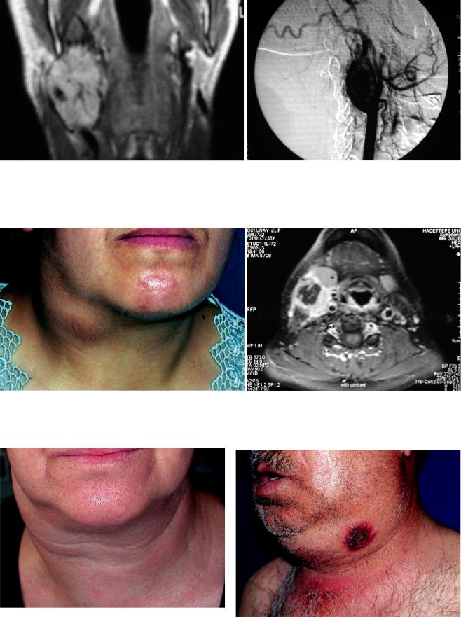
a |
b |
Fig. 3.10.9 (a, b) Carotid body tumor. Carotid body tumors arise from the carotid body located at the bifurcation of the internal carotid artery. Pulsation may be palpated and a bruit can be heard by stethoscope. The tumor can be moved in the horizontal plane but
not in the vertical plane. On MR examination, the highly vascularized tumor located between the external and internal carotid arteries confirms the diagnosis
a |
b |
Fig. 3.10.10 (a, b) Tuberculosis. Cervical lymph node enlargement due to tuberculosis is not common but is becoming more frequent. Due to the chronicity of the disease they may be confused with neoplasms, especially lymphomas. They are multiple and coalesce.
Pulmonary tuberculosis may associated with tuberculous lymphadenopathy. Node biopsy, if necessary for histological confirmation, should always be excisional. Incisional biopsy may result in fistula and chronic discharge
Fig. 3.10.11 Very large lymphadenopathy located at the right upper |
|
neck in a patient with lymphoma |
Fig. 3.10.12 Deep neck infection |

3.10 Neck Masses 167
Fig. 3.10.13 Anaplastic thyroid carcinoma (courtesy of Dr. Ş. Hosal) Fig. 3.10.14 Recurrent giant malignant fibrosarcoma on the left side of the face
K C DENN ATA O R H E S O N R A E

168 Chapter 3 |
Throat & Neck |
3.11 |
a |
Neck Malignancies
a
b
c
Fig. 3.11.1 (a) Peristomal recurrence that developed at the site of tracheotomy in a patient with laryngeal carcinoma with subglottic extension. (b) Recurrent laryngeal carcinoma with skin invasion causing respiratory obstruction
Fig. 3.11.2 (a) Recurrence of a malignant tumor that invaded the skin in the neck. (b) Specimen after excision of the lesion. (c) The defect in the neck after excision of the lesion. Common, internal, and external carotid arteries, vagus nerve, and hypoglossal nerve crossing these structures are seen