

Учебники / Diagnosis in Otorhinolaryngology Onerci 2009
.pdf2.3 Allergic Rhinitis
a
Fig. 2.3.9 Allergic shiners, dark discoloration of the lower lids
b
Fig. 2.3.8 (a, b) Giant papillary conjunctivitis, generally seen in patients using contact lenses. Tarsal conjunctiva is sensitized to the allergen adhered to the outer surface of the lens. Due to continuous irritation, the upper tarsal conjunctiva develops giant papillae. The cornea is never involved. Discontinuing lens use together with some topical antiallergic eye drops such as sodium cromoglycate helps the situation (courtesy of Kıratli)
Fig. 2.3.10 Stuffed animals with artificial fur are major dust reservoirs and should not be kept in the room of an allergic child
If either parents is atopic
33% of the offsprings is atopic
If both parents are atopic, 2/3 of the offsprings atopic
67
A E R
O N
E S
H T
O R
AT
K C E N D N A
Fig. 2.3.11 Inheritance of atopic diseases

68 |
Chapter 2 Nose |
Table 2.3.1 Pollen avoidance
Avoid going on picnics during the pollen season
Wear sunglasses outdoors
Stay inside in the late afternoona
Keep the windows closed in the late afternoona
Keep the windows closed in the car
Use pollen filters if possible
aPollensrisewiththeheatduringthedayandcomedownastheairstarts cooling during late afternoon. Therefore, in the late afternoon exposure is highest
Table 2.3.2 Dust mite allergen avoidancea
Indoors
Avoid humidity
Avoid warm environment
Ventilate adequately
Reducing the burden of allergens
Avoid wall-to-wall carpeting; tile floors with small rugs are preferable
Remove dust reservoirs such as stuffed animals with artificial fur, soft toys, woollen blankets, old mattresses, silk flowers, mounted animals, books on open shelves, feather pillows or bedding, upholstered furniture
Use pillow covers and mattress covers impermeable to dust mites
Removing the allergens
Superfiltering or HEPA filter vacuum cleaners preferable (at least twice a week)
Clean with a damp cloth every week
Wash the laundry with water hotter than 60°C every week; if possible expose to sunlight
aAgeneralunderstandingofallergyandtreatmentisimportant.Fulfilling these measures only partially may not be enough to prevent the symptoms. For example, killing the mites with chemicals may not eradicate the allergen. It may persist for months or even years. Since the mites live in deep pores and attach to the fabric, deep vacuum cleaning is needed to remove the allergen
2.4 Nasal Vestibulitis and Nasal Furunculosis and Mucormycosis
2.4
Nasal Vestibulitis and Nasal Furunculosis
and Mucormycosis
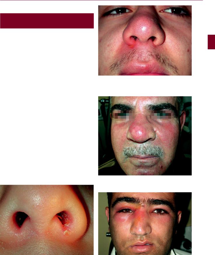
Infection of the skin of the nasal vestibule is termed nasal vestibulitis. It may be secondary to constant rhinorrhea, nose picking, or viral infections such as herpes simplex and herpes zoster. Foreign bodies frequently cause vestibulitis in children due to purulent discharge. Nasal furunculosis is Staphylococcus aureus infection of the hair follicles. Nose picking is a frequent cause of furunculosis. Topical and if necessary systemic antibiotics are prescribed. The patient should be instructed not to squeeze out pus from this area. Since the veins draining this area are valveless and directly join the cavernous sinus, there is a potential risk of spreading infection to the cavernous sinus via these facial veins. Eczema may also mimic vestibulitis. In these cases steroid base ointment may help the patient. In persistent vestibulitis, neoplastic diseases such as basal cell or squamous cell carcinoma should be kept in mind.
Fig. 2.4.2 If not treated, the infection in the nasal vestibule may spread to the upper lip. The upper lip on the right side appears hyperemic and edematous
Fig. 2.4.3 Furunculosis in the nasal vestibule with spread of the infection to the nasal tip and dorsum
69
A E R
O N
E S
H T
O R
AT
K C E N D N A
Fig. 2.4.1 Nasal vestibulitis on the left side. Note the slight edema and hyperemia as well as excoriation of the skin on the left side
Fig. 2.4.4 Infection starting as nasal vestibulitis with spread of infection to the nasal dorsum and right periorbital area

70 |
Chapter 2 Nose |
a
Fig. 2.4.6 Constant rhinorrhea and the need to wipe the nose due to allergic rhinitis have resulted in vestibulitis
b
Fig. 2.4.5 Venous drainage of the nose. (a) frontal view, (b) Lateral view. Since the veins draining this area are valveless and directly join the cavernous sinus, there is a potential risk of spreading infection to the cavernous sinus via these facial veins. This area of the nose is termed the danger triangle. Squeezing the pus from this area should be avoided
Fig. 2.4.7 Right alar rim. Basal cell carcinoma with slight hyperemia around it. In persistent vestibulitis, neoplastic diseases such as basal cell or squamous cell carcinoma should be kept in mind
Fig. 2.4.8 Hyperemia and edema in the columella and nasal tip mimicking severe vestibulitis secondary to squamous cell carcinoma infiltration. The neoplastic lesion is filling the left nasal passage

2.4 Nasal Vestibulitis and Nasal Furunculosis and Mucormycosis
a
|
Fig. 2.4.10 Mucormycosis in a child with leukemia. Gross tissue |
b |
necrosis with a black eschar is characteristic of mucormycosis |
|
a |
c
b
Fig. 2.4.9 Mucormycosis in a diabetic patient. Mucormycosis may infect different areas of the body, but the most frequent fatal form is the rhinocerebral form. (a) Necrotic areas on the face, (b) black necrotic areas in the nasal mucosa, (c) and after removal of necrotic area in the nose
Fig. 2.4.11 (a, b) Mucormycosis in an immunocompromised child. The disease progresses rapidly with extension of tissue necrosis out of the nose into the orbit and face. Local management requires wide debridement of necrotic tissue with a margin of normal-appearing tissue (Courtesy of TESAV)
71
A E R
O N
E S
H T
O R
AT
K C E N D N A

72 |
Chapter 2 Nose |
2.5
Sinusitis
According to the duration of the disease, sinusitis is divided into two categories: acute and chronic.
Acute rhinosinusitis (ARS) is defined as the sudden onset of symptoms lasting less than 12 weeks (with sypmtom free intervals-complete resolution of symptoms, if the problem is recurrent).ARScanoccuronceormorethanonceinadefined time period. This is usually expressed as episodes/year, but
a
b
there must be complete resolution of symptoms between episodes for it to constitute recurrent ARS.
Chronic rhinosinusitis (CRS) is defined as disease lasting more than 12 weeks without complete resolution of symptoms. CRS may also be subject to exacerbations.
The presence of polypoid degeneration in the maxillary sinus deserves special attention. If the polyp is on the floor of the antrum, dental disease should be suspected. If the polyp is on the roof of the antrum, carcinoma should be ruled out in elderly patients. Any evidence of bone erosion should raise the possibility of carcinoma. In patients over 40 years of age, the possibility of carcinoma should always be kept in mind and intrasinus exploration should be performed if necessary.
Fig. 2.5.2 Ostiomeatal complex area (OMC) (blue). The frontal, maxillary, and anterior ethmoidal cells drain to the OMC. It is a narrow area. Any edema may cause contact of the mucosal surfaces, which may lead to impaired mucociliary activity
Fig. 2.5.1 Developmental anatomy of the ethmoidal, frontal, and maxillary sinuses (a) and sphenoidal sinus (b). The ethmoidal and maxillary sinuses are present at birth. The sphenoidal sinus is in the form of a small invagination and develops later. The frontal sinus develops from the anterior ethmoidal cells and moves from an infraorbital position to a supraorbital position, starting to develop at the age of 7. Yellow, 6 months; red, 1 year; green, 3 years; blue, 8 years; maroon, 12 years of age, adult size

a |
c |
b
2.5 Sinusitis 73
A E R
O N
E S
H T
O R
AT
K C E N D N A
Fig. 2.5.3 (a) Maxillary sinus ostium from nasal side, (b) OMC lateral to the middle turbinate. Superior and posterior to the middle turbinate, the superior turbinate and sphenoid sinus ostium are seen;
(c) superior turbinate. Lateral to the superior turbinate are the posterior ethmoidal cells, and medial to the superior turbinate is the sphenoidal sinus ostium
Fig. 2.5.4 Maxillary sinus ostium from the maxillary sinus side. Mucociliary activity is toward the ostium

74 |
Chapter 2 Nose |
a
c
d
b
Fig. 2.5.5 Nasal discharge. (a) Mucoid drainage after common cold; (b) purulent drainage in the inferior meatus; (c) postnasal purulent drainage; (d) allergic mucin: viscid, thick yellow–green drainage, generally eosinophilic
a
b
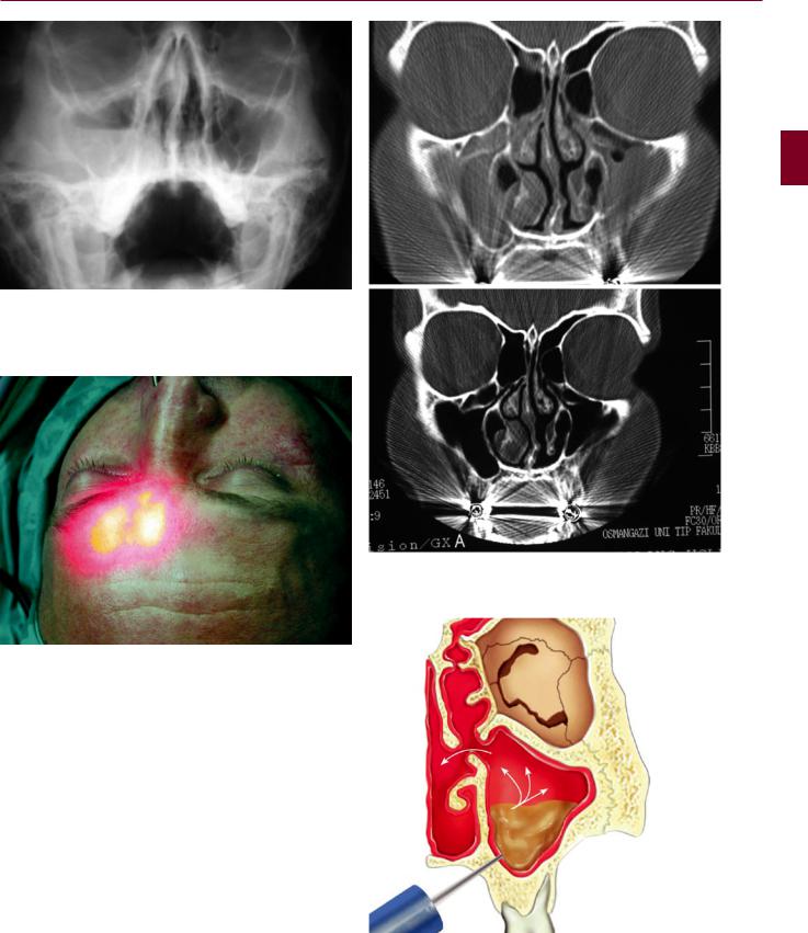
Fig. 2.5.6 Waters view showing right acute maxillary sinusitis. There is an air–fluid level in the right maxillary sinus
2.5 Sinusitis 75
A E R
O N
E S
H T
O R
AT
K C E N D N A
Fig. 2.5.8 (a) Coronal CT shows bilateral maxillary sinusitis; (b) 15 days after starting medical treatment the sinuses appear to be normal
Fig. 2.5.7 Transillumination of the frontal sinus. In frontal sinusitis the frontal sinus fails to transilluminate
Fig. 2.5.9 Maxillary sinus irrigation. An opening is created via the inferior meatus between the nose and maxillary sinus or via canine fossa

76 |
Chapter 2 Nose |
Fig. 2.5.10 The purpose of endoscopic sinus surgery is to restore ventilation and drainage of the paranasal sinuses. (a) Preoperative and (b) postoperative view
a
b
Fig. 2.5.11 (a) Epithelized sinuses and normal mucosa of the sinuses after endoscopic sinus surgery. (b) Coronal CT after surgery shows that all sinuses are clean and ostia are open
Fig. 2.5.12 Coronal paranasal sinus CT showing previously performed bilateral Caldwell-Luc operation. Note that both nasoantral windows in the inferior meatus are widely open