

Учебники / Diagnosis in Otorhinolaryngology Onerci 2009
.pdf3.3 Snoring 129
3.3
Snoring
Fig. 3.3.1 Snoring is the sound created by vibrations of the pharyngeal structures. Apnea is the cessation of breathing for more than 10 s. Sleep apnea is defined as more than five episodes of apnea per hour. Apnea may be central or obstructive. Central apnea is caused by a lack of drive in the central nervous system (CNS). The majority of apneas are obstructive. Some patients may have a mixed type of apnea. Since severe apnea may have serious cardiac and CNS complications, sleep apnea is a disease to be treated. The gold standard in the diagnosis of sleep apnea is polysomnography
Fig. 3.3.2 Hypertrophic tonsils may be the only cause of snoring and apnea. Tonsillectomy may solve the problem
a
b
Fig. 3.3.3 (a) Tongue base is at its normal position. (b) Tongue base can be displaced posteriorly, narrowing the oropharynx and causing snoring and apnea
K C DENN ATA O R H E S O N R A E

130 Chapter 3 Throat & Neck
a
b
Fig. 3.3.5 Postoperative appearance of a patient who has undergone laser-assisted uvulopalatoplasty (LAUP)
Fig. 3.3.4 Radiosurgery of the palate. (a) Radiosurgery produces fibrosis in the soft palate and increases the stiffness of the soft palate.
(b) Applications at the base of the uvula may cause edema of the uvula. (c) If too much energy is delivered at the same point it may cause perforation of the soft palate
Fig. 3.3.6 Schematic view of laser-assisted uvulopalatoplasty and classic uvulopalatopharyngoplasty (UPPP) with tonsillectomy

3.3 Snoring 131
Fig. 3.3.7 Uvulopalatal flap (courtesy of Çelikoyar). After the mucosa of the lingual surface of the uvula and some of the soft palate are stripped away, the uvula is reflected back toward the soft palate and fixed into its new position
Fig. 3.3.8 Appearance of soft palate after classic UPPP
Table 3.3.1 The questions to be asked in a patient with obstructive sleep apnea syndrome
Do you snore?
Does your family complain about your snoring?
Does your spouse say that your breathing stops at certain intervals?
Do you wake up during your sleep with air hunger?
Do you have morning headaches?
Do you feel tired in the morning when you wake up?
Do you have daytime somnolence?
Do you feel sleepy at work?
Do you fall asleep while watching TV?
Do you fall asleep while sitting?
Fig. 3.3.9 Hyoid suspension operation. The hyoid bone is fixated on the upper edge of the thyroid cartilage with a steel wire suture. This brings the tongue base anteriorly and caudally. The hyoid suspension operation is believed to prevent the hypopharyngeal collapse of the tongue muscles
Fig. 3.3.10 Genioglossus advancement technique. By advancing the genial tubercle and genioglossus muscle, the hypopharyngeal airway is enlarged and the collapse is prevented
K C DENN ATA O R H E S O N R A E

132 Chapter 3 Throat & Neck
3.4
Temporomandibular Joint
Temporomandibular joint (TMJ) problems are very common |
a |
inthepopulation.Patientsmaypresentwiththesymptomsof |
|
pain, joint noises, locking, and limited opening. Pain may be |
|
due to either the joint problem itself or the related muscles of |
|
mastication. Pain localized in front of the tragus is probably |
|
TMJ pain. Pain localized to the mandibular or temple area |
|
may be myofascial pain. Myofascial pain is more severe in |
|
the mornings because of the activities of the muscle during |
|
the night, such as clenching or grinding the teeth. Clicking is |
|
related to the anterior dislocation of the disc. Only clicking |
|
does not require any treatment. |
|
TheprimarymanagementofTMJdisordersisreassuranceof |
|
thepatientthatthereisnoseriousunderlyingcondition.Muscle |
|
relaxants,nonsteroidalanti-inflammatorydrugs,andheatappli- |
b |
cationmayimprovethesymptoms.Thepatientshouldbeevalu- |
|
ated by a dentist regarding the occlusion, and occlusal or ante- |
|
rior repositioning splints may be advised to be worn at night. |
|
Fig. 3.4.1 Cadaver dissection. Temporomandibular joint (TMJ) disc and condyle with attached superior pterygoid muscle. The disc of the TMJ is continuous with the superior pterygoid muscle fibers. Some of the fibers of the pterygoid muscle attach to the condyle as well. This allows the joint to work in a synchronous way
Fig. 3.4.3 (a) Schematic representation of TMJ anterior disc dislocation with reduction. The disc is normally placed between the condyle and the articular fossa. (b) MR examination; sagittal view of the TMJ. The disk is located anterior to the articular fossa
|
Fig. 3.4.4 Occlusal splints help to keep the jaws in normal |
Fig. 3.4.2 Normal occlusion is necessary for normal TMJ function |
occlusion |

3.4 Temporomandibular Joint 133
a
b
Fig. 3.4.5 (a) Surgical arthroscopy. (b) Appearance of the inside of the TMJ and avascular disc in white color
A E
R
O N E S
b HR
AAT O
ENN
D
K C
Fig. 3.4.6 (a) In TMJ ankylosis, opening of the mouth is limited. (b) On coronal CT, left-sided ankylosis of the TMJ is seen
Fig. 3.4.7 Arthroscopic view of TMJ anterior disc dislocation. Avascular white disc has escaped to the anterior recess and the posterior ligament lies between the condyle and the articular fossa

134 Chapter 3 Throat & Neck
3.5
Airway Obstructions
Table 3.5.1 Differential diagnosis in acute epiglottitis and acute laryngotracheobronchitis
|
Acute epiglottitis |
Acute laryngotra- |
|
|
cheobronchitis |
Pathogen |
Haemophilus influenzae |
Viruses |
|
type B |
|
Age group |
2–6 |
1–3 |
Incidence |
10% |
90% |
Onset |
Rapid (hours) |
Slow (days) |
General |
Severe systemic |
Satisfactory |
condition |
toxicity |
|
Cough |
Absent |
Barking cough |
Dysphagia |
Severe |
Absent |
Drooling |
Marked |
Absent |
Stridor |
Inspiratory |
Biphasic |
Fever |
>39°C |
<39°C |
Posture |
Sitting up |
Lying back |
Voice |
Muffled (not hoarse) |
Hoarse |
X-ray |
Thumbprint sign |
Steeple sign |
|
|
|
Spasmodic croup must be differentiated from croup. It is seen between the ages of 1–5 years. Its onset is rapid, starting usually in the evening. There is no associated infection. Humidity relieves the symptoms.
Laryngomalacia is the most common cause of stridor in infants and is the most common congenital laryngeal abnormality.Theepiglottisisomegashaped.Thearyepiglotticfoldsare tall, foreshortened, and thin and the arytenoids are large with redundant mucosa. The main theory of its cause is cartilaginous immaturity. The other theory is neuromuscular control abnormalities.Thediseaseisgenerallyself-limiting.However, in severe laryngomalacia surgical therapy may be necessary.
Croup is infection of the larynx resulting in airway obstruction and stridor. Stridor is the high-frequency sound created during inspiration and expiration. Narrowing of the trachea or the larynx produces inspiratory stridor. Bronchial narrowing produces expiratory stridor. Stridor is not a disease or a diagnosis but only a symptom. Under the term croup, acute epiglottitis,acutelaryngotracheobronchitis,andbacterialtracheitis can be mentioned. Acute laryngotracheobronchitis is the most common infection causing airway obstruction in children. Acute epiglottitis has a very rapid onset and is highly lethal if not diagnosed and treated immediately. Introduction of the HIB vaccine has reduced the incidence of acute epiglottitis by more than 90%. Clinical features of thedifferentialdiagnosisbetweenacuteepiglottitisandacute laryngotracheobronchitis can be seen in Table 3.5.1.
Bacterial tracheitis is generally seen from infancy to adulthood.ThemostcommonlyisolatedpathogenisStaphylococcusaureus.Theinitialclinicalcourseissimilartothatseenin croup.Thetrachealmucosaisedematousanddiffuselyulcerated causing tracheal narrowing. Thick purulent secretions partially obstruct the tracheal lumen. Thick purulent secretions in the trachea sometimes cause severe airway obstruction. When needed, endotracheal intubation and repeated suction of the secretions should be performed.
Fig. 3.5.1 Steeple sign in a child with acute laryngitis
Fig. 3.5.2 Neonates with webs present with respiratory obstruction. The degree of obstruction is related to the size of the web (courtesy of Dr. Unal)

3.5 Airway Obstructions 135
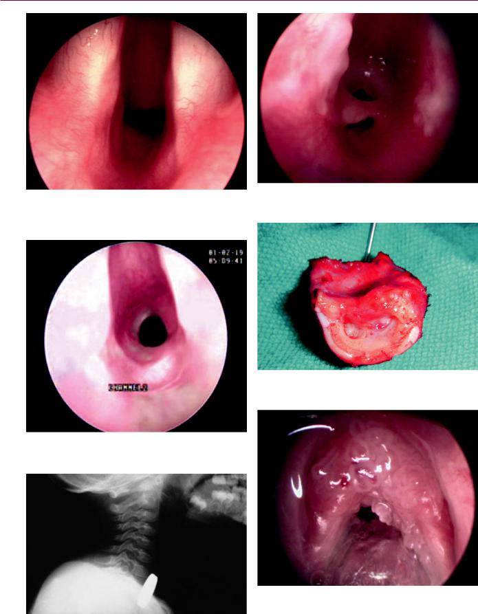
Fig. 3.5.3 Laryngeal web almost completely obstructing the airway. In severe respiratory distress, the airway must be secured either by intubation or tracheotomy. Thin webs can be opened by CO2 laser. For thicker webs open surgery may be needed (courtesy of Dr. Unal)
Fig. 3.5.4 Subglottic hemangiomas are the most common congenital tumors causing stridor. Subglottic and tracheal hemangiomas are generally asymptomatic at birth. Later they may become symptomatic. These tumors may regress spontaneously. In obstructing cases tracheotomy may be considered (courtesy of Dr. Unal)
a
b
Fig. 3.5.5 (a) Tracheal hemangioma before and (b) after steroid treatment. Steroid treatment for young patients under 1 year of age may be useful (courtesy of Dr. Unal)
K C DENN ATA O R H E S O N R A E
136 Chapter 3 Throat & Neck
Fig. 3.5.6 Congenital subglottic stenosis obstructing the airway. Subglottic stenosis may be congenital, but it is usually due to trauma of prolonged endotracheal intubation (courtesy of Dr. Unal)
Fig. 3.5.8 Subglottic stenosis due to prolonged intubation (courtesy of Dr. Unal)
Fig. 3.5.7 Early-stage subglottic stenosis due to prolonged intubation (courtesy of Dr. Unal)
Fig. 3.5.9 Tracheal resection due to subglottic stenosis (courtesy of Dr. Unal)
Fig. 3.5.10 A foreign body (a coin) in the esophagus (courtesy of Dr. Unal)
Fig. 3.5.11 Juvenile laryngeal papillomatosis is the most common benign tumor of the larynx. It is caused by the human papilloma virus. CO2 laser is of great help in the excision of papillomas. Many cases resolve by early adulthood

3.6 Hoarseness 137
3.6
Hoarseness
a |
b |
Fig. 3.6.1 (a) Vocal nodules are bilateral lesions at the vibratory |
is the main reason for nodules. Voice therapy is the main treatment |
surface of the vocal cords at the junction of the anterior one-third |
modality. With voice therapy, 80% of vocal nodules resolve. If voice |
and posterior two-thirds. This is the area of maximum trauma at |
therapy fails, vocal nodules may be excised by a direct microlaryn- |
higher pitch levels during shouting and singing. Misuse of the voice |
goscopic approach. (b) After excision of the nodules |
a b
K C DENN ATA O R H E S O N R A E
Fig. 3.6.2 (a) Right hemorrhagic vocal cord nodule at the junction of the anterior one-third and posterior two-thirds of the vocal cord.
(b) Cyst at the middle of the left vocal cord. Vocal cord cysts can
occur at any location on the vocal cord, and vocal abuse and gastroesophageal reflux play an important role. The cyst is evacuated through an incision without removing any mucosa

138 Chapter 3 Throat & Neck
Fig. 3.6.3 Vocal polyps move in |
a |
b |
and out during respiration. (a) |
|
|
Inspiration, (b) expiration (cour- |
|
|
tesy of Dr. Yılmaz) |
|
|
a |
a |
b |
b |
Fig. 3.6.4 (a) Right vocal cord polyp a few millimeters behind the anterior commissure. (b) After excision. Mucosa was preserved as much as possible and anterior commissure was not touched
Fig. 3.6.5 (a–c) Vocal cord polyps are usually single lesions which can occur anywhere on the vocal cord. The treatment is microlaryngoscopic removal of the polyps