

- •Preface
- •Acknowledgments
- •Contents
- •Contributors
- •1. Introduction
- •2. Evaluation of the Craniomaxillofacial Deformity Patient
- •3. Craniofacial Deformities: Review of Etiologies, Distribution, and Their Classification
- •4. Etiology of Skeletal Malocclusion
- •5. Etiology, Distribution, and Classification of Craniomaxillofacial Deformities: Traumatic Defects
- •6. Etiology, Distribution, and Classification of Craniomaxillofacial Deformities: Review of Nasal Deformities
- •7. Review of Benign Tumors of the Maxillofacial Region and Considerations for Bone Invasion
- •8. Oral Malignancies: Etiology, Distribution, and Basic Treatment Considerations
- •9. Craniomaxillofacial Bone Infections: Etiologies, Distributions, and Associated Defects
- •11. Craniomaxillofacial Bone Healing, Biomechanics, and Rigid Internal Fixation
- •12. Metal for Craniomaxillofacial Internal Fixation Implants and Its Physiological Implications
- •13. Bioresorbable Materials for Bone Fixation: Review of Biological Concepts and Mechanical Aspects
- •14. Advanced Bone Healing Concepts in Craniomaxillofacial Reconstructive and Corrective Bone Surgery
- •15. The ITI Dental Implant System
- •16. Localized Ridge Augmentation Using Guided Bone Regeneration in Deficient Implant Sites
- •17. The ITI Dental Implant System in Maxillofacial Applications
- •18. Maxillary Sinus Grafting and Osseointegration Surgery
- •19. Computerized Tomography and Its Use for Craniomaxillofacial Dental Implantology
- •20B. Atlas of Cases
- •21A. Prosthodontic Considerations in Dental Implant Restoration
- •21B. Overdenture Case Reports
- •22. AO/ASIF Mandibular Hardware
- •23. Aesthetic Considerations in Reconstructive and Corrective Craniomaxillofacial Bone Surgery
- •24. Considerations for Reconstruction of the Head and Neck Oncologic Patient
- •25. Autogenous Bone Grafts in Maxillofacial Reconstruction
- •26. Current Practice and Future Trends in Craniomaxillofacial Reconstructive and Corrective Microvascular Bone Surgery
- •27. Considerations in the Fixation of Bone Grafts for the Reconstruction of Mandibular Continuity Defects
- •28. Indications and Technical Considerations of Different Fibula Grafts
- •29. Soft Tissue Flaps for Coverage of Craniomaxillofacial Osseous Continuity Defects with or Without Bone Graft and Rigid Fixation
- •30. Mandibular Condyle Reconstruction with Free Costochondral Grafting
- •31. Microsurgical Reconstruction of Large Defects of the Maxilla, Midface, and Cranial Base
- •32. Condylar Prosthesis for the Replacement of the Mandibular Condyle
- •33. Problems Related to Mandibular Condylar Prosthesis
- •34. Reconstruction of Defects of the Mandibular Angle
- •35. Mandibular Body Reconstruction
- •36. Marginal Mandibulectomy
- •37. Reconstruction of Extensive Anterior Defects of the Mandible
- •38. Radiation Therapy and Considerations for Internal Fixation Devices
- •39. Management of Posttraumatic Osteomyelitis of the Mandible
- •40. Bilateral Maxillary Defects: THORP Plate Reconstruction with Removable Prosthesis
- •41. AO/ASIF Craniofacial Fixation System Hardware
- •43. Orbital Reconstruction
- •44. Nasal Reconstruction Using Bone Grafts and Rigid Internal Fixation
- •46. Orthognathic Examination
- •47. Considerations in Planning for Bimaxillary Surgery and the Implications of Rigid Internal Fixation
- •48. Reconstruction of Cleft Lip and Palate Osseous Defects and Deformities
- •49. Maxillary Osteotomies and Considerations for Rigid Internal Fixation
- •50. Mandibular Osteotomies and Considerations for Rigid Internal Fixation
- •51. Genioplasty Techniques and Considerations for Rigid Internal Fixation
- •52. Long-Term Stability of Maxillary and Mandibular Osteotomies with Rigid Internal Fixation
- •53. Le Fort II and Le Fort III Osteotomies for Midface Reconstruction and Considerations for Internal Fixation
- •54. Craniofacial Deformities: Introduction and Principles of Management
- •55. The Effects of Plate and Screw Fixation on the Growing Craniofacial Skeleton
- •56. Calvarial Bone Graft Harvesting Techniques: Considerations for Their Use with Rigid Fixation Techniques in the Craniomaxillofacial Region
- •57. Crouzon Syndrome: Basic Dysmorphology and Staging of Reconstruction
- •58. Hemifacial Microsomia
- •59. Orbital Hypertelorism: Surgical Management
- •60. Surgical Correction of the Apert Craniofacial Deformities
- •Index
48
Reconstruction of Cleft Lip and Palate Osseous Defects and Deformities
Klaus Honigmann and Adrian Sugar
Embryological Development
Primary Embryological Palate (Lip, Alveolus)
Embryologically, a cleft is a non-union of facial growth centers, these being ridges and tubercles conditioned by the growth of mesenchyme.1,2 At the end of the first month of pregnancy, by the activity of mesodermal cells, the medial nasal tubercle and maxillary tubercle join each other forming the primary embryological palate. This provides the base for the upper lip and the premaxilla. A more recent view suggests that the material for the premaxilla does not come down from the frontal tubercle but comes forward from the base of the skull.3 In the middle of the second month of pregnancy, fusion takes place between the premaxillary and maxillary centers.4 Non-union between the premaxilla and the maxillary alveolar process leads to a unior bilateral alveolar cleft.
Secondary Embryological Palate (Hard and
Soft Palate)
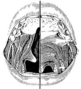
By the end of the second month of pregnancy, the mandible has grown so far that the tongue now finds enough space and can descend to the mandibular level. In consequence the palatal tubercles, at first positioned lateral to the tongue, can rise up, turn medially, and join each other and the downwardgrowing vomer in the midline. Non-union of the palatal shelves and the vomer results in a palatal cleft. A palatal cleft as a part of cleft lip and palate may be unior bilateral. In an isolated cleft of hard and soft palate, the palatal cleft is always bilateral.
Incidence and Etiology
The incidence of clefting in Europeans varies according to source, from 1 cleft child in 500 newborn,5 1 in 530,6 1 in 580,7 to 1 in 630.8 In the Japanese, the incidence varies from 1 in 3709 to 1 in 470,10 and in white Americans, from 1 in
530 in Washington,11 1 in 750 in Pennsylvania, to 1 in 1050 in Philadelphia.12 More rare are clefts in the African population, 1 in 2400,10 and in the black American population, 1 in 3300 to 1 in 4400.12
The etiology of cleft lip and palate is explained by a majority of authors as a multifactorial genetic system with additive polygenia and threshold effect.13,14 This means that the combination of defects in different genes lowers the threshold for a negative influence of environmental factors. As a pathologic principle, these environmental factors cause a deficiency in oxygen supply to the fetus15,16 just at the time when the facial tubercles are transformed and close the fetal cleft.
Classification and Diagnosis
Davis and Ritchie17 introduced an anatomic classification with group 1 for prealveolar clefts, group 2 for postalveolar clefts, and group 3 for complete preand postalveolar clefts. The cleft extension in these anatomic sections is added in thirds (1/3, 2/3, 3/3). Veau18 classified into four groups: group A represents clefts of the soft palate, group B clefts of the hard palate, group C complete unilateral clefts of lip, alveolus, and palate, and group D complete bilateral clefts of lip, alveolus, and palate. Based on practical considerations, FoghAndersen19 divided clefts into three groups: group 1 is for cleft lip, group 2 for cleft lip and palate, and group 3 for cleft palate. The Cleft Lip and Palate Subcommittee of the International Confederation of Plastic Surgical Societies supplements this with a group 4 for rare clefts (median, oblique, horizontal, and other very rare clefts).
Kernahan and Stark20 approach classification from the view of embryological development. They differentiate group 1 for clefts anterior to the incisive foramen (that is, clefts of the primary embryological palate, the anatomic sections of lip and premaxilla), group 2 for clefts posterior to the incisive foramen (that is, clefts of the secondary embryological palate, the anatomic sections of hard and soft palate), and group 3 for clefts that are both anterior and posterior to the incisive fora-
539
540
men (that is, clefts of the primary and secondary embryological palate, the anatomic sections of lip, alveolus, and hard and soft palate). This latter classification has been adopted by the World Health Organization into the International Classification of Diseases.
For clinical practice, Koch21 proposed division into the four anatomic sections, that is, lip, alveolus, hard palate, and soft palate, and the subdivision of these sections into thirds. He uses the initials of the sections and adds the number of the third to which the cleft extends. Arabic numbers stand for the open cleft parts and Roman numbers for the submucous cleft parts. For visualization of cleft types, Pfeifer22 introduced a diagram of primary and secondary palate, the cleft extension being given by outfilling of the respective fields. A 90° rotation of this symbol allows the diagnosis to be written. Kernahan23 permits visualization with a striped Y, modifications of which have been proposed.24–26 For use on a computer, Kriens27 introduced LAHSHAL. Honigmann28 uses the initials of the four sections and adds the number of the third to which the cleft extends. Reflecting the x-ray situation, this method begins with the right side of the lip and ends with the left side. As an example, the diagnosis of a complete rightsided unilateral cleft is written as L3 A3 H3 S3 and that of a complete bilateral cleft L3 A3 H3 S3 H3 A3 L3. Submucous parts of the cleft are written by a Roman number; for example, HI SIII1 is the short diagnosis of a submucous cleft in the posterior third of the hard palate and in the complete soft palate with a bifid uvula. Unfortunately, there is no agreement among these classifications, making comparison of cases between the different centers difficult.
Dysmorphology
Nonseparation of Nasal and Oral Cavity
An alveolar as well as a palatal cleft means no separation of the nasal and oral cavities. A histological examination of the border between these two cavities shows a transition zone of squamous cell epithelium in the mouth and pseudostratified ciliated columnar epithelium in the nose.
Movable Premaxilla in Bilateral Alveolar Cleft
In bilateral alveolar clefts, the premaxilla is fixed only at the nasal septum and the vomer. No osseous connection between the premaxilla and the maxilla exists. As a result, the premaxilla is movable: it can protrude, swing, or descend and end in a malposition.
Variations of Cleft-Adjacent Teeth
The lateral incisor on the cleft side is usually harmed. This can be manifested by an absence of the tooth germ or a malformed tooth. It may be found medial or lateral to the cleft and two lateral incisors may even be present, each on one side
K. Honigmann and A. Sugar
of the cleft. In the second dentition, the canine and central incisor roots may be immediately adjacent to the cleft and the central incisor is commonly rotated and may be ectopic.
Support of Alar
The alveolar crest normally gives support to the overlying soft tissues. In an alveolar cleft, the support for the overlying alar of the nose is lacking, and the alar base tends to drop into the cleft. This contributes to the nasal asymmetry.
A Cleft Palate Is Also a Nasal Malformation
In every case, a palatal cleft means a nasal malformation too. In bilateral cleft palate, the vomer is hypoplastic and both nasal meati are open. In unilateral cleft palate, the vomer deviates to the closed side and one nasal meatus is open.
Is a Cleft a Deficiency?
We are used to considering this birth defect as involving a lack of tissue. Kriens29 has analyzed plaster models of 251 untreated cleft infants and, especially in 91 of them with a complete unilateral cleft lip and palate, he has found no actual deficiency of tissue. His three-dimensional reflex microscope measurements explain the visible clinical cleft as a displacement and a distortion of the bony cleft segments caused by volume, tonus, and action of the tongue and its imbalance with the action of the facial soft tissues. Thus, there is some reason to question the presence of an actual tissue deficiency, at least in the alveolar cleft.
Functional Consequences
To regard a cleft as a deformity only is inadequate. This malformation causes a number of functional disturbances that indispensably should be taken into consideration in determining appropriate treatment.
Nutrition Difficulties
Nutrition in cleft infants has often been described as difficult because of their incompetence in sucking.30–32 In reality, babies need to suck only to position the nipple, and for drinking they “milk” the nipple with their tongue.33 Nutrition difficulties are caused by the tongue position in the cleft palate, occluding the nasal airway.34,35 Therefore, cleft babies are unable to drink and breathe at the same time.
A palatal obturator, once introduced as an orthopedic device for bringing the maxillary segments into a better position36 and for steering growth of maxillary segments,37,38 improves nutrition.35,39–41 With its separation of nasal and oral cavities, it brings the tongue out of the nasal airway and into a more anterior position in which the tongue can reach the nipple.34 Based on this knowledge, breast-feeding with all its advantages, especially in cleft infants,42–44 becomes
48. CLP Osseous Defects and Deformities |
541 |
possible and should be recommended to all who desire to do so.
Hearing Disorders
By swallowing, the Eustachian tubes are opened and the secretions of the middle ear can flow off. In the presence of a cleft palate, however, the velopharyngeal ring muscle system is interrupted45 and the insertion of the tensor veli palatini muscles at the tube cartilage is abnormal.46,47 In consequence, the opening mechanism of the Eustachian tubes cannot work. The middle ear secretions become thickened and are congested. The resulting seromucotympanon48 hinders sound conduction and promotes middle ear infections. Speech development relies on hearing49,50 and in cleft infants, who usually suffer some speech problems anyway, defective hearing will mean a double handicap. Moreover, in hearing disorders the maturation of the hearing tracts to the central nervous system is retarded.51
The management of these problems depends on careful otologic examination of the tympanic membrane with the microscope. In the presence of retraction of the ear drum, paracentesis with suction (myringotomy) of any middle ear secretions is indicated. If seromucous or putrid secretions are present, a tube (gromet) should be inserted. An active approach to early middle ear drainage, as well as closure of the soft palate during the first year of life, is desirable to reduce the incidence of middle ear disease in cleft children.52,53
Speech Problems
Nasality, articulation problems, suprapalatal resonance, voice diseases, myofunctional imbalances, and mimic movements may all be manifested in the speech problems of cleft patients. Nasality is the result of velopharyngeal incompetence caused by shortness or inadequate activity of the soft palate, which itself is based on the displaced muscle insertions (Figure 48.1). Articulation problems originate from a posterior tongue position but malposition of teeth and gaps in the dental arches may contribute.
An increase in suprapalatal resonance tends to occur because of a wider nasopharynx, which is caused by interruption of the velopharyngeal ring muscle system by the cleft of the soft palate. Measurement of the distance between the pterygoid processes54 and its expression in the pterygomandibular index and the pterygoid abduction angle55 provide evidence for this. Vomerian hypoplasia and unreconstructed nasal meati contribute to the nasopharyngeal width and thus to the increased suprapalatal resonance.
Voice diseases may occur accidentally or following attempts to compensate for speech disturbances. Myofunctional imbalances are the result of the displaced tongue with its dislocated functional pressure and the dysharmonious interaction of tongue and orofacial musculature.56 Mimic movements are the attempt by cleft patients to compensate for velopharyn-
FIGURE 48.1 Shortening of the soft palate by displaced muscle insertions in a cleft.
geal incompetence by grimace with labial, nasal, or frontal muscles. Speech therapy actually starts with breast-feeding, which demands considerable effort by the child and provides practice for the musculature. Later on, stimulation and myofunctional therapy create favorable general conditions for speech development.57,58 Treatment of articulation problems comes at the end of the speech therapy program. In this way, it is hoped that normal colloquial speech will be achieved by the time of school entry.
Growth Disturbances
Maxillary hypoplasia59 and scarring from surgery40,59–62 are said to be responsible for growth disturbances. Often the influence of muscle action on growth of bony structures is not considered, although it plays an important role.63,64 This was recognized in 1961 by Rosenthal,65 who described the functional stimulation on the clefted maxilla by early reconstruction of soft palate muscles. In our experience, reduction of scarring after palatal repair is possible if the creation of a dead space between the palatal flaps and the underlying bone surface can be avoided. This can be achieved by a palatal dressing applied at the end of the operation66,67 and the exact reconstruction of the nasal meati with intact epithelial layers on both sides of the cleft.
Orthodontic supervision, modern methods of orthodontic treatment, and continuous follow-up can help to reduce the incidence of major incongruences of the maxilla and mandible. If it should become necessary, operative correction of dysgnathia can be carried out without major problems, as we describe later. However, the significance of growth dis-
542
turbances today is much less in comparison to the other problems associated with a cleft (such as speech problems, hearing disorders, and psychological disturbance) in those areas where good primary surgery and careful multidisciplinary fol- low-up are the norm.
Psychological Problems
A number of studies68–70,72 have examined the influence that a cleft lip and/or palate has on the affected children themselves, their families, and the general public including health care professionals. It seems clear that children with repaired clefts tend to perceive themselves as being less socially adept and more frequently sad and angry than their peers. On the whole, they tend to identify their problems as relating both to their facial deformity and to difficulties with and abnormalities of speech. This poor self-concept has the potential for influencing performance in school, but not all such children are affected.
As Lansdown71 put it, it is not that such children are likely to be neurotic or delinquent. Their problems are however likely to lead to increased psychological strain. For them life is just that little bit harder, and those of us looking after such children may help by being constantly aware of this and offering support to children and families when it is needed.
Alveolar Cleft Defect Bone Grafting
Wolff73 and Veau74 considered that closure of a cleft lip and palate should involve repair and reconstruction of each of the clefted and abnormal tissues/layers corresponding to normal anatomy. They did not realize this aim themselves. Wassmund75 in particular emphasized the importance of achieving the complete closure of the alveolar cleft with a nasal and an oral layer. The requirements of Wolff and Veau however were not fulfilled until Schmid76 in 1951 proposed and carried out bone grafting into the alveolar cleft. Since then a number of surgeons have initiated different methods of alveolar cleft bone grafting, including Nordin and Johansson,77 Schrudde and Stellmach,78 and Schuchardt and Pfeifer,79 as well as Boyne and Sands.80
Aims of Bone Grafting
Continuous Maxillary Alveolus
At one time, bone grafting tended to be performed only in bilateral alveolar clefts to fix a mobile premaxilla to the maxilla. This reason remains valid.81 It has however now been extended to include the achievement of a continuous alveolus in all cleft patients with an alveolar defect. For those patients who will subsequently need a maxillary osteotomy, it creates the possibility of that procedure being carried out more safely, and perhaps with more stability, in one piece.
K. Honigmann and A. Sugar
Tooth Eruption and Support, Orthodontic
Alignment, and Prosthetics
In the overwhelming majority of cases, a cleft alveolar osseous defect prevents the normal eruption of teeth adjacent to the cleft. It is therefore one of the principal aims of alveolar bone grafting to enable the eruption of those teeth.82–84 Such a tooth passing through the graft brings with it its own supporting structures including periodontal membrane and bone, and this is applicable to both dentitions.85 Not only should an appropriately timed alveolar graft allow teeth to erupt through it, but it should also give improved bony support to other teeth adjacent to the graft, even those which have already erupted. This makes possible what would otherwise be quite difficult orthodontic movements, for example, derotation of incisors or orthodontic alignment of palatally erupted canines,86 without compromising their support.
The permanent maxillary canine on the side of the cleft is foremost among the teeth concerned.86 Successful bone grafting carries with it the potential for allowing the eruption of this tooth in such a way that there is a continuous dental arch.81,87 This may avoid the need for prosthetic rehabilitation. When gaps persist in the maxillary dental arch in the cleft area (either because of hypodontia, failure, or absence of a graft), a graft when inserted should give good support to dental replacements such as a denture or fixed bridge. If it has sufficient volume, it may also make prosthetic rehabilitation possible with titanium implants supporting a crown or fixed bridge in the cleft area.83 If placed after the maxillary segments have been expanded and moved in other directions, a graft in the alveolar defect(s) will to a considerable degree give support to that orthopedic movement.
Support for Alar Base
The grafted alveolar cleft gives support to the base of the alar on the cleft side.81,83 Although not a substitute for correction of the cleft nasal deformity, by correcting the skeletal dysplasia it brings the patient closer to normality.
Closure of Oronasal Fistulae
The closure of oronasal fistulae demands the recreation of the original anatomy and tension-free repair of a nasal and oral layer. The presence of a graft between those layers not only restores normal anatomy but assists successful fistula clo-
sure.81,83,88
General Considerations
Preoperative Orthopedics
The pioneer in the field of preoperative orthopedics in cleft infants is McNeil.36,89 The stated aims of such treatment are arch alignment with reduction of cleft width before the first
48. CLP Osseous Defects and Deformities
surgical intervention, with postoperative retention and stimulation of bone formation through functional impulses. Among the many followers of McNeil’s concept, Hotz and GrafPinthus90,91 consider the tongue as a factor of essential influence on the cleft width. For them, the crucial effect of the plate is in keeping the tongue away from the cleft. Up to the present in bilateral clefts, the principal argument for preoperative orthopedics is the movement of the premaxilla and the prolabium to a more favorable position for surgical re- pair.92–96 A variety of techniques has been proposed, including extraoral traction97–100 and oral pinning with trac-
tion.101–103
Although most clinical reports defend the advantages of presurgical orthopedics, there is reason to doubt whether neonatal orthopedics is worth undertaking in the majority of cases104 and certainly to question whether it achieves what is claimed. These questions will not be answered satisfactorily until sufficiently large longitudinal studies based on serial dental casts, lateral cephalometric, and photographic records beyond the postpubertal growth period have been published.105
In our unit, Honigmann’s107 experience until the mid-1980s had been with preoperative orthopedics in most of the bilateral and some unilateral clefts. Subsequently, this inconvenience for the babies, especially in the treatment with extraoral devices, was abandoned. Today the policy in Basel is to trust in the effect of the repaired labial muscles to approximate the maxillary segments.106 We have never seen a dehiscence of the repaired lip, even in wide bilateral clefts with extreme protrusion of the premaxilla, nor have the aesthetic results appeared worsened.
General Conditions
The general condition of the patient should be as good as possible before surgical repair is carried out. Local infections, general infections, and vascular and metabolic diseases increase the risk of failures and should be treated before the operation. In the presence of infection, surgery should be postponed. A prophylactic perioperative antibiotic regimen seems to be helpful. Nevertheless, in both primary and secondary cleft repair we apply antibiotics only when the intervention includes bone grafting and/or osteotomy.
Nomenclature
The nomenclature of alveolar bone grafting with regard to timing is quite confusing. The terms ‘primary,’ ‘secondary,’ and ‘tertiary’ osteoplasty are in use, as well as ‘early’ and ‘late’ as a supplement. Some authors are referring to dental age, others to a surgically operated or unoperated alveolar cleft.
Honigmann proposed in 1992 an alternative nomenclature.107 Accepting the terms “plasty” for primary repair and “correction” for secondary repair, one can call a first alveo-
543
lar bone grafting an ‘alveolo-osteoplasty’ and a repeated grafting an ‘alveolo-osteocorrection.’ By adding the patient’s age at operation, a clear basis for comparison with other centers is possible.
At the congress of the German Association for Oral and Maxillofacial Surgery in 1992 at Munich, a nomenclature commission recommended the following terms:
Primary osteoplasty: bone grafting during the first dentition, independently of a oneor two-stage intervention
Secondary osteoplasty: bone grafting during the mixed dentition
Tertiary osteoplasty: bone grafting after the end of the second dentition.108,109
Operating Technique
Recipient Site
The condition of the recipient site is far more important than the type of graft material used. Criteria for the quality of the recipient site are blood supply and complete coverage of the graft by soft tissue.
For a good blood supply, the recipient site should ideally be free of scars. This situation exists only in a surgically unoperated cleft. Delayed closure of the alveolar cleft after previous repair of the lip or the hard palate certainly involves dissection in a scarred area. The graft should have close contact to surrounding tissues, which means to the alveolar process stumps as well as to the soft tissue cover. Dead space around the grafted material will fill with hematoma, which through its organization by connective tissue leads to a thicker scar.
Complete coverage of the graft by soft tissue is an important factor for achieving a good blood supply and is essential for the protection of the graft against infection. Infection produces necrosis and with it graft failure. The ideal soft tissue cover is that which corresponds completely to the normal anatomy of this area. The nasal mucosa should be horizontal (axial) without any transposed oral mucosa. The palatal mucosa should be adjacent to the alveolus at its palatal inclination, and the buccal mucosa should similarly lie on the inclination of the buccal alveolus. Over the alveolar crest, there should be fixed gingivae, with mobile mucosa away from the crest.
Bone Grafts
A wide variety of materials has been proposed for alveolar grafting including homologous bone, allogeneic freeze-dried bone marrow, homologous cartilage, and various bone substitutes. Good, or at least satisfactory, results with these materials have been claimed. However, there is widespread acceptance that autologous bone grafts have the lowest risk in primary healing and give the best results.110,111 We therefore limit ourselves to considering them only.
544
Different donor sites for autologous bone grafts have been proposed and used including anterior and posterior iliac crest,112,113 rib,113 mandible,114–117 calvarium,82,118 tibia,83 periosteal flaps,119–121 and periosteal grafts.120,122,123 The decision in favor of one or other of the different donor sites depends among other things on the age of the patient at operation, that is, the quantity of cancellous bone at the different donor sites in different ages. In an optimal recipient site, one can obtain good results with every graft, although autologous cancellous bone is the most proven successful graft. With it one can fill out the defect completely. It allows vessels to grow into the graft from the recipient site and to transform the graft into the locally adapted bone in the easiest and most rapid way. Moreover, cancellous bone has the highest resistance against infection.
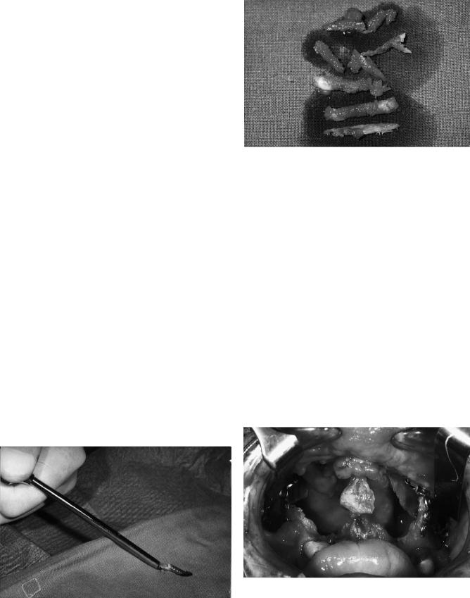
In patients older than 2 years, cancellous bone can be harvested from the iliac crest with the help of a trocar (Figures 48.2 and 48.3). This procedure diminishes the extension of the secondary intervention and the pain at the donor site, and the resultinggraftiscompressed. Alternatively, andespecially when large quantities are required, the iliac crest itself can be raised as an osteoplastic flap, cancellous bone chips removed, and the lid replaced. The key to prevention of postoperative morbidity at this site is the avoidance of any muscle stripping in particular on the lateral aspect of the crest and the use of a longacting local anesthetic agent (e.g., bupivicaine) titrated over 24 hours postoperatively into the wound via an epidural cannula.
Adequate stability is always important especially in bilateral alveolar clefts. During the first postoperative weeks, bone grafting cannot abolish the mobility of the premaxilla. Indeed, mobility of the fragments may well prevent bone union between the fragments and across the cleft(s). Some form of fixation of the fragments is needed, for example, by external devices such as dentally fixed splints or arch wires. Internal fixation methods such as plates and screws can be applied in a simultaneous osteotomy of the premaxilla or in a secondary intervention with the need for bigger grafts (Figures
K. Honigmann and A. Sugar
FIGURE 48.3 Bone harvested from the iliac crest by trocar.
48.4–48.6). Some authors describe a simultaneous palatoosteoplasty. Their intention is to reconstruct all the layers corresponding to the normal anatomic situation. We have no personal experience with this procedure because we cannot see the functional need.
The Basel Approach
In 1983, Honigmann described a method that had been adopted in 1980.124 This technique involved closure of the soft palate and the lip in one stage in uniand bilateral complete clefts at the age of 6 months. The alveolar and the hard palate cleft were closed in a second intervention at the age of 3 to 5 years with bone grafting into the alveolar cleft. The bone graft was harvested from the iliac crest, and from 1985 onward using a trocar. In some cases the bone chips were mixed with a granulate of tricalcium phosphate.125 The aims of the timing were to construct the labial and velar muscle systems as soon as possible for optimal functional development, to re-
FIGURE 48.2 Bone collection from the iliac crest by trocar.
FIGURE 48.4 A bilateral cleft lip and palate with a big bone defect; status after Le Fort I osteotomy and fixation with 2.0-mm plates and screws.
48. CLP Osseous Defects and Deformities
FIGURE 48.5 Same patient as in Figure 48.4 grafted with corticocancellous bone from the iliac crest; fixation with 2.0-mm plates and screws.
FIGURE 48.6 Same patient as in Figure 48.6; oral cover of the graft with a tongue flap.
545
duce the number of interventions for primary cleft repair, and to enable the children to enter school with a completely closed cleft and normal colloquial speech. The failure rate in bone grafting at that time was 11.3%. Normal colloquial speech at school entrance was achieved in 91.6% of the children.28
In 1991, this concept was changed with the aim of obtaining a completely closed cleft at the end of the first year of life for a better functional and psychological development of the cleft child. Based on the aim of reducing the number of surgical interventions and thus hospitalizations, an attempt was made to try to close all forms of clefts in one stage at least by the age of 6 months. Because of modern methods of pediatric anaesthesia, there were no significant problems even in a 4-hour operation, which was needed in complete bilateral clefts. Subsequently it was found that this all-in-one procedure for unilateral cleft lip and palate patients had been proposed in 1966.126 The late results of that work were reported at the 7th International Congress on Cleft Palate and Related Craniofacial Anomalies in 1993 at Broadbeach, Australia.127 The operative steps in detail are as follows.
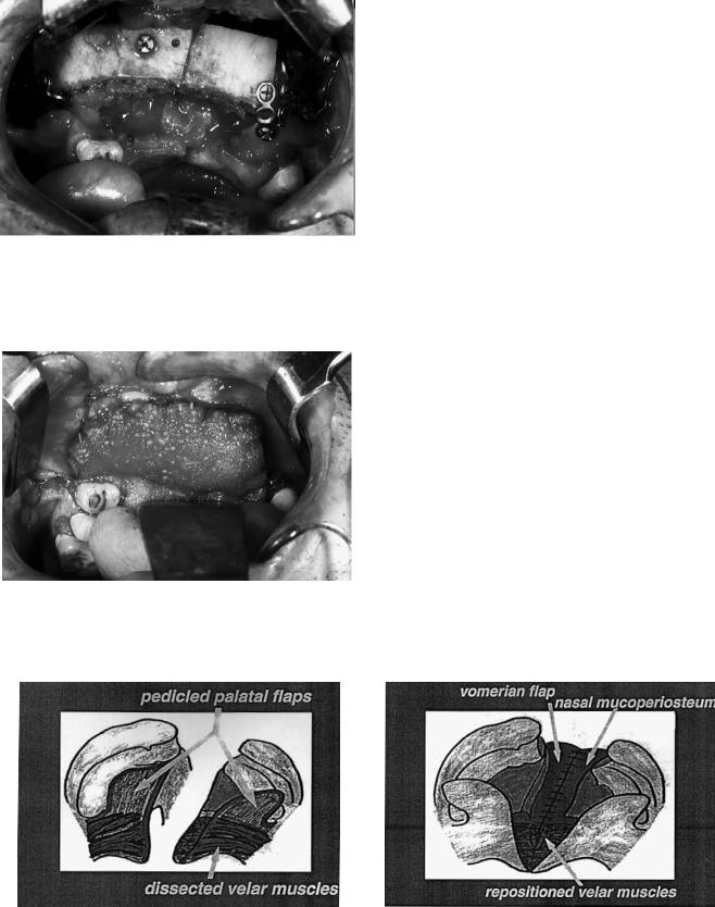
The child’s head is placed in the ‘Rose’ position, that is, the surgeon is seated with the child’s head on his/her knees. The mouth is opened by a Rosenthal retractor (the widely used Dingman retractor covers the lip and the alveolar cleft with its extraoral frame, so it is impossible to get the view needed for the alveolo-osteoplasty). The incision of the soft palate edges continues with the dissection of pedicled palatal flaps including the preparation and mobilization of the palatal vessels (Figure 48.7). This provides a good view for the intravelar muscle dissection. With the aid of mucoperiosteal vomerine flaps and the mobilized lateral nasal mucoperiosteum, the nasal meatus can be formed in the complete alveolar and palatal cleft (Figure 48.8), and in bilateral clefts the two nasal meati can be separated (Figure 48.9).
Suture of the mobilized and posteriorly directed soft palate muscle stumps and pushback of the totally mobile palatal soft
FIGURE 48.7 Dissection of the soft palate muscles and the pedicled palatal flaps.
FIGURE 48.8 Formation of the nasal meatus in the unilateral alveolar and palatal cleft.

546
FIGURE 48.9 Separation of the two nasal meati in a bilateral cleft.
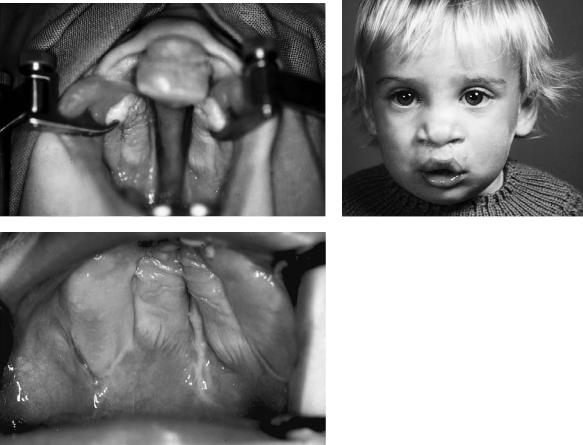
tissues lengthens the soft palate into a normalized anatomic situation (see Figure 48.1). The palatal flaps are sutured only in the midline and then lightly pressed against the palatal bone with the aid of a palatal dressing. Thus a dead space between the palatal bone and soft tissues can be avoided, and with it a hematoma and the resulting thicker scar. After repositioning the child onto the table, a rib bone graft is harvested (Figure 48.10) and the alveolar cleft(s) filled with the cancellous bone (Figure 48.11). Integrated into the final lip repair is cover of the bone graft by mucoperiosteum advanced from the vestibular side of the lesser maxillary segment and its suturing with the tips of the palatal flaps.
In this manner, alveolar bone grafting is a part of an all- in-one closure of all clefts. More than 80 complete uniand bilateral clefts have been closed in this all-in-one procedure (case 1: Figure 48.12 and case 2: Figure 48.13). At this time, the rate of healing complications is 5.9% (3 partial hard palate dehiscences, 2 bone graft losses), and the first functional results with regard to speech development and hearing disorders are very encouraging.
FIGURE 48.10 Rib graft resection.
K. Honigmann and A. Sugar
FIGURE 48.11 Primary alveolar cleft bone grafting.
The Swansea Approach
By contrast, Sugar’s approach to alveolar bone grafting in Swansea (and until 1994 in Chepstow) has been unchanged since 1985. Grafting has been carried out ideally in the mixed dentition shortly before the eruption of the permanent maxillary canine teeth, the classic secondary graft. This approach has varied little from the method proposed by Boyne and Sands80 and reported by Abyholm and colleagues.81 However, in our patients, operating on children whose primary surgery has been carried out by a number of surgeons, there has been a clear need for a significant amount of orthodontics, primarily to correct collapsed or misplaced alveolar segments, before grafting can take place. Only cancellous bone harvested from the anterior iliac crest has been used and with consistently good results.
During this period, a significant number of cleft patients presented who had, for various reasons, missed the opportunity of receiving a graft into their alveolar clefts during the mixed dentition phase. In most cases these have been managed with careful orthodontic preparation with fixed bands and tertiary alveolar grafting in exactly the same way as mentioned.128 This has applied equally to those patients who have not required orthognathic surgery, the graft not only facilitating closure of fistulae but also giving support to dental restorations with or without osseointegrated implants. Whenever grafting is carried out during orthodontic therapy, the orthodontist places in advance either lateral retaining arms from molar bands or rigid arch wires to maintain arch width. This is usually reinforced by a transpalatal bar, positioned sufficiently far posteriorly and relieved from the mucosa to enable any required palatal surgery to be performed.
In all cases the complete alveolar cleft is identified. Any labial fistula is excised and this excision incorporated into the mucoperiosteal flap(s) of the lesser segment(s) (see Figures 48.14a–g–48.22). These flaps critically include keratinized gingivae. In unilateral cases, a mucoperiosteal flap is also raised up to one unit on the greater segment. In bilateral cases, virtu-
48. CLP Osseous Defects and Deformities |
547 |
|||
a |
b |
|||
|
|
|
|
|
|
|
|
|
|
c |
|
|
|
d |
|
|
|
|
|
|
|
|
|
|
|
|
|
|
|
e
FIGURE 48.12 Case 1. (a) Five-month-old boy with a unilateral cleft lip and palate (CLP). (b) Intraoral aspect. (C) Two years old, after the one-stage closure. (d) Intraoral aspect. (e) X-ray of the grafted alveolar cleft, 18 months postoperative.
548 |
|
|
|
K. Honigmann and A. Sugar |
a |
|
b |
||
|
|
|
|
|
|
|
|
|
|
c
FIGURE 48.13 Case 2. (a) Bilateral complete CLP in a 6-month-old boy. (b) Same boy, aged 1 year and 6 months, after the one-stage closure. (c) Intraoral aspect.
ally no dissection is permitted on the premaxilla, whose blood supply is perilous. The closure of anterior palatal fistulae in two layers at this stage is mandatory. The repair of posterior palatal fistulae away from the alveolar cleft is optional, but the opportunity to do this simultaneously is difficult to resist.
Scar tissue within the alveolar cleft is excised and the nasal mucosa repaired. It is important that this repair is carried out in such a way that the nasal floor lies at the same height as the normal side. This, together with excision of the scar tissue in the cleft, redefines the complete alveolar deficit into which are then packed the cancellous bone chips. The lateral flaps are then advanced, aided by appropriate division of periosteum, and closed with keratinized fixed gingivae over the alveolar crest. These flaps are sutured across the crest to the palatal oral mucosa. The posterior deficits of mucoperiosteum over the alveolus buccally from where the flaps have been advanced are allowed to heal by secondary epithelialization. Antibiotics are administered intravenously during the operation. Even when large fistulae have been present we have always been able to use local flaps, although on occasion the palatal flaps have had to be ‘islanded’ (i.e., Millard island flaps) when advancement has been required. We have never needed or used a Burion flap in this situation.
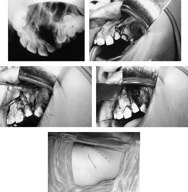
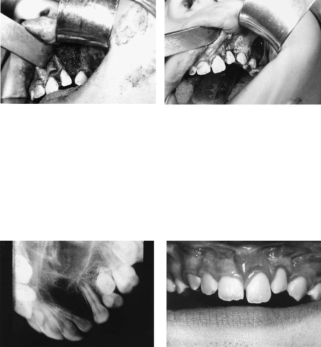
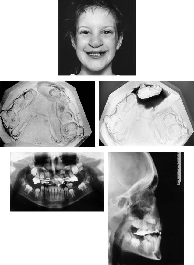
Case 3 (Figure 48.14)
A 10-year-old with left unilateral complete cleft of lip and alveolus.
Treatment:
1.Raising of mucoperiosteal flaps
2.Excision of sinus and scar tissue within cleft
3.Removal of supernumerary tooth
4.Repair of nasal mucosa at level of normal nasal floor
5.Harvesting of cancellous bone from anterior iliac crest
6.Insertion of graft into alveolar defect
7.Flap advancement and closure over graft
The Role of Osseointegrated Implants
Although modern cleft surgery aims to create a dentition without gaps, this aim is not always achieved. The incidence of hypodontia in cleft patients is higher than in the noncleft population, and it is not always possible for this to be disguised with the help of grafting, orthodontic treatment, and orthognathic surgery alone. There are also many patients who have not received alveolar bone grafts and also those who have lost
48. CLP Osseous Defects and Deformities |
549 |
a |
b |
c |
d |
e
FIGURE 48.14 Case 3. (a) X-ray of secondary alveolar defect. (b) Incisions for alveolar bone grafting outlined with excision of labial fistula. (c) Scar tissue within the alveolar cleft. (d) Alveolar defect af-
ter excision of scar tissue and repair of the nasal mucosa. (e) Incision (continuous line) marked lateral to the left anterior iliac crest (interrupted line) for harvesting of cancellous bone.
Continued.
550 |
K. Honigmann and A. Sugar |
f |
g |
h
i |
j |
FIGURE 48.14. Case 3. Continued. (f) Alveolar defect packed with cancellous bone chips harvested from the anterior iliac crest. (g) Flap closure over the bone graft; note the advancement of the flap from the lesser segment including gingivae and leaving a posterior defect
over the lateral maxilla, which is left to epithelialize by secondary intention. (h) Diagram of procedure. (i) X-ray of the alveolus in the grafted area in the same patient 6 months after surgery. (j) Oral view of the same patient 6 months after surgery.
48. CLP Osseous Defects and Deformities
teeth early and whose conventional dental restorative treatment is problematic.
The restoration of gaps in the dentition is ultimately the responsibility of the restorative dentist. Their options include dentures and fixed bridgework supported by teeth. The availability of titanium osseointegrated implants now adds to this repertoire the possibility of crowns or bridges supported by implants, as well as implant-supported overdentures.
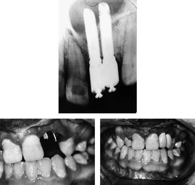
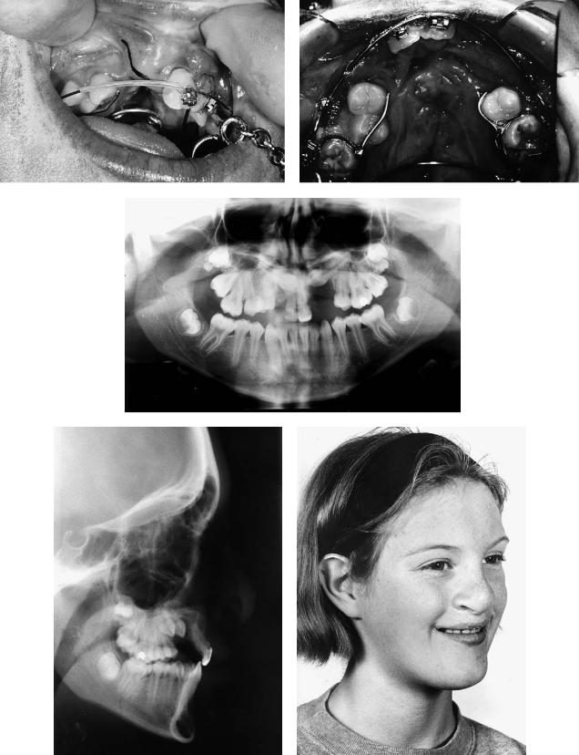
Case 4 (Figure 48.15)
A 25-year-old with left unilateral complete cleft lip and palate, not having received an alveolar bone graft and missing the left maxillary lateral incisor.
Treatment:
1.Alveolar bone grafting with autogenous cancellous iliac bone as described in Figure 48.14
2.Orthodontic arch alignment
3.Insertion of Bränemark titanium fixture into grafted area with additional small bone graft for labial defect provided from suction filter during the drilling process and covered with resorbable membrane (two-stage implant procedure)
4.Construction of implant-retained crown
(Restorative treatment courtesy of Will McLaughlin, Consultant in Restorative Dentistry, University Dental Hospital, Cardiff, Wales)
Case 5 (Figure 48.16)
A 16-year-old with bilateral complete cleft lip and palate assessed following orthodontics and bilateral alveolar bone grafting and with regard to two missing teeth in the left cleft.
Treatment:
1.Insertion of two Bränemark titanium fixtures (two-stage procedure) into maxillary alveolus, previously grafted in conjunction with orthodontics
2.Construction of implant-retained bridge
(Restorative treatment courtesy of Arshad Ali, Consultant in Restorative Dentistry, Morriston Hospital, Swansea, Wales)
Maxillary Osteotomies
Secondary deformities in patients with repaired cleft lip and palate present an interesting, if not difficult, surgical challenge. Careful assessment of the patient in the years following primary repair needs to take into consideration speech, hearing, facial growth, and dental development. The presence of fistulae, lip scars, and poor lip function, as well as residual nasal deformity and nasal resistance, needs to be assessed for correction. Alveolar defects and occlusion should be considered along with dental overcrowding, missing, malformed
551
and misplaced teeth, caries, and periodontal health. The ability and desire of the patient (and in the case of children, their family) to comply with what can often be prolonged treatment needs to be determined and taken into account.
This heterogeneity of problems requires the cooperation of a number of different specialties, foremost of which are a surgeon, speech therapist/pathologist, hearing specialist, and orthodontist, all preferably with a special interest in cleft problems. In late adolescence, a specialist in restorative dentistry is a valuable addition to the team. It is particularly useful to attempt to identify at as early an age as possible those children with significant midface hypoplasia that may require later surgical correction. If orthognathic surgery is to be delayed until approximately 16 years of age when most jaw growth is complete, early identification of those children is helpful.
Timing
In most cases speech patterns will have developed by the age of 4, and it should be possible to assess the need for a pharyngoplasty to correct velopharyngeal incompetence. Speech assessment and recording, anenometry, nasendoscopy, and video-fluoroscopy all assist in that decision. Ideally this should be carried out before school entry.
At the age of 8 years, and with the aid of orthopantomogram (OPT) and oblique occlusal and lateral cephalometric radiographs, it is useful to start to consider the need for dental extractions for orthopedic alignment of displaced and collapsed arches and for grafting of alveolar defects. When facial growth appears to be essentially normal, definitive orthodontics can then continue.
A clinical evaluation of facial form, noting the presence or absence of midface hypoplasia, a class III malocclusion, and dental compensation, may lead the team to the conclusion that jaw osteotomies are indicated in due course. This in turn allows the decision that orthodontics should be limited at that stage to the orthopedic alignment of segments and perhaps the correction of minor anterior incisal discrepancies. Definitive presurgical fixed-band orthodontics can then be delayed until the approximate age of 14 years when the patient can be prepared for orthognathic correction by osteotomies at 16. This has the merit of saving the child from 6 to 8 years of continuous orthodontic treatment with the inconvenience and almost inevitable lack of compliance that can result.
The Role of Alveolar Bone Grafting
Primary Grafting
We have described in our previous section the purpose of considering and carrying out alveolar bone grafting as well as a number of different approaches to it. Primary alveolar bone

a
b
c |
d |
e |
f |

48. CLP Osseous Defects and Deformities |
553 |
g
h
i
FIGURE 48.15 Case 4. (a) X-ray of alveolar defect. (b) Diagram of alveolar defect. (c) X-ray of grafted alveolar defect. (d) Diagram of grafted alveolar defect. (e) Intraoral x-ray of implant in grafted alve-
olar defect. (f) Lateral cephalogram showing position of implant. (g) Oral view with implant/abutment in situ. (h) Diagram showing implant in situ. (i) Oral view showing implant retained crown in situ.
554 |
K. Honigmann and A. Sugar |
a
b |
c |
FIGURE 48.16 Case 5. (a) X-ray of implants in grafted alveolus. (b) Oral view of abutments. (c) Implant-retained bridge in situ.
grafting is that which is carried out during the primary dentition or even before the eruption of the deciduous teeth. We do not yet have available from Basel mediumor long-term results of this approach, and much of the hostility to primary grafting has come from the apparently poor effect on maxillary growth.129 However, others130 have reported very encouraging results in this respect more recently. Rosenstein et al.130 have presented the long-term results in a regimen of cleft repair that has included primary bone grafting of the alveolar cleft at 4 to 6 months of age. This remains an area of considerable controversy.
Secondary Grafting
Secondary alveolar bone grafting, by which we mean grafting shortly before the eruption of the permanent maxillary canine teeth, has by contrast become very widely accepted. The method described by Boyne and Sands80 was popularized by the reporting of large series by Abyholm and his colleagues.81 It has undoubtedly made an important difference to the management of cleft patients. It makes the simultaneous repair of residual fistulae easier and by producing a one-piece maxilla facilitates a future maxillary osteotomy if needed. The pro-
48. CLP Osseous Defects and Deformities
duction is facilitated by well-aligned and continuous dental arches, with good bone support for the maxillary permanent canine and adjacent teeth. If there are gaps in the dental arch, it produces a stable base for the construction of fixed bridgework and implant-retained crowns and bridges. The overwhelming majority of compliant cleft children with an alveolar defect that has not been previously grafted will benefit from secondary alveolar bone grafting provided that the preparation and timing are carefully considered and the surgery well executed.
The popularizing of this technique in Norway was based, in the main, on children who did not have grossly collapsed dental arches. It has been the experience of the authors that secondary bone grafting of alveolar clefts without prior correction of misplaced segments creates significant difficulties. The segments may become fixed in an abnormal position with
a
b
555
orthopedic movement no longer possible or at best very difficult (Figure 48.17).
Case 6 (Figure 48.17)
Patient with bilateral complete cleft lip and palate.
Treatment:
1.The alveolar clefts had been bone grafted before orthopedic expansion and alignment of segments.
2.The premaxilla was thus fixed in its position significantly displaced inferiorly and to the right as were the lateral segments in their contracted position.
3.Later orthodontics was thus made very difficult. In some cases the problem can only be resolved with the help of multipiece osteotomies (see case 8, Figure 48.19).
c
FIGURE 48.17 Case 6. (a–c) Result of grafting of bilateral alveolar clefts before orthopedic alignment of the segments.
556
Careful assessment with an orthodontist experienced in the management of clefts is therefore essential to determine the presurgical needs, which should include alignment of any misplaced segments. After this, the orthodontist will design an appliance that will both retain the parts which have been moved and not impede surgery. Because the latter may well involve the repair of residual palatal oronasal fistulae, the appliance in situ during surgery must not cover any part of the palate to which access is required.
K. Honigmann and A. Sugar
some of which are particularly designed for analysis of the patient with a jaw deformity. While these can be useful, allowance does need to be made for the different values that are observed in cleft patients. A particularly relevant example is the cranial base to which the position of the maxilla and mandible is usually related. When the cranial base angle is abnormal (that is, it is outside the normal range of values), the angles of SNA and of SNB also vary widely, and this needs to be taken into consideration.
Tertiary Grafting
Patients who present after the eruption of the permanent canine teeth and at the end of the mixed dentition phase of development sometimes have not received any form of alveolar bone graft. Others have poor results from earlier grafting attempts and have inadequate bone for orthodontic movement of teeth, for support for prostheses, or for carrying out a maxillary osteotomy in one piece. In these cases, and notwithstanding the allegedly poor results that have been claimed for such late grafting by some authors (relative to secondary grafting), it has been our reported experience that excellent results can still be obtained.128 We therefore always consider, in conjunction with our multidisciplinary team, tertiary grafting in such cases.
Investigation
Facial Appearance
The principal tool in the diagnosis of residual facial deformity is clinical evaluation by an experienced surgeon. It is useful to document those parts of the upper, middle, and lower face that show anteroposterior, vertical, and transverse deficiencies or excesses. Dysmorphology and abnormality should be noted in all areas and in particular of the nasal bones, septum, tip, columella, and alar bases, as well as of the philtrum and upper lip.
Measurement of some aspects of the face in both frontal and profile views and comparison with norms is of value. The exposure at rest and when smiling of the upper incisor teeth, as well as measurement of the clinical crown height, are just a few examples. These enable the surgeon to determine the vertical movements needed of the anterior maxilla to create an ideal relationship with the upper lip, but consideration needs to be given to the need for lip revision in this respect and any of the resultant effects on lip–tooth relationship.
The interalar distance needs to be known if only to avoid making it worse after maxillary advancement; sometimes simultaneous revision of this distance needs to be built into the treatment plan. The intercanthal distance and nasofrontal angle may also increase in Le Fort II or Le Fort III osteotomies and should be recorded. The relationship between the maxillary and mandibular dental centers and the facial midline and chin needs to be known so that attempts at creating symmetry may be made. The presence of missing teeth in the cleft patient may make this particularly difficult.
Many forms of cephalometric measurement are available,
Occlusion
Dental study casts are essential in the overall analysis. In this way, the precise needs of presurgical orthodontics can be determined and results monitored.
Speech
It is always desirable that the cleft patient should be managed in coordination with a speech therapist/pathologist with experience of and interest in cleft patients. Children should be assessed at regular intervals during their development. The axiom that treatment should aim at producing an individual who “looks well and speaks well” remains valid today.
In relation to midface osteotomies, it is well recognized that these have the significant potential for improving the articulatory aspects of speech by correcting malocclusion and skeletal disproportion. However they also carry the unwanted risk of producing, or making worse, velopharyngeal incompetence (VPI). Consequently all cleft patients should have a thorough speech assessment immediately before undergoing midface advancement. This should involve a standard form of assessment with speech recording and anenometry. Nasendoscopy and videofluoroscopy may be valuable but can usually be reserved for those cases with problems postoperatively. The experienced speech therapist/pathologist, especially working in the same team and with the same surgeon, should be able to identify those patients most at risk of developing VPI.
Deformities/Diagnosis
Maxillary hypoplasia in cleft patients has a clear relationship to both the original deformity and the consequences of early surgical repair. We now describe the principal forms.
Unilateral Complete Cleft Lip and Palate (UCLP)
In this cleft defect, when midfacial hypoplasia is present it is manifested predominantly by an anteroposterior deficiency of the maxilla with lack of support to the nasal tip. There is often a vertical deficiency producing a lack of exposure of the upper incisor teeth at rest, influenced by any distortion of the upper lip. There will usually be an alveolar defect on the side of the cleft unless it has been grafted previously. Even without a previous periosteoplasty,119 bone bridging across the alveolar defect is sometimes seen. Transverse collapse of the alveolar segments may also occur, perhaps the most common
48. CLP Osseous Defects and Deformities
being displacement inward (palatally) and upward (cranially) of the lesser segment.
Several studies have shown that the mandible often lacks some forward growth in the repaired UCLP patient. In relation to surgery, it is questionable whether this usually requires correction. There is, however, often a lack of chin prominence but an excess of chin height. These contribute to an unesthetic and often drooping or ptotic appearance of the lower lip and warrant intervention.
Although the principal secondary nasal deformities are predominantly cartilage and soft tissue, the lack of support to the nasal tip may be severe. The dorsum of the nose is usually described as being essentially normal, but cases are seen where it is retropositioned and asymmetry is not uncommon. Labial or palatal fistulae may be present, communicating with the nasal cavity. The septum is usually deviated to the noncleft side and is often quite wide. In the authors’ experience, septa more than 1 cm wide can occur with complete blockage of the nasal airway. The inferior turbinate on the cleft side is usually hypertrophied.
Bilateral Complete Cleft Lip and Palate (BCLP)
Although class III malocclusions are seen in BCLP patients, very much depending on the method of primary repair, the principal finding is prominence of the premaxilla (and prolabium), especially vertically. In ungrafted cases, the premaxilla is usually mobile, poorly inclined (retroclined), and to one side or the other. The patient will often have, or with the aid of orthodontics be capable of having, a class I incisor relationship.
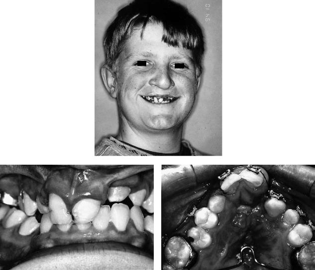
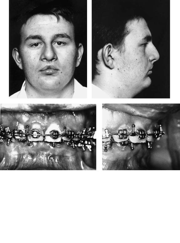
Class II-based deformities with mandibular retrognathia or retrogenia are seen in BCLP patients (Figure 48.18), and sometimes this is the only skeletal defect that requires correction. Occasionally bimaxillary advancement is indicated.
Case 7 (Figure 48.18)
Patient with bilateral complete cleft lip and palate and anteroposterior deficiency of the mandible.
Treatment:
1.Fixed-band orthodontics commenced in both arches to remove dental compensation, align teeth, and produce compatible arches on the basis of three-point contact following orthognathic surgery.
2.Before the movement of teeth adjacent to the alveolar clefts, these clefts were bone grafted in the way that we have described.
3.Following completion of the presurgical phase of orthodontics, mandibular advancement was carried out using bilateral sagittal split osteotomies of the mandibular rami, fixation being by four 2.7-mm titanium position screws (two on each side) inserted transbuccally.
4.Orthodontics was then completed.
557
(Orthodontics courtesy of Jeremy Knox, Dept. of Child Dental Health, University Dental Hospital, Cardiff, Wales)
It is common for teeth in the premaxilla of bilateral cleft patients to be poorly formed and prone to caries or crumbling; such teeth are not a good support for orthodontic devices. Nevertheless, malposition of the premaxilla and lateral segments can usually be corrected by the orthodontist before cleft bone grafting. Jones and Sugar128 have reported one case in whom this was carried out with an orthodontic device when the patient had no teeth on the premaxilla. There are, however, instances in which repositioning of the premaxilla can be difficult or impossible. In such occasional cases, surgical repositioning of the premaxilla before grafting should be considered (see case 9, Figure 48.20). The nose in the bilateral cleft patient may be broad at the alar bases and often also at the bridge with a short columella. Anteroposterior deficiency of the dorsum is rare.
Cleft Palate (CP)
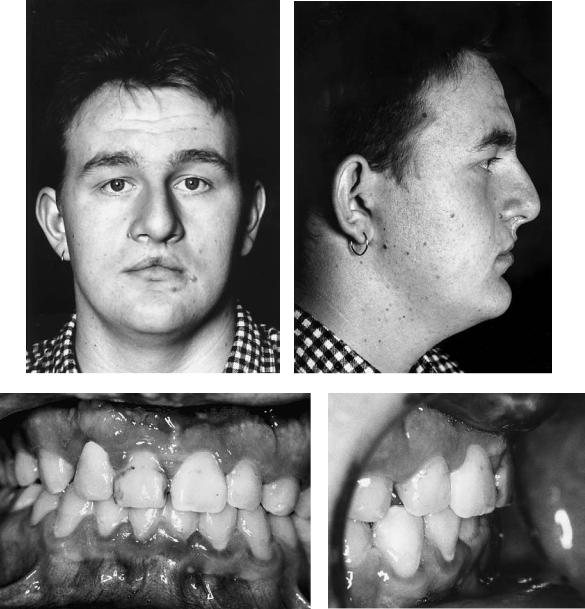
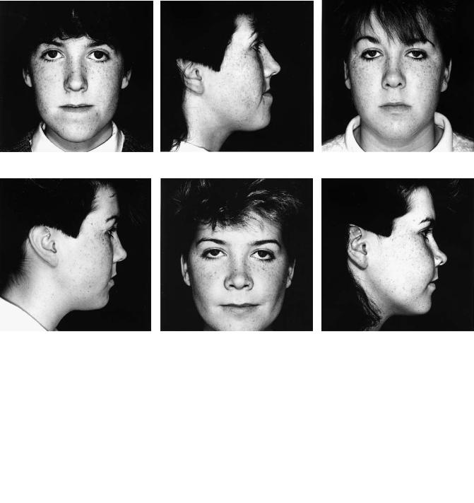
The patient with a repaired isolated cleft of the palate may also exhibit anteroposterior and sometimes vertical deficiency of the maxilla. It has been argued that many deformities of this kind in these and complete cleft lip and palate patients are not necessarily cleft related. Undoubtedly instances of class III skeletally based malocclusion of familial rather than cleft origin do occur, but the relative rarity of class II deformities in cleft patients is food for thought. The patient in case 8 (Figure 48.19a) has a repaired cleft palate with maxillary hypoplasia. Figure 48.19(c) shows her “identical” twin sister who has no cleft.
Case 8 (Figure 48.19)
Patient in Figure 48.19(a) has a repaired cleft of the secondary palate with anteroposterior and vertical deficiency of the maxilla. Figure 48.19(c) shows her identical twin sister who had no cleft, the photographs being taken on the same day as those of her sister. The principal difference noticeable between the sisters is the maxillary hypoplasia exhibited by the sister with a repaired cleft.
Surgery:
1.One-piece Le Fort I maxillary advancement and downward movement
2.Fixation using four L-shaped titanium 2-mm miniplates
3.Augmentation of the anterior maxillary bone steps only with corticocancellous blocks harvested from the medial aspect of the anterior iliac crest
Indications for Orthognathic Surgery
The principal indications for carrying out orthognathic surgery in patients with repaired cleft lip and/or palate are as follows.
1.To improve facial aesthetics and in particular the appearance of the midface, including the upper lip and nose
558 |
K. Honigmann and A. Sugar |
a |
|
|
|
|
|
b |
|
|
|
|
|
|
|
|
|
|
|
|
|
|
|
|
|
|
|
|
|
c |
d |
e
48. CLP Osseous Defects and Deformities |
559 |
|||||
f |
|
|
|
g |
||
|
|
|
|
|
|
|
|
|
|
|
|
|
|
|
|
|
|
|
|
|
|
|
|
|
|
|
|
|
|
|
|
|
|
|
h |
i |
FIGURE 48.18 Case 7. Anteroposterior mandibular deficiency in a pa- |
cedure (sagittal split advancement). (f–i) Following surgery and |
tient with bilateral cleft lip and palate (BCLP). (a–d) After ortho- |
completion of orthodontics. |
dontic preparation but before surgery. (e) Diagram of surgical pro- |
|
560 |
|
|
|
|
|
|
K. Honigmann and A. Sugar |
|||
a |
|
|
|
b |
|
c |
||||
|
|
|
|
|
|
|
|
|
|
|
|
|
|
|
|
|
|
|
|
|
|
|
|
|
|
|
|
|
|
|
|
|
d |
|
e |
|
|
|
f |
|
|
|
|
|
|
|
|
|
|
|
|
|
|
|
|
|
|
|
|
|
|
|
|
|
|
|
|
|
|
|
|
|
|
|
|
|
|
|
|
|
g
FIGURE 48.19 Case 8. (a,b) Patient with repaired cleft palate before orthognathic surgery. (c,d) Identical twin sister of patient in a,b who had no cleft. (e,f) Patient in a,b after Le Fort I maxillary advancement and correction of vertical deficiency. (g) Diagram of procedure.
48. CLP Osseous Defects and Deformities
2.To permit the full correction of skeletally based malocclusions
3.To improve the nasal airways by reducing nasal resistance
4.To improve speech, especially the articulatory aspects
Orthodontic Requirements
To achieve these aims optimally, orthodontic management is required to accomplish these aims:
1.Correct major displacement of segments by orthopedic movements
2.Permit the ideal choice of timing for alveolar bone grafting
3.Correct crowding and adopt a rational approach to tooth position where teeth are missing (hypodontia)
4.Remove dental compensation, especially abnormal inclinations of upper and lower incisors
5.Produce well-coordinated dental arches that will be compatible after surgery
6.Fine-tune tooth positioning and occlusion following surgery
Planning, Soft Tissue Effects, and Predictions
Planning in orthognathic surgery131 is the process by which the assessment, investigation, and resulting diagnosis are translated into a coherent treatment plan. It should be based predominantly on a clinical determination of treatment objectives. In the typical case with moderate to severe anteroposterior and vertical deficiency of the maxilla, and provided that the alveolar segments were aligned before bone grafting, it will probably involve the advancement of the maxilla at the Le Fort I level. Although every patient needs to be assessed individually, there is a tendency in some quarters to avoid large maxillary advancements by “splitting the difference” and moving the mandible back simultaneously. There are undoubtedly cases of true mandibular prognathism in which this is called for, but it is still necessary to carry out full correction of a retropositioned maxilla. Advancements of more than 2 cm may be necessary.
Model surgery is an absolute requirement in all cases. Models should be set up on a semiadjustable anatomic articulator after face bow recording. Reference lines are drawn and various distances in three planes are recorded. The desired movements are then carried out and the measurements retaken and recorded. These movements need to relate to the clinical treatment objectives, and it is valuable to test the achievement of those objectives against a predictive computer program. Once the movements have been finalized, acrylic occlusal wafers should be constructed, one in the case of a single-jaw osteotomy and two (including an intermediate position) in the case of bimaxillary procedures. These at least will remove some of the guesswork from the operating room, although vertical determinations will still need to be made.
561
Most computer packages for orthognathic surgery planning are based on surgery on a digitized lateral cephalometric radiograph. They are not infallible but can be a remarkably valuable indication of what will happen. We have analyzed two of the most commonly used such packages in the United States and U.K. specifically for internally fixed Le Fort I osteotomies including clefts.132,133 Soft tissue changes in cleft patients have a tendency to differ from those in noncleft patients, probably because of the lack of elasticity of the enveloping tissues. It is hoped that in the future such programs will be able to take this into consideration and thus give more accurate predictions. It is questionable whether more sophisticated (and expensive) techniques of three-dimensional prediction are of much value in the average case. However, video capture techniques with color print predictions of the result of surgical movements allow the patient to see a reasonable simulation of what surgery can achieve. They may also be helpful to the surgeon.
Treatment planning should take into consideration the views of the speech therapist or speech pathologist on the likelihood of the development or worsening of velopharyngeal incompetence. When very large advancements are considered, this may dictate a modification of surgical technique.
Surgical Procedures
Premaxillary Osteotomy
Osteotomies of the maxilla of cleft patients have to be tailored to the different anatomy, to the blood supply of the different parts of the maxilla, and to the nature and effects of previous surgery. This is especially the case when the part to be moved is the premaxilla. In the bilateral cleft patient, the bone of the premaxilla is attached very narrowly to the nasal septum. Its blood supply is derived principally from the labial mucoperiosteum. These need to be taken into consideration when designing the surgical approach and osteotomy technique if the premaxilla is not to become a free graft.
Premaxillary osteotomies will be needed only rarely because orthodontic methods are quite good at guiding this bone into the correct position. When needed, it will usually be because the bone would not move in this way. The bony attachment of the premaxilla may be approached from the palatal side or laterally (Figure 48.20), in both cases from within the cleft. It is also possible to use a midline labial vertical mucoperiosteal incision. Following osteotomy of the narrow attachment, the bone may then be moved digitally and fixed in its new position with the guidance of an occlusal wafer.
Fixation is best achieved with a strong arch wire within preexisting fixed orthodontic bands. It is unlikely, however, that this premaxilla will then become stable without the arch wire. It will eventually be stabilized by bilateral alveolar bone grafting, and the authors consider that this is visually best carried out as a separate procedure a few months later.
562
Case 9 (Figure 48.20)
A 10-year-old girl with bilateral complete cleft lip and palate. The premaxilla is misplaced and would not move with orthodontic appliances.
Treatment:
1.Model surgery to reposition the premaxilla and fabricate an occlusal wafer
2.Noted that this was only possible with the surgical removal of part of the premaxilla including a developing supernumerary (or abnormal lateral incisor) tooth germ
3.Securing of orthodontic fixed bands and fabrication of a strong arch wire that would support the premaxilla in its new position
4.Surgery in which the premaxilla was approached through a small lateral incision, permitting the removal of both the required amount of the premaxilla and division with a small osteotome of its bony attachment
5.Digital movement of the premaxilla into its new position, temporary fixation into the preformed occlusal wafer, and stabilization with a strong arch wire. The wafer was then removed
6.Three months later, bilateral alveolar bone grafting was carried out with simultaneous repair of the palatal fistula
7.Continued orthodontics
(Orthodontics courtesy of Prof. Malcolm Jones, Consultant Orthodontist and Head of Department of Child Dental Health, University Dental Hospital, Cardiff, Wales)
Le Fort I Osteotomy
The Le Fort I osteotomy is the most valuable procedure in cleft adolescents with maxillary hypoplasia. We consider that the most important aims must be full mobilization and good fixation.
Nasal airway obstruction, a severely retropositioned maxilla, and previous pharyngoplasty may all conspire to make nasal endotracheal intubation in these patients difficult. However, it is most unusual for the nares to prevent passage of an endotracheal tube at least on one side. Forewarning of the problem of the tube hitting the posterior pharyngeal wall enables the anesthetist to carefully redirect it inferiorly. This can sometimes be helped by a finger placed in the mouth above and behind the soft palate, where the tube can be palpated and brought forward and downward. Pharyngoplasties, especially superiorly or inferiorly based pharyngeal flaps, may limit access for intubation. The presence of such flaps should be noted preoperatively; most can be bypassed without damage but the patient should be warned of the risk of the pharyngoplasty being damaged or in extreme cases of it having to be divided and repaired. Fortunately, dynamic pharyngoplasties have become more popular and they present much less restriction to intubation.
In the past, multipiece and segmental procedures were effectively forced on surgeons with what was then the stage of
K. Honigmann and A. Sugar
development of orthodontic support and before the common use of alveolar bone grafting. The work of Tideman et al. is particularly recognized in this context,134 with his innovative use of substantial closure of the alveolar cleft by advancement of the lesser or lateral segments. Posnick135 has also developed a closely related approach based on orthodontics and multipiece osteotomies in the ungrafted cleft patient. Case 10 (Figure 48.21) demonstrates an adaptation of these techniques in a previously bone-grafted bilateral cleft patient where presurgical orthodontic preparation could not be completed to permit a one-piece osteotomy.
Case 10 (Figure 48.21)
An 18-year-old with bilateral complete cleft lip and palate. The occlusion was mildly class III with the premaxillary teeth proclined. Further orthodontic preparation was not possible because of the very short roots on the upper central incisors. Successful bilateral alveolar bone grafting had been carried out elsewhere.
Treatment:
1.Following presurgical orthodontics, sectional arch wires were placed
2.Le Fort I osteotomy carried out from a lateral approach attempted to preserve the maximum mucoperiosteal attachment to the premaxilla both palatally and labially
3.Ostectomies carried out in the previously grafted clefts bilaterally
4.Positioning of the three bone segments of the maxilla into a preformed occlusal wafer and wiring of a prefabricated arch wire across all segments fixed to the orthodontic brackets. The proclined premaxilla was retroclined, and the lateral segments advanced to close off the gaps in the dental arch coinciding with the alveolar clefts
5.Internal bone fixation with titanium 2-mm L-shaped miniplates was followed by removal of the wafer and intermaxillary fixation (IMF)
6.Grafting of the anterior bone steps with corticocancellous blocks harvested from the medial aspect of the anterior iliac crest, and of the interdental bone cuts with cancellous bone chips
(Orthodontics courtesy of David Howells, Consultant Orthodontist, Morriston Hospital, Swansea, Wales)
With these particular methods, special care is required for blood supply, and tunneling incisions are usually advisable anteriorly. Difficulty may be encountered because of the presence of scar tissue from the primary palate repair and poor access to break it down; this can be a particular problem for large advancements. Loss of part of the maxilla is rarely reported but is not unknown when carrying out maxillary osteotomies in cleft patients, and it is arguable that segmental procedures increase the risk. Although demonstrating good results, it has been shown136 that grafting the cleft at the time
48. CLP Osseous Defects and Deformities |
563 |
a
b |
|
|
|
c |
|
|
|
d |
e |
FIGURE 48.20 Case 9. (a) A 10-year-old girl with BCLP and a malpositioned premaxilla. (b) Model of maxillary arch. (c) Model surgery to reposition the premaxilla. (d) OPT before premaxillary surgery. (e) Lateral cephalogram before premaxillary surgery.
Continued.
f |
g |
|
h
i |
|
|
|
j |
|
|
|
|
|
|
|
|
|
|
|
|
|
|
|
|
|
|
|
|
FIGURE 48.20 Case 9. Continued. (f) Surgical approach to the premaxilla marked. (g) Repositioned premaxilla after osteotomy. (h) OPT of repositioned premaxilla with bilateral alveolar bone grafts.
(i) Lateral cephalogram taken at same time as h. (j) Patient following this treatment.

48. CLP Osseous Defects and Deformities |
565 |
a |
b |
c
d
FIGURE 48.21 Case 10. Multipiece maxillary Le Fort I osteotomy in a bilateral cleft lip and palate (CLP) patient using a modified Tideman/Posnick technique. This patient had been grafted previously elsewhere, but the proclination of the premaxilla and condition of the roots of the upper incisor teeth prevented complete orthodontic decompensation and correction of the position of the premaxilla (a,b). Consequently, osteotomies were carried out through the grafted alveolus on each side. The premaxilla was then retroclined, the lateral segments advanced to close off the alveolar clefts, and the whole maxilla advanced. Fixation was aided by a temporary acrylic wafer and intermaxillary fixation (IMF), both during surgery only, and was maintained with an arch bar wired to the orthodontic brackets and four 2-mm titanium L-shaped miniplates (c,d).

566 |
K. Honigmann and A. Sugar |
of osteotomy is not quite as effective as grafting as a separate procedure. Our own experience in South Wales (before 1985 when the present approach was adopted) was of a much greater difference in the results with much more successful graft take in the alveolar cleft when this was performed as a procedure separate from maxillary osteotomy.
Three developments have permitted us to change our approach:
1.Improved primary surgery, leaving the maxilla in a better developed condition with less hypoplasia and less arch collapse.
2.Improved dental health and sophisticated orthodontics so that cleft patients can now expect to have a complete dentition (with the exception of those teeth that have not developed) with oral hygiene and tooth condition such that they can be offered fixed-band orthodontics.
3.Alveolar bone grafting which, in a good multidisciplinary team, can be timed to fit in with bone and tooth development and with other procedures, and will usually produce a continuous maxillary dental arch.
It is the view of the authors, therefore, that segmental osteotomies in the cleft maxilla can usually be avoided. Most maxillae will present in one piece following successful secondary (or primary) alveolar grafting. In those rare cases when the grafting has been less than totally successful, it can be repeated and other patients who have not received a primary or secondary graft at all can be prepared for tertiary grafting in the way that we have described.128 Having taken this approach, it is not very logical or sensible to follow with sectioning of the maxilla into multiple pieces. Consequently, we try to carry out all cleft osteotomies with a one-piece maxilla using a downfracture approach. To date only one case (a BCLP case bone grafted in another unit) has shown signs of the maxilla failing to remain in one piece, and the minor cracks that occurred in the grafted area did not compromise the result, the segments being held in a strong arch wire.
The incision is placed anteriorly (Figure 48.22), being modified from the standard Le Fort I approach. It commences high in the cheek, just above and anterior to the openings of the parotid ducts. It is then continued down across the inside of the upper lip. This permits a broader posterolateral pedicle to supplement the palatal supply and still gives good access to the pterygoid area. This incision is used for grafted unilateral and bilateral cases alike, and since it was adopted 11 years ago not a single instance of compromised blood supply has been encountered.
The osteotomy is carried out using saws and fine osteotomes and with separation of tuberosities from pterygoid plates with a chisel. The cuts are placed high to facilitate internal miniplate fixation. Mobilization is carried out digitally and with disimpaction forceps and mobilizers. The nasal mucosa is preserved on both sides but in places may have to be cut to separate it from the oral (principally palatal) mucosa.
FIGURE 48.22 Modified incision for one-piece Le Fort I osteotomies in all grafted cleft patients.
There is often very little space for disimpaction forceps in the palate, especially with rubber protection for the blades. There is also a small risk of damaging previous palate repairs. We therefore always use a purpose-constructed metal palatal coverage plate (Figure 48.23) first designed in our unit by Ross and Bocca that permits use of the forceps without rubber covers and protects the palate effectively during mobilization. We always break down digitally the palatal scar tissue holding the maxilla back, and we do this from above through the opening created by the downfracture.
With the maxilla displaced downward, it is then possible to assess the internal nasal structures. A broad septum may be reduced, and inferior (partial) turbinectomy carried out if indicated. A preformed acrylic wafer is attached to the teeth by orthodontic powerchain and intermaxillary fixation (IMF) placed with more powerchain.137 The maxilla is fixed using
FIGURE 48.23 Palatal protection plate for mobilizing cleft maxillae with disimpaction forcep (Designed by Ross and Bocca).
48. CLP Osseous Defects and Deformities
L-shaped 2-mm miniplates, long L-shaped plates being particularly valuable for large advancements (Case 11, Figure 48.24).
Case 11 (Figure 48.24)
A 17-year old with bilateral complete cleft lip and palate and treated hypertelorbitism.
Treatment:
1.Orthopedic expansion and alignment of segments
2.Bilateral alveolar bone grafting and palatal fistula repair
3.Presurgical orthodontic preparation
4.One-piece Le Fort I osteotomy as described in the text, internally fixed with long L-shaped titanium 2-mm miniplates and bone grafted. The advancement in this case was 22 mm and the downward movement anteriorly was 10 mm
5.Completion of orthodontics. The stability of the result is demonstrated in the lateral cephalometry in Figure 48.24(h) 2 years after surgery
(Orthodontics courtesy of Prof. Malcom Jones, Consultant Orthodontist and Head of Dept. of Child Dental Health, University Dental Hospital, Cardiff, Wales, and David Bachmeyer, Sydney, New South Wales, Australia)
We always bone graft these cleft osteotomies, using autogenous corticocancellous blocks harvested from the medial aspect of the anterior iliac crest. The grafts are placed anterolaterally and occasionally are fixed with screws. Grafts are never placed into the region behind the maxilla, where they are in any event unstable. The wounds are closed primarily and without tension with no attempt to use the so-called V to Y single or multiple advancements. We consider that these closures, designed to produce vertical lip lengthening, actually produce increased anteroposterior lip projection and a tight wound and lip. In our hands, IMF is always removed at the end of the operation and before extubation. We have never encountered instability in these cases, even for the largest maxillary advancements (more than 2.5 cm in some cases), and have never had to resort to later IMF.
Case 12 (Figure 48.25)
A 21-year-old with repaired complete unilateral cleft lip and palate, anteroposterior and vertical midface deficiency and retrogenia, and secondary alveolar bone graft having been inserted previously.
Treatment:
1.Presurgical orthodontics
2.One-piece Le Fort I maxillary osteotomy as described above with miniplate fixation
567
3.Advancement genioplasty (horizontal sliding osteotomy) with 2-mm miniplate fixation
4.Completion of orthodontics
(Orthodontics courtesy of Russell Samuels, Consultant Orthodontist, Glenfield Hospital, Leicester, England)
Case 13 (Figure 48.26)
A 16-year-old with right unilateral complete cleft lip and palate. Secondary alveolar bone graft inserted after arch expansion at the age of 9 years with simultaneous closure of a large palatal fistula. Presented with significant anteroposterior and vertical maxillary deficiency.
Treatment:
1.Presurgical orthodontic preparation
2.Le Fort I maxillary advancement (1.5 cm) and downward movement (5 mm) as described above, fixation being with 2-mm titanium L-shaped miniplates and anterior maxillary grafting with corticocancellous blocks harvested from the medial aspect of the anterior iliac crest
3.Completion of orthodontics
(Orthodontics courtesy of Simon James, Consultant Orthodontist, Withybush General Hospital, Haverfordwest, Wales)
This approach has of course made these procedures much more popular with our anesthetic colleagues with safer postoperative airway management. Expensive intensive or high dependency care management in the immediate postoperative period can almost always be avoided. It has also made the postoperative period much more comfortable for the patient, who has often had to endure many operations.
The downfracture approach has been criticized in some quarters because of the risk of making velopharyngeal function worse. Using the technique described here, this has not been our experience. Velopharyngeal incompetence only seems to be present postoperatively in patients in whom it was present before surgery. It has been reported that the development of VPI can be avoided if a palatal approach to the osteotomy is adopted, the intention being to leave the palatal musculature and soft palate behind when the maxilla is ad- vanced.138–141 This certainly has merit but unfortunately also has some disadvantages. Intraoperatively, there is reduced access anteriorly to the nose and for fixation, and among the postoperative complications there is a high incidence of residual oronasal fistulae that require further surgery. Average skeletal relapse in the position of the maxilla anteroposteriorly has been reported as high as 29% in one series.140
The literature and experience indicate that Le Fort I osteotomies in cleft patients can be associated with particularly high incidences of relapse in the opposite direction to the movements carried out. It is our clear impression that this is no longer the case with our approach.142 This is discussed further later.

a |
b |
|
c |
|
d |
|
|
|
|
|
|
e |
|
f |
|
|
|
|
|
|
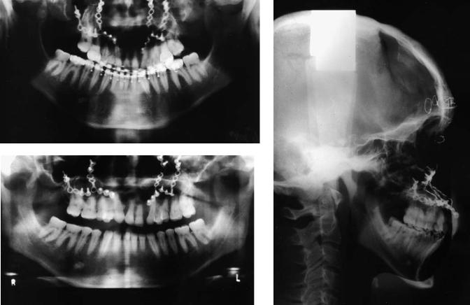
48. CLP Osseous Defects and Deformities |
569 |
g |
h |
i
FIGURE 48.24 Case 11. Use of long cantilevered L-shaped 2-mm |
eral cephalogram before maxillary advancement. (e) Profile after |
miniplates to fix and maintain a large one-piece maxillary ad- |
large maxillary advancement (22 mm) and downward movement |
vancement (22 mm) and anterior downward movement (10 mm) |
(10 mm). (f) Lateral cephalogram demonstrating the use of long |
in a patient with bilateral complete cleft lip and palate. (a) Maxil- |
cantilevered titanium L-shaped miniplates for fixation of this large |
lary dental arch before orthodontics and grafting. (b) Maxillary |
movement. (g) OPT taken at the same time as f. (h) Lateral |
dental arch after orthodontics and bilateral alveolar bone grafting. |
cephalogram showing stability of the movement 2 years later. (i) |
(c) Profile of this patient before maxillary advancement. (d) Lat- |
OPT taken at the same time as (h). |
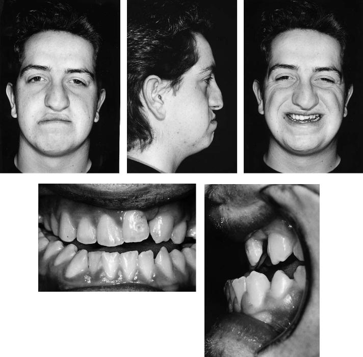
570 |
|
|
|
|
|
K. Honigmann and A. Sugar |
||||
a |
|
|
|
b |
|
c |
||||
|
|
|
|
|
|
|
|
|
|
|
d |
e |

48. CLP Osseous Defects and Deformities |
|
571 |
|||||||||
f |
g |
|
h |
||||||||
|
|
|
|
|
|
|
|
|
|
|
|
|
|
|
|
|
|
|
|
|
|
|
|
i |
j |
FIGURE 48.25 Case 12. One-piece maxillary and chin advancement osteotomies in a patient with a repaired unilateral complete cleft lip and palate. (a,b,c) Facial views before orthognathic surgery. (d,e) Occlusion before surgery. (f–h) Facial views after Le Fort I onepiece maxillary advancement osteotomy fixed internally with four
2-mm titanium L-shaped miniplates, grafting anteriorly with corticocancellous autogenous bone blocks from the medial aspect of the anterior iliac crest placed, and advancement genioplasty also fixed with miniplates. Views taken before rhinoplasty. (i,j) Occlusion after the surgery.
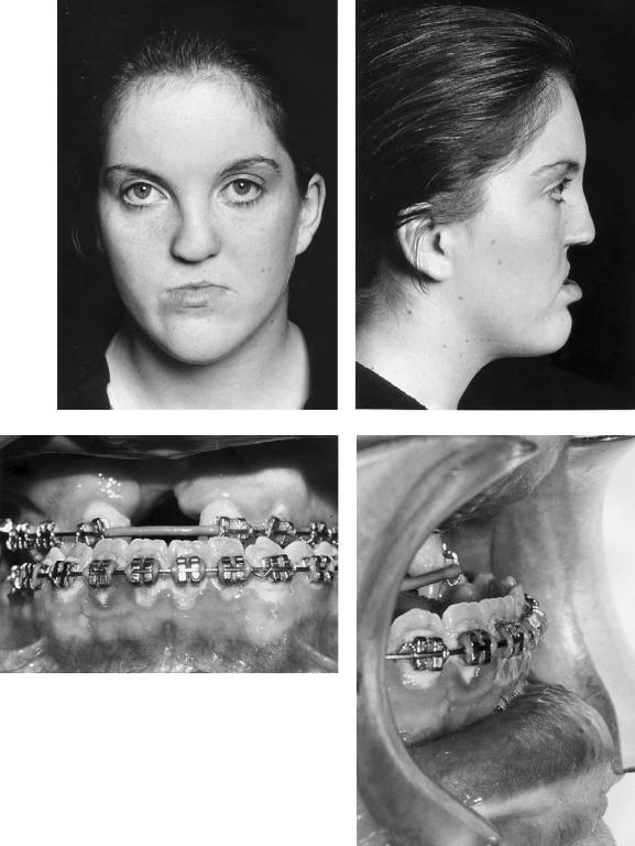
572 |
|
|
|
|
K. Honigmann and A. Sugar |
|
a |
|
|
|
|
|
b |
|
|
|
|
|
|
|
|
|
|
|
|
|
|
|
|
|
|
|
|
|
c |
d |
FIGURE 48.26 Case 13. One-piece maxillary advancement and downward movment in a patient with a repaired unilateral complete cleft lip and palate. (a,b) Facial views before orthognathic surgery. (c,d) Occlusion before surgery. (e,f) Facial views after large Le Fort I ad-
vancement osteotomy, with fixation by four 2-mm titanium L-shaped miniplates, and bone grafting anteriorly with bone harvested from the anterior iliac crest. (g,h) Occlusion after the surgery with temporary prosthesis in situ.
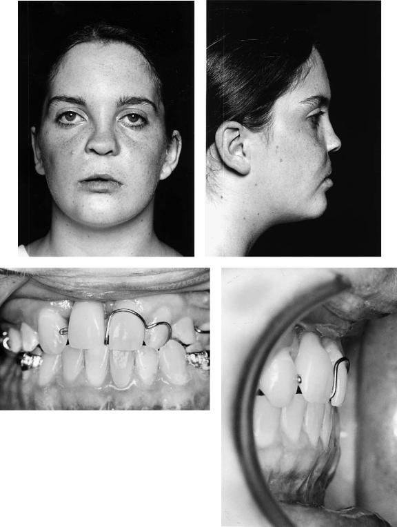
48. CLP Osseous Defects and Deformities |
573 |
e |
|
|
|
|
|
f |
|
|
|
|
|
|
|
|
|
|
|
|
|
|
|
|
|
|
|
|
|
g |
h |
FIGURE 48.26 Case 13. Continued.
574 |
K. Honigman and A. Sugar |
Le Fort II Osteotomy
Nasomaxillary hypoplasia is seen in some cleft patients with genuine retroposition of both the maxilla and entire nose. When the shape of the nose is otherwise normal, Le Fort I osteotomies may well make nasal appearance worse. The best approach is to carry out a Le Fort II osteotomy as described by Henderson and Jackson.143
We carry out this procedure through coronal and oral incisions because this gives the best access for the osteotomy, fixation, and grafting, and avoids further scars on the face. It is never possible to achieve the same degree of mobility as with the Le Fort I downfracture osteotomy. This seems to be compensated by the much larger block of bone tissue mobilized and the good opportunity for rigid fixation. Miniplate fixation is always used, usually with 2-mm plates and screws, and we prefer two L-shaped plates across the sides of the nose and two at the zygomatic buttress. The gaps are filled with autogenous corticocancellous or cancellous blocks of bone harvested from the anterior iliac crest.
When carrying out Le Fort II osteotomies it is important to consider carefully the nasofrontal angle, which can become too obtuse, and the intercanthal distance, which can increase with displacement anteriorly of the canthi. The former can be avoided by judicious bone removal to reduce the nasofrontal angle at osteotomy. The latter often requires transnasal canthopexy through the osteotomy gap so that the ligaments can be approximated and moved into a more posterior position.
Case 14 (Figure 48.27)
An 18-year-old with unilateral complete cleft lip and palate and nasomaxillary hypoplasia.
Treatment:
1.Presurgical orthodontics
2.Le Fort II osteotomy carried out through combined coronal and oral approaches
3.Fixation at four sites (nasofrontal and malar-maxillary) on both sides with 2-mm L-shaped titanium miniplates
4.Completion of orthodontics
5.Result also shown 6 years postoperatively with complete stability
(Orthodontics courtesy of Prof. Malcolm Jones, Consultant Orthodontist and Head of Department of Child Dental Health, University Dental Hospital, Cardiff, Wales)
A related approach to these nasomaxillary problems in cleft patients has been described by Tideman144 and is only really feasible because of internal plate fixation. The different needs in terms of advancement of the nose and maxilla are addressed in appropriate cases by carrying out a Le Fort II osteotomy to place the nose in its correct position and a few weeks later a Le Fort I to reposition the maxilla for the occlusion.
Malar Maxillary Le Fort III Osteotomy
Occasionally the nature of the midface deformity suggests the need for a Le Fort III or modified Le Fort III procedure. It is rare that these can be accomplished at one level, the needs for malar advancement usually being different from those at a dentoalveolar and occlusal level. We have therefore carried out these procedures at two levels at the same operation (i.e., simultaneous Le Fort III and Le Fort I).
Genioplasty
Retrogenia and increased chin height are common in cleft patients and are very amenable to correction. We favor a horizontal genioplasty osteotomy, sometimes with the excision of a slice of bone above the osteotomy to permit upward positioning. The attachment of the periosteum and suprahyoid musculature to the chin point is preserved, and the mental nerves carefully identified and avoided. Fixation is with two L-shaped 2-mm miniplates, one being placed on each side. Plates placed in the midline in this area are often palpable later. Although we have tried to use smaller plates and screws in this site, we have found that the titanium screw heads tend to shear off in this quite dense bone unless the holes are pretapped.
Bimaxillary Procedures
True mandibular prognathism is rare in cleft patients but when present needs to be corrected in a conventional orthognathic manner. Even more rarely, and predominantly in bilateral cleft patients, there is an indication for bimaxillary advancement. We favor, for the mandibular movement in whichever direction, bilateral sagittal split osteotomies with fixation using bicortical 2.4-mm screws. Where bone is in contact we will sometimes insert lag screws but in most cases, and especially where there are gaps, position screws are more appropriate. We used to insert three on each side but two good rigid screws at the upper border are probably sufficient. A transbuccal approach is used as we have described145 and is greatly facilitated by more recent improved instrumentation. Incisions of only 3 to 5 mm are required and, perhaps surprisingly, we have never seen a poor scar in more than 200 patients treated in this way.
Stability
It is widely recognized that midfacial advancement osteotomies in cleft patients are potentially less stable than in noncleft patients. Much of the responsibility for this has been ascribed to the presence of scar tissue in the region of the previous palate repair posteriorly, the common need for very large advancements, and the difficulty in achieving good fixation.
We therefore decided to study140 a carefully controlled group of our cleft patients. These all underwent consecutive
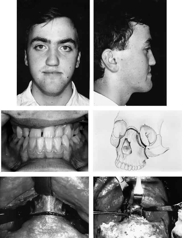
a |
b |
|
c |
|
|
|
|
|
d |
|
|
|
|
|
||
|
|
|
|
|
||
|
|
|
|
|
e |
f |
FIGURE 48.27 Case 14. Patient with repaired unilateral complete cleft lip and palate and nasomaxillary hypoplasia managed by a Le Fort II osteotomy. (a–c) Facial and occlusal views before
surgery. (d) Drawing of the Le Fort II nasomaxillary osteotomy.
(e) The nasofrontal exposure and osteotomy. (f) The nasofrontal fixation before grafting.
Continued.
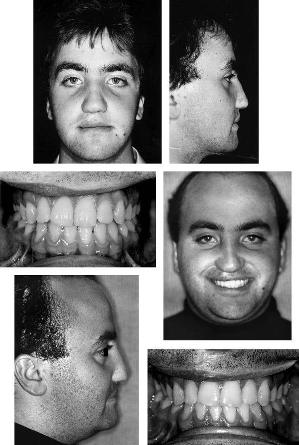
g |
|
|
|
|
|
h |
|
|
|
|
|
|
|
|
|
|
|
|
|
|
|
|
|
|
|
|
|
i |
|
|
|
j |
|
|
|
|
|
|
|
|
|
|
k
l
FIGURE 48.27 Case 14. Continued. (g–i) Facial and occlusal views after surgery and orthodontics. (j–l) Facial and occlusal views 6 years after surgery.
48. CLP Osseous Defects and Deformities
Le Fort I osteotomies to our prescribed protocol as described. Presurgical fixed-band orthodontics was always carried out, with secondary or tertiary bone grafting of any alveolar defect before or during that orthodontic phase. One-piece osteotomy from an anterior downfracture approach with anterior maxillary grafting using autogenous corticocancellous blocks and internal fixation was carried out by the same surgical team. Neither intermaxillary fixation nor external fixation was used in any case.
We have compared this test group with a control group of noncleft patients being treated by the same surgeons and to the same protocol. The study has been carried out with serial lateral cephalometric radiographs taken on the same machine by the same radiographer and at the same time intervals up to a minimum of 1 year. These were digitized by the same calibrated individual on two occasions, with at least 2 months between determinations, using Dentofacial Planner 4.32 software. Error measurement using paired t-tests showed no statistically significant difference between the two digitizations for both hard and soft tissue points.
The mean maxillary hard tissue advancement was similar in both groups (10.3 mm with a SD of 3.4 in the cleft group, and 10.5 with a SD of 2.9 in the nonclefts). The mean vertical movements were downward in the cleft group and upward in the nonclefts. The hard tissue changes up to 1 year, reflecting relapse or remodeling, were very small in both groups. The clefts moved posteriorly by 1.2 mm or 11.5% (SD 0.7) and the nonclefts by 0.7 mm or 6.5% (SD 0.8). The difference between the operated cleft and noncleft relapse rates was not statistically significant. The vertical changes were barely measurable and were all less than 0.5 mm. There was no statistically significant difference in the horizontal surgical soft tissue changes between the two groups, but the vertical soft tissue changes were different. The upper lip tended to go up in the nonclefts and down in the clefts. Upper-lip thickness decreased in both groups.
This study continues, and is presently based on relatively small numbers (10–15) in each group. However, there is good reason to believe at this stage that there is little difference in the way that cleft and noncleft osteotomies heal up to 1 year later after they have been performed in the way described. The one-piece cleft maxillary osteotomies also seem to be quite stable, and relapse or remodeling is well within clinically acceptable limits.
References
1.Streeter GL. Developmental horizons in human embryos. Contrib Embryol (Carnegie Inst Wash). 1951;11:165–196.
2.Töndury G. Über die Genese der Lippen-, KieferGau- menspalten. Fortschr Kiefer Gesichts Chir. 1955;1:1–8.
3.Kriens O. Kommt der Zweischenkiefer vom Stirnfortsatz oder? Vortrag 7. Symposium Interdiszipl. Arbeitskreis LKG-Spalten, 1994, Mainz.
4.Jirasek JE. The development of the face and mouth cavity. Acta Chir Plast. 1966;8:237–248.
577
5.Klaskova O. An epidemiological study of cleft lip and palate in Bohemia. Acta Chir Plast. 1973;15:258–262.
6.Jensen BL, Kreiborg S, Dahl E. Cleft lip and palate in Denmark, 1976–1981: epidemiology, variability, and early somatic development. Cleft Palate J. 1988;25:258–269.
7.Saxen I, Lahti A. Cleft lip and palate in Finland: incidence, secular, seasonal, and geographical variation. Teratology. 1974; 9:217–223.
8.Muehler, G. Die Bedeutung der Humangenetischen Beratung von Gesichtsspalttraegern und ihren Familien. Stomatol DDR. 1982;32:861–867.
9.Neel JK. A study of major congenital defects in Japanese infants. Am J Hum Genet. 1958;10:398–403.
10.Coerdt I. Erkrankungen der Kiefer und der Zaehne. In: Betke K, Kuenzer W, Schaub J, eds. Keller/Wiskott. Lehrbuch der Kinderheilkunde. Stuttgart: Thiemes; 1991.
11.Emanuel I, Culver BH, Erickson JD, Guthrie B, Schuldberg D. The further epidemiological differentiation of cleft lip and palate: a population study of clefts in King County, Washington, 1956–1965. Teratology 1973;7:271–282.
12.Ivy RH. The influence of race on the incidence of certain congenital anomalies, notably cleft lip–cleft palate. Plast Reconstr Surg. 1962;30:581–585.
13.Jörgensen G. Zur Aetiologie der Lippen-, Kieferund Gaumenspalten. Med Heute. 1969;18:293–298.
14.Fraser FC. The multifactorial/threshold concept—uses and misuses. Teratology. 1976;14:267–280.
15.Poswillo D. Mechanisms and pathogenesis of malformation. Br Med Bull. 1976;32:59–64.
16.Millicovsky G, Johnston MC. Hyperoxia and hypoxia in pregnancy: simple experimental manipulation alters the incidence of cleft lip and palate in CL/Fr mice. Proc Natl Acad Sci USA. 1981;78:5722–5723.
17.Davis JS, Ritchie HP. Classification of congenital clefts of lip and palate. JAMA. 1922;79:1323–1328.
18.Veau V. Division Palatine. Paris: Masson et Cie, 1931.
19.Fogh-Andersen P. Documentation. Discussion by invitation. In: Schuchardt K, ed. Treatment of Patients with Clefts of Lip. Alveolus and Palate. Stuttgart: Thieme; 1966.
20.Kernahan DA, Stark RB. A new classification for cleft lip and cleft palate. Plast Reconstr Surg. 1958;22:435–441.
21.Koch J. Zur Diagnostik der Lippen-, Kiefer-, Gaumenspalten. Dtsch Stomatol. 1963;13:660–666.
22.Pfeifer G. Classification of clefts of lip, alveolus and palate. Discussion by invitation. In: Schuchardt K, ed. Treatment of Patients with Clefts of Lip, Alveolus and Palate. Stuttgart: Thieme; 1966:224–226.
23.Kernahan DA. The striped Y. Plast Reconstr Surg. 1971;47: 469–470.
24.Elsahy NI. The modified striped Y—a systematic classification for cleft lip and palate. Cleft Palate J. 1973;10:247–50.
25.Millard DR. Cleft Craft. Vol. I. Boston: Little, Brown; 1976.
26.Friedman HI, Sayetta RB, Coston GN, Hussey JR. Symbolic representation of cleft lip and palate. Cleft Palate Craniofac J. 1991;28:252–259.
27.Kriens O. LAHSHAL. A concise documentation system for cleft lip, alveolus and palate diagnosis. In: Kriens O, ed. What Is a Cleft Lip and Palate? Stuttgart: Thieme; 1989:30–34.
28.Honigmann K. Die Pirmärbehandlung von Lippen-Kiefer- Gaumen-Segelspalten. Basel: Habil.-Schrift University; 1995.
578
29.Kriens O. Data-objective diagnosis of infant cleft lip, alveolus, and palate. Morphologic data guiding understanding and treatment concepts. Cleft Palate Craniofac J. 1991;28:157–168.
30.Paradise JL. Primary care of infants and children with cleft palate. In: Bluestone CD, Stool SE, Arjona SK, eds. Pediatric Otolaryngology: Philadelphia: WB Saunders; 1983.
31.Schmitt BD, Berman S. Ear, nose & throat. In: Kempe CH, Silver HK, O’Brien D, Fulginiti VA, eds. Current Pediatric Diagnosis & Treatment. Norwalk: Appleton & Lange; 1987.
32.Biesalski P, Collo D. Hals-Nasen-Ohren-Krankheiten im Kindesalter. Aufl. 2. Stuttgart: Thieme; 1991.
33.Clarren SK, Anderson B, Wolf L. Feeding infants with cleft lip, cleft palate, or cleft lip and palate. Cleft Palate J. 1987; 24:244–249.
34.Koppenburg P, Leidig E, Bacher M, Dausch-Neumann D. Die Darstellung von Lage und Beweglichkeit der Zunge bei Neugeborenen mit oralen Spaltfehlbildungen durch transorale Ultraschallsonographie. Dtsch Zahnärztl Z. 1988;43:806–809.
35.Kriens O. Neuere Aspekte in der Behandlung von Lippen- Kiefer-Gaumenspalten. Spaltträger Forum. 1990;2:25–30.
36.McNeil CK. Orthodontic procedures in the treatment of congenital cleft palate. Dent Rec. 1950;70:126–132.
37.Hotz M. Preand early postoperative growth guidance in cleft lip and palate cases by maxillary orthopedics. Cleft Palate J. 1969;6:368–372.
38.Gnoinski WM. Early maxillary orthopedics as a supplement to conventional primary surgery in complete cleft lip and palate cases—long-term results. J Maxillofac Surg. 1982;10:165–172.
39.Behrman RE, Vaughan VC. Nelson Textbook of Pediatrics. 13th ed. Philadelphia: WB Saunders; 1987.
40.Gnoinski WM. Third international symposium on the early treatment of cleft lip and palate. Zürich 1984. Schweiz Monatsschr Zahnmed. 1985;95:462/18–469/25.
41.Saunders IDF, Geary L, Fleming P, Gregg TA. A simplified feeding appliance for the infant with a cleft lip and palate. Quintessence Int. 1989;20:907–910.
42.Weatherly-White RCA, Kuehn DP, Mirrett P, Gilman JI, Weatherly-White CC. Early repair and breast-feeding for infants with cleft lip. Plast Reconstr Surg. 1987;79:879–885.
43.Duncan B, Ey J, Holberg CJ. Exclusive breast-feeding for at least 4 months protects against otitis media. Pediatrics. 1993; 91:867–872.
44.Honigmann K, Herzog C. Stillen von Kindern mit einer Lippen-Kiefer-Gaumen-Segelspalte. Paediatrica. 1993;4:16–22.
45.Tasaka Y, Kawano M, Honjo I. Eustachian tube function in OME patients with cleft palate. Acta Otolaryngol Suppl (Stockh). 1990;471:5–8.
46.Moss ALH, Piggott RW, Jones KJ. Submucous cleft palate. Br Med J. 1988;297:85–86.
47.Matsune S, Sando I, Takahashi H. Insertion of the tensor veli palatini muscle into the eustachian tube cartilage in cleft palate cases. Ann Otol Rhinol Laryngol. 1991;100:439–446.
48.Stool SE, Randall P. Unexpected ear diseases in infants with cleft palate. Cleft Palate J. 1967;4:99–103.
49.Fria TJ, Paradise JL, Sabo DL, Elster BA. Conductive hearing loss in infants and young children with cleft palate. J Pediatr. 1987;111:84–87.
50.McMillan JA, Oski FA, Stockman JA, Nieburg PI. Pädiatrische Daten und Fakten. Stuttgart: Ferdinand Enke Verlag; 1988.
51.Koch J, Schiel H, Koch H. Neue Gesichtspunkte zur kausalen
K. Honigmann and A. Sugar
Therapie der Hörund Sprachentwicklungsstörungen durch Gaumenspalten. Monatsschr Kinderheilkd. 1987;135:75–80.
52.Godbersen GS. Die mechanische Verlegung der Tubenostien. Folia Phoniatr. 1990;42:105–110.
53.Braganza RA, Kearns DB, Burton DM, Seid AB, Pransky SM. Closure of the soft palate for persistent otorrhea after placement of pressure equalization tubes in cleft palate infants. Cleft Palate Craniofac J. 1991;28:305–307.
54.Malek R, Psaume J, Mousset MS, Trichet CH. A new sequence and a new technique in complete cleft lip and palate repair. In: Hotz M, Gnoinski W, Perko M, Nussbaumer H, Hof E, Haubensack R, eds. Early Treatment of Cleft Lip and Palate. Stuttgart: Hans Huber; 1986.
55.Oesch IL, Looser C, Bettex MC. Influence of early closure of soft palatal clefts on the pharyngeal skeleton: Observation by CT scan. Cleft Palate J. 1987;24:291–298.
56.Garliner D. Myofunctional Therapy. Philadelphia: WB Saunders; 1976.
57.Codoni S. Anwendung der myofunktionellen Diagnostik und Therapie bei de Behandlung des LKG-Spalten-Kindes. Spaltträger Forum. 1992;4:22–28.
58.Graf-Pinthus B, Campiche M. Die Suche nach einem holistischen Behandlungskonzept bei Lippen-Kiefer-Gaumen-Spalten.
Myofunktion Ther. 1994;1:58–66.
59.Obwegeser H. Chirurgische Behandlungsmöglichkeiten von Sekundärdeformierungen bei Spaltpatienten. Fortschr Kieferorthop. 1988;49:272–296.
60.Markus AF, Smith WP, Delaire J. Primary closure of cleft palate: a functional approach. Br J Oral Maxillofac Surg. 1993;31:71–77.
61.Henderson D, Poswillo D. A Colour Atlas and Textbook of Orthognathic Surgery. London: Wolfe; 1985.
62.Bardach J, Salyer KE. Surgical Techniques in Cleft Lip and Palate. St. Louis: Mosby-Year Book; 1991.
63.Fränkel R. Die Bedeutung der Weichteile für die Induktion und Formorientierung des Kieferwachstums unter Zugrundelegung der Behandlungsergebnisse mit Funktionsreglern. Fortschr Kieferorthop. 1964;25:413.
64.Koch J. Jährige Erfahrungen mit der primären Knochentransplantation beim Verschluss der Kieferund Gaumenspalte im 4. Lebensjahr. In: Pfeifer G. ed. Lippen-Kiefer-Gaumenspal- ten. 3. Internationales Symposium, Hamburg 1979. Stuttgart: Thieme; 1982.
65.Rosenthal W. Die primäre orthopädische Beeinflussung des Spaltkiefers beim Säugling. Dtsch Stomatol. 1961;11:622–630.
66.Rosenthal W. Spezielle Zahn-, Mundund Kieferchirurgie. Leipzig: Johann Ambrosius Barth; 1951.
67.Honigmann K. The celluloid-acetone-dressing in palatoplasty.
Cleft Palate Craniofac J. 1994;31:228–229.
68.Kapp-Simon K. Self-concept of primary-school-age children with cleft lip, cleft palate, or both. Cleft Palate J. 1986;23: 24–27.
69.Strauss RP, Broder H, Helms RW. Perceptions of appearance and speech by adolescent patients with cleft lip and palate and by their parents. Cleft Palate J. 1988;25:335–342.
70.Broder H, Strauss RP. Self-concept of early primary school age children with visible or invisible defects. Cleft Palate J. 1989;26:114–117.
71.Lansdown R. Psychological problems of patients with cleft lip and palate: discussion paper. J R Soc Med. 1990;83:448–450.
48.CLP Osseous Defects and Deformities
72.Eliason MJ, Hardin MA, Olin WH. Factors that influence ratings of facial appearance for children with cleft lip and palate.
Cleft Palate Craniofac J. 1991;28:190–193.
73.Wolff J. Das Gesetz der Transformation der Knochen. Berlin: Hirschwald; 1892.
74.Veau V. Plastie palatine. J Chir. 1936;48:465–481.
75.Wassmund M. Der Verschluss des Kieferspaltes und Nasenbodens bei vollständiger Spaltibildung—Zeitpunkt und Methoden. Fortschr Kiefer Gesichts Chir. 1955;1:27–36.
76.Schmid E. Die Annäherung der Kieferstümpfe bei Lippen- Kiefer-Gaumenspalten; ihre schädlichen Folgen und Vermeidung. Fortschr Kiefer Gesichts Chir. 1955;1:37–39.
77.Nordin KE, Johansson B. Freie Knochentransplantation bei Defekten im Alveolarkamm nach kieferorthopädischer Einstellung der Maxilla bei Lippen-Kiefer-Gaumenspalten. Fortschr Kiefer Gesichts Chir. 1955;1:168–171.
78.Schrudde J, Stellmach R. Die primäre Osteoplastik der Defekte des Kieferbogens bei Lippen-Kiefer-Gaumenspalten beim Säugling. Zentrabl Chir. 1958;83:849–859.
79.Schuchardt K, Pfeifer G. Erfahrungen Über primäre Knochentransplantationen bei Lippen-Kiefer-Gaumenspalten. Langenbecks Arch Klin Chir. 1960;295:881–884.
80.Boyne P, Sands N. Secondary bone grafting of residual alveolar and palatal clefts. J Oral Surg. 1972;30:87–92.
81.Abyholm FE, Bergland O, Semb G. Secondary bone grafting of alveolar clefts. A surgical orthodontic treatment enabling a non-prosthodontic rehabilitation in cleft lip and palate patients.
Scand J Plast Reconstr Surg. 1981;15:127–140.
82.Wolfe SA, Berkowitz S. The use of cranial bone grafts in the closure of alveolar and anterior palatal clefts. Plast Reconstr Surg. 1983;72:659–671.
83.Kalaaji A, Lilja J, Friede H. Bone grafting at the stage of mixed and permanent dentition in patients with clefts of the lip and primary palate. Plast Reconstr Surg. 1994;93:690–696.
84.Dado DV. Primary (early) alveolar bone grafting. Clin Plast Surg. 1993;20:683–689.
85.HoltgraveEA.DieosteoplastischeVersorgungdesKieferspaltes— ein Fortschritt für die kieferorthopädische Behandlung des Spaltpatienten? Fortschr Kieferorthop. 1991;52:237–244.
86.Müssig D. Die Einstellung spaltnaher Eckzähne in Abhängigkeit vom Zeitpunkt der spätprimären Osteoplastik.
Fortschr Kieferorthop. 1991;52:245–251.
87.Blanchard-Moreau P, Breton P, Lebescond Y, Beziat JL, Freidel M. L’osteoplastie secondaire dans les fentes congenitales du palais primaire. Technique et resultats a propos de 43 observations. Rev Stomatol Chir Maxillofac. 1989;90:84–88.
88.Witsenburg B, Remmelink H-J. Reconstruction of residual alveolo-palatal bone defects in cleft patients. J Cranio-Max- illo-Fac Surg. 1993;21:239–244.
89.McNeil CK. Oral and Facial Deformity. London: Pitman & Sons; 1954.
90.Hotz R, Graf-Pinthus B. Zur kieferorthopädischen Frühbehandlung der Lippen-Kiefer-Gaumenspalten nach McNeil.
Schweiz Monatsschr Zahnheilkd. 1960;70:1–9.
91.Hotz R, Graf-Pinthus B. Weitere Erfahrungen mit der präoperativen kieferorthopädischen Frühbehandlung von totalen Lippen-Kiefer-Gaumenspalten. Schweiz Monatsschr Zahnheilkd. 1962;72:583–590.
92.Peat JH. Early orthodontic treatment for complete clefts. Am J Orthodont. 1974;65:28–38.
579
93.Larson O, Nordin KE, Nylen B, Eklund G. Early bone grafting in complete cleft lip and palate cases following maxillofacial orthopedics. II. The soft tissue development from seven to thirteen years of age. Scand J Plast Reconstr Surg. 1983;17:51–62.
94.Reisberg DJ, Figueroa AA, Gold HO. An intraoral appliance for management of the protrusive premaxilla in bilateral cleft lip. Cleft Palate J. 1988;25:53–57.
95.Asher-McDade C, Shaw WC. Current cleft lip and palate management in the United Kingdom. Br J Plast Surg. 1990; 43:318–321.
96.Cutting C, Grayson B. The prolabial unwinding flap method for one-stage repair of bilateral cleft lip, nose, and alveolus.
Plast Reconstr Surg. 1993;91:37–47.
97.Cronin TD, Penoff JH. Bilateral clefts of the primary palate. Cleft Palate J. 1971;8:349–363.
98.Hellquist R. Early maxillary orthopedics in relation to maxillary cleft repair by perioplasty. Cleft Palate J. 1971;8:36–55.
99.Vargervik K. Growth characteristics of the premaxilla and orthodontic treatment principles in bilateral cleft lip and palate. Cleft Palate J. 1983;20:289–302.
100.Rutrick R, Black PW, Jurkiewicz MJ. Bilateral cleft lip and palate presurgical treatment. Ann Plast Surg. 1984;12:105–117.
101.Georgiade NG, Mladick RA, Thoren FL. Positioning of the premaxilla in bilateral cleft lips by oral pinning and traction.
Plast Reconstr Surg. 1968;4:240–243.
102.Georgiade NG, Latham RA. Maxillary oral alignment in the bilateral cleft lip and plate infant, using the pinned coaxial screw appliance. Plast Reconstr Surg. 1975;56:52–60.
103.Latham RA. Orthopedic advancement of the cleft maxillary segment: a preliminary report. Cleft Palate J. 1980;17:227–233.
104.Stassen LFA. The management of patients with a cleft lip and palate deformity. Br J Oral Maxillofac Surg. 1994;32:1–2.
105.Berkowitz S. Commentary. Cleft Palate J. 1990;27:423–424.
106.Stellmach R. Discussion by invitation. In: Schuchardt K, ed.
Treatment of Patients with Clefts of Lip, Alveolus and Palate. Stuttgart: Thieme; 1966.
107.Honigmann K, Prein J. Nomenklaturvorschlag zur Knochentransplantation in eine Kieferspalte. Dtsch Z Mund Kiefer Gesichts Chir. 1992;16:272.
108.Stellmach R. Historische Entwicklung und derzeitiger Stand der Osteoplastik bei Lippen-Kiefer-Gaumen-Spalten. Fortschr Kiefer Gesichts Chir. 1993;38:11–14.
109.Mühling J. Die Osteoplastik bei Lippen-Kiefer-Gaumen-Spal- ten und zum Lasereinsatz in der Mund-Kiefer-Gesichts- Chirurgie. Schweiz Monatsschr Zahnmed. 1993;103:82–84.
110.Witsenburg B. The reconstruction of anterior residual bone defects in patients with cleft lip, alveolus and palate. A review. J Maxillofac Surg. 1985;13:197–208.
111.Mullerova Z, Brousilova M, Jiroutova O. The use of bone grafts in orofacial clefts. Acta Chir Plast. 1993;35:3–4.
112.Koch J. The closure of the bony part of the cleft lip and palate malformation: the problem of primary osteoplasty. In: HjortingHansen E, ed. Proceedings from the 8th International Conference on Oral and Maxillofacial Surgery. Chicago: Quintessence; 1985.
113.Helms JA, Speidel TM, Denis KL. Effect of timing on longterm clinical success of alveolar cleft bone grafts. Am J Orthod Dentofac Orthop. 1987;92:232–240.
114.Bosker H, van Dijk L. Het bottransplantaat van de mandibular voor herstel na de gnatho-palatoschisis. Ned Tijdschr Tandheelkd. 1980;87:383–389.
580
115. Koole R, Bosker H, van der Dussen FN. Late secondary autogenous bone grafting in cleft patients comparing mandibular (ectomesenchymal) and iliac crest (mesenchymal) grafts.
J Craniomaxillofac Surg (Suppl). 1989;17:28–30.
116.Borstlap WA, Heidbüchel KLWM, Freihofer HPM, KuijpersJagtman AM. Early secondary bone grafting of alveolar cleft defects. A comparison between chin and rib grafts. J. Craniomaxillofac Surg. 1990;18:201–205.
117.Sindet-Pedersen S, Enmark H. Reconstruction of alveolar clefts with mandibular or iliac crest bone grafts. J Oral Maxillofac Surg. 1990;48:554–558.
118.Maviglio P, De Santis P, Mavilio D, Fiume D. Cranial bone graft in treatment of alveolar cleft. Riv Ital Chir Plast. 1990; 22:335–340.
119.Skoog T. The use of periosteal flaps in the repair of clefts of the primary palate. Cleft Palate J. 1965;24:332–339.
120.Rintala AE, Ranta R. Periosteal flaps and grafts in primary cleft repair: a follow-up study. Plast Reconstr Surg. 1989;83:17–22.
121.Smahel Z, Mullerova Z. Facial growth and development in unilateral cleft lip and palate during the period of puberty: comparison of the development after periosteoplasty and after primarybonegrafting.Cleft Palate Craniofac J.1994;31:106–115.
122.Stricker M, Chancholle AR, Flot F, Malka G, Montoya A. Periosteal grafting for the repair of complete primary cleft repair. Ann Chir Plast. 1977;22:117–125.
123.Azzolini A, Riberti C, Bertani A. Tibial periosteal graft in the repair of the primary cleft palate: preliminary report of a new technique. Ateneo-Parmasense Acta Biomed. 1980;51:473–480.
124.Honigmann K. Die kombinierte Segelund Lippenplastik als neues Behandlungskonzept in der Spaltchirurgie. Dtsch Zahn Mund Kieferheilkd. 1983;71:600–604.
125.Honigmann K, Prein J. Die Kieferosteoplastik als Teil des operativen Gesamtkonzeptes zum LKG-Spaltenverschluss.
Fortschr Kiefer Gesichts Chir. 1993;38:69–70.
126.Davies D. The one-stage repair of unilateral cleft lip and palate: a preliminary report. Plast Reconstr Surg. 1966;38:129–136.
127.Fernandes D, Davies D. The radical one-stage repair of cleft lip and palate in the neonate. Presentation at the 7th International Congress on Cleft Palate and Related Craniofacial Anomalies, Broadbeach, Australia, 1993.
128.Jones ML, Sugar AW. The late management of cleft lip and palate problems: a joint orthodontic surgical approach. J R Coll Surg Edinb. 1990;35:376–386.
129.Jolleys A, Robertson NR. A study of the effect of early bone grafting in complete clefts of the lip and palate. Br J Plast Surg. 1972;25:229–237.
130.Rosenstein S, Dado DV, Kernahan D, Griffith BH, Grasseschi M. The case for early bone grafting in cleft lip and palate: a second report. Plast Reconstr Surg. 1991;87:644–654.
131.Sugar AW. Orthognathic surgery. In: Jones ML, Oliver RG,
K. Honigmann and A. Sugar
eds. Walther and Houston’s Orthodontic Notes. London: Wright (Butterworth-Heinemann Ltd.); 1994: ch 19.
132.Eales EA, Newton C, Jones ML, Sugar AW. The accuracy of computerized prediction of the soft-tissue profile: a study of 25 patients treated by the Le Fort 1 osteotomy. Int J Adult Orthod Orthognath Surg. 1994;9:141–152.
133.Eales EA, Jones ML, Newton C, Sugar AW. A study of the accuracy of predicted soft tissue changes produced by a computer software package (COG 3.4) in a series of patients treated by the Le Fort I osteotomy. Br J Oral Maxillofac Surg. 1995;33:362–369.
134.Tideman H, Stoelinga P, Gallia L. Le Fort I advancement with segmental palatal osteotomies in patients with cleft palates. J Oral Surg. 1980;38:196–199.
135.Posnick JC, Tompson B. Modification of the maxillary Le Fort I osteotomy in cleft-orthognathic surgery: the unilateral cleft lip and palate deformity. J Oral Maxillofac Surg. 1992;50:666–675.
136.Samman N, Cheung LK, Tideman H. A comparison of alveolar bone grafting with and without simultaneous maxillary osteotomies in cleft palate patients. Int J Oral Maxillofac Surg. 1994;23:65–70.
137.Smith AT. The use of orthodontic chain elastic for temporary intermaxillaryfixation.Br J Oral Maxillofac Surg.1993;31:250–251.
138.Converse JM, Shapiro HH. Treatment of developmental malformations of the jaws. Plast Reconstr Surg. 1952;10:473–510.
139.Wake M. Paper read to the British Association of Oral Surgeons, April 1976, Royal College of Surgeons of England, London.
140.Poole MD, Robinson PP, Nunn ME. Maxillary advancement in cleft lip and palate patients. A modification of the Le Fort I osteotomy and preliminary results. J Maxillofac Surg. 1986; 14:123–127.
141.James DR, Brook K. Maxillary hypoplasia in patients with cleft lip and palate deformity—the alternative surgical approach. Eur J Orthod. 1985;7:231–247.
142.Samuels R, Sugar AW, Jones ML, Newton C. Hard and soft tissue changes of internally fixed cleft and non-cleft Le Fort I osteotomies. In: Abstracts of the 12th Congress of the European Association for Cranio-Maxillo-Facial Surgery, The Hague, The Netherlands, September 5–10, 1994. J Craniomaxillofac Surg. 1994;22(suppl 1):abstract 127, 44.
143.Henderson D, Jackson IT. Naso-maxillary hypoplasia: the Le Fort II osteotomy. Br J Oral Surg. 1973;11:77–93.
144.Tideman H. Staged Le Fort II/Le Fort I osteotomies in cleft patients with naso-maxillary hypoplasia. Norman Rowe Lecture, Conference of British Association of Oral and Maxillofacial Surgeons, Cardiff, Wales, April 1993.
145.Llewelyn J, Sugar AW. Lag screws in sagittal split os- teotomies—should they be removed? Br J Oral Maxillofac Surg. 1992;30:83–86.