

- •Preface
- •Acknowledgments
- •Contents
- •Contributors
- •1. Introduction
- •2. Evaluation of the Craniomaxillofacial Deformity Patient
- •3. Craniofacial Deformities: Review of Etiologies, Distribution, and Their Classification
- •4. Etiology of Skeletal Malocclusion
- •5. Etiology, Distribution, and Classification of Craniomaxillofacial Deformities: Traumatic Defects
- •6. Etiology, Distribution, and Classification of Craniomaxillofacial Deformities: Review of Nasal Deformities
- •7. Review of Benign Tumors of the Maxillofacial Region and Considerations for Bone Invasion
- •8. Oral Malignancies: Etiology, Distribution, and Basic Treatment Considerations
- •9. Craniomaxillofacial Bone Infections: Etiologies, Distributions, and Associated Defects
- •11. Craniomaxillofacial Bone Healing, Biomechanics, and Rigid Internal Fixation
- •12. Metal for Craniomaxillofacial Internal Fixation Implants and Its Physiological Implications
- •13. Bioresorbable Materials for Bone Fixation: Review of Biological Concepts and Mechanical Aspects
- •14. Advanced Bone Healing Concepts in Craniomaxillofacial Reconstructive and Corrective Bone Surgery
- •15. The ITI Dental Implant System
- •16. Localized Ridge Augmentation Using Guided Bone Regeneration in Deficient Implant Sites
- •17. The ITI Dental Implant System in Maxillofacial Applications
- •18. Maxillary Sinus Grafting and Osseointegration Surgery
- •19. Computerized Tomography and Its Use for Craniomaxillofacial Dental Implantology
- •20B. Atlas of Cases
- •21A. Prosthodontic Considerations in Dental Implant Restoration
- •21B. Overdenture Case Reports
- •22. AO/ASIF Mandibular Hardware
- •23. Aesthetic Considerations in Reconstructive and Corrective Craniomaxillofacial Bone Surgery
- •24. Considerations for Reconstruction of the Head and Neck Oncologic Patient
- •25. Autogenous Bone Grafts in Maxillofacial Reconstruction
- •26. Current Practice and Future Trends in Craniomaxillofacial Reconstructive and Corrective Microvascular Bone Surgery
- •27. Considerations in the Fixation of Bone Grafts for the Reconstruction of Mandibular Continuity Defects
- •28. Indications and Technical Considerations of Different Fibula Grafts
- •29. Soft Tissue Flaps for Coverage of Craniomaxillofacial Osseous Continuity Defects with or Without Bone Graft and Rigid Fixation
- •30. Mandibular Condyle Reconstruction with Free Costochondral Grafting
- •31. Microsurgical Reconstruction of Large Defects of the Maxilla, Midface, and Cranial Base
- •32. Condylar Prosthesis for the Replacement of the Mandibular Condyle
- •33. Problems Related to Mandibular Condylar Prosthesis
- •34. Reconstruction of Defects of the Mandibular Angle
- •35. Mandibular Body Reconstruction
- •36. Marginal Mandibulectomy
- •37. Reconstruction of Extensive Anterior Defects of the Mandible
- •38. Radiation Therapy and Considerations for Internal Fixation Devices
- •39. Management of Posttraumatic Osteomyelitis of the Mandible
- •40. Bilateral Maxillary Defects: THORP Plate Reconstruction with Removable Prosthesis
- •41. AO/ASIF Craniofacial Fixation System Hardware
- •43. Orbital Reconstruction
- •44. Nasal Reconstruction Using Bone Grafts and Rigid Internal Fixation
- •46. Orthognathic Examination
- •47. Considerations in Planning for Bimaxillary Surgery and the Implications of Rigid Internal Fixation
- •48. Reconstruction of Cleft Lip and Palate Osseous Defects and Deformities
- •49. Maxillary Osteotomies and Considerations for Rigid Internal Fixation
- •50. Mandibular Osteotomies and Considerations for Rigid Internal Fixation
- •51. Genioplasty Techniques and Considerations for Rigid Internal Fixation
- •52. Long-Term Stability of Maxillary and Mandibular Osteotomies with Rigid Internal Fixation
- •53. Le Fort II and Le Fort III Osteotomies for Midface Reconstruction and Considerations for Internal Fixation
- •54. Craniofacial Deformities: Introduction and Principles of Management
- •55. The Effects of Plate and Screw Fixation on the Growing Craniofacial Skeleton
- •56. Calvarial Bone Graft Harvesting Techniques: Considerations for Their Use with Rigid Fixation Techniques in the Craniomaxillofacial Region
- •57. Crouzon Syndrome: Basic Dysmorphology and Staging of Reconstruction
- •58. Hemifacial Microsomia
- •59. Orbital Hypertelorism: Surgical Management
- •60. Surgical Correction of the Apert Craniofacial Deformities
- •Index
50
Mandibular Osteotomies and Considerations for Rigid Internal Fixation
Victor Escobar, Alex M. Greenberg, and Alan Schwimmer
Introduction |
Over the years, many modifications to mandibular os- |
||
|
|
teotomies have been reported,20,28–30 but the most stable was |
|
Since the mandibular osteotomy described by Hullihen,1 max- |
the step osteotomy30 (Figure 50.6) used for advancement of |
||
illomandibular fixation (MMF) during the healing period has |
the body of the mandible. During this time, the procedures |
||
been the fixation method of choice (Figure 50.1). New sur- |
have shifted from the body to the ramus. It was Blair in 190731 |
||
gical techniques include the use of rigid internal fixation (RIF) |
who first performed a subcondylar vertical ramus osteotomy |
||
with plates2 and/or screws3,4 (Figure 50.2). The avoidance of |
(Figure 50.7). Since then, many variations of this osteotomy |
||
MMF allows increased stability and improved postoperative |
have been described including several types of sliding os- |
||
4,5 |
6 |
teotomies for the repositioning of segments in multiple planes |
|
care |
and accelerates psychological recovery. |
of space.20,32–34 A transcutaneous approach was used until |
|
Over time, clinicians have critically reviewed their results, |
|||
and as techniques have developed, it has been suggested that |
196433 despite Ernst’s35 description in 1938 of a transoral ap- |
||
immobilization of the mandible is accompanied by a decrease |
proach. As with the anterior mandibular segmental os- |
||
in mandibular range of motion7–9 secondary to degenerative |
teotomy,36 the subcondylar osteotomy has undergone many |
||
changes in the temporomandibular joint (TMJ).10 Rigid inter- |
changes that finally resulted in the intraoral vertical ramus os- |
||
nal fixation shortens immobilization time11–13 and minimizes |
teotomy (IVRO) which is used today for the treatment of |
||
the reduction in mandibular range of motion after orthognathic |
mandibular prognathism (Figure 50.8).37 The intraoral verti- |
||
surgery.7–9,14 Improvements in diagnosis, treatment planning, |
cal ramus osteotomy was performed originally at the neck of |
||
and osteotomy techniques15,16 allow patients to open and close |
the condyle21,33,38 (Figure 50.8e). Because of the lack of sta- |
||
the mandible immediately after surgery17 and to maintain oral |
bility of this osteotomy postoperatively (presumably because |
||
nutritional uptake, and reduce postoperative apprehension |
of the lack of medial pterygoid attachment), longer cuts were |
||
|
18,19 |
suggested beginning at the sigmoid notch and ending at dif- |
|
caused by respiratory limitation, nausea, and vomiting. |
ferent levels behind the angle of the mandible39,40 close to |
||
|
|
||
|
|
the gonial notch.41 In patients requiring mandibular advance- |
|
Mandibular Osteotomies |
ment, an anterior iliac crest bone graft block was inserted be- |
||
tween the segments42,43 and in many cases wire osteosynthe- |
|||
Historical Review |
sis was utilized.44 Other variations of the subcondylar |
||
osteotomy have included the inverted L osteotomy45 and its |
|||
|
|
||
Mandibular osteotomies for the correction of dentofacial de- |
modifications,46,47 including the C osteotomy48,49 (Figures |
||
formities have been performed since the first report of an an- |
50.8b,c). |
||
terior subapical mandibular osteotomy by Hullihen in 1849 |
As pointed out by Bloomquist,29 perhaps the greatest de- |
||
(Figure 50.3). For the remainder of the century, management |
velopment in mandibular osteotomies of the vertical ramus is |
||
of malocclusion secondary to prognathism was mainly a trans- |
the sagittal split ramus osteotomy (SSRO),50 which was in- |
||
cutaneous resection of a piece of mandibular bone20 (Figure |
troduced by Perthes in 1924 and later popularized by Ob- |
||
50.4) to achieve shortening of the mandible.21,22 A variation |
wegeser36 and Trauner in 1955 for the management of prog- |
||
of Hullihen’s procedure1 was used to advance the anterior |
nathism (Figure 50.9). Two years later, the same authors51 |
||
lower segment23 and to close anterior open bites.24 These pro- |
proposed correction of both prognathic and retrognathic |
||
cedures resembled the anterior mandibular segmental subapi- |
mandibles with the sagittal split ramus osteotomy and simul- |
||
cal osteotomy in use today.25 Two other modifications are |
taneous genioplasty. With time, several modifications and im- |
||
still in use—single tooth osteotomies26 and total mandibular |
provements to the SSRO have been developed52,53 (Figure |
||
alveolar osteotomy27 (see Figure 50.5). |
50.9). While Dal Pont54 suggested that the lateral cut be made |
||
606
50. Mandibular Osteotomies and Considerations for Rigid Internal Fixation |
607 |
FIGURE 50.1 Panoramic radiograph reveals bilateral mandibular intraoral vertical ramus osteotomies with maxillomandibular fixation. Note circumandibular and piriform rim skeletal wire fixation to prevent extrusion of orthodontically treated teeth.
a |
b |
c |
d |
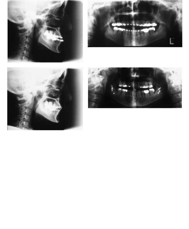
FIGURE 50.2 Patient with skeletal class II malocclusion having undergone bilateral mandibular sagittal split ramus osteotomies with miniplate fixation. Note circumandibular and piriform rim skeletal wire fixation to prevent extrusion of orthodontically treated teeth:
(a) preoperative lateral cephalometric radiograph; (b) preoperative panoramic radiograph; (c) postoperative lateral cephalometric radiograph; (d) postoperative panoramic radiograph.
Continued.
608 |
V. Escobar, A.M. Greenberg, and A. Schwimmer |
e |
f |
g |
h |
FIGURE 50.2 Continued. Patient with skeletal class III malocclusion having undergone bilateral mandibular sagittal split ramus osteotomies with three bilateral superior border 2.0-mm lag screws. (e)
Preoperative lateral cephalometric radiograph; (f) preoperative panoramic radiograph; (g) postoperative lateral cephalometric radiograph; (h) postoperative panoramic radiograph.
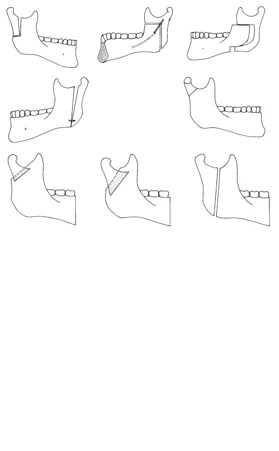
FIGURE 50.3 Subapical osteotomy similar to the one performed by FIGURE 50.4 Body osteotomy as performed by Blair.21 (From Bell102) Hullihen in 1849. (From Bell102)
50. Mandibular Osteotomies and Considerations for Rigid Internal Fixation |
609 |
FIGURE 50.5 Diagram of subapical mandibular osteotomy. (From Powell and Riley103)
forward along the lateral aspect of the second molar to increase the area of bone contact [Figure 50.9(e)], Hunsuck55 suggested decreasing the extent of the medial osteotomy to just posterior to the mandibular foramen to produce a smaller mucoperiosteal reflection and reduce trauma to the neurovascular bundle [Figure 50.9(f)]. To reduce the complications associated with the SSRO and the unpredictable manner in which the inferior border of the mandible may split (Hunsuck effect), an inferior border osteotomy has been proposed.56 However, this increases the extent of pterygomasseteric sling stripping, the risk of intraoperative bleeding, and the frequency of inferior alveolar nerve damage.57 One of the most important innovations to SSROs is the use of lag screw fixation introduced by Spiessl.58 It was theorized that screw fixation would result in more rapid healing, osteosynthesis, decreased relapse, increased patient comfort, and improved condylar control.59
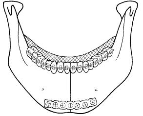
Four major types of osteotomies are performed today:
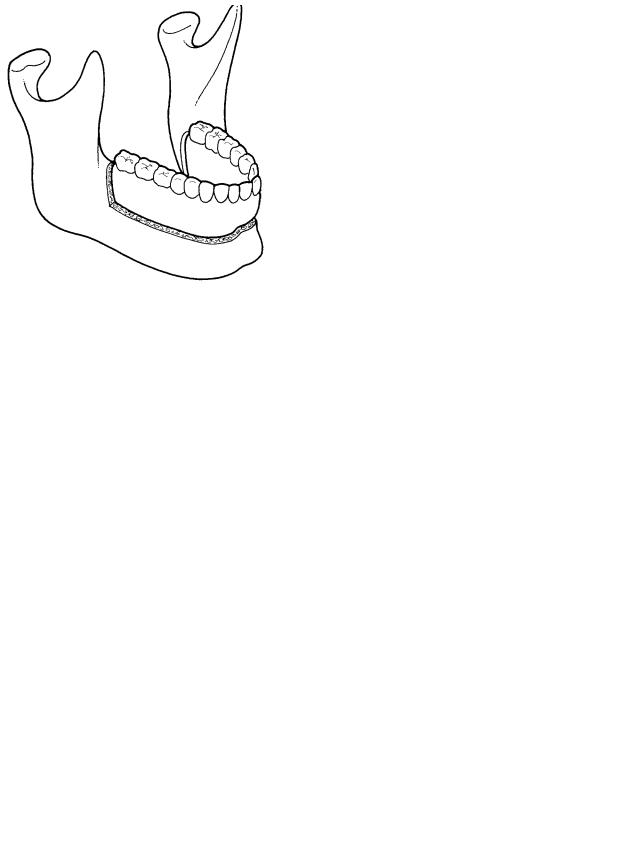
(1)anterior mandibular segmental (subapical) osteotomies,
(2)anterior horizontal mandibular osteotomy (genioplasty),
(3)sagittal split ramus osteotomy (SSRO), and (4) intraoral vertical ramus osteotomy (IVRO).
FIGURE 50.6 Von Eiselberg step osteotomy. (From Bloomquist29)
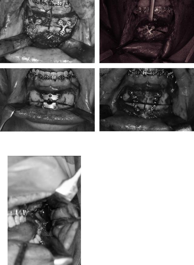
thetic containing epinephrine and a labial vestibular incision made approximately 5 mm labial to the depth of the vestibule.60 The incision extends through the mentalis muscles, through periosteum, and down to bone. The extent of the incision should be at least 10 mm beyond the planned vertical osteotomies to prevent undesirable laceration of soft tissue. The mental nerves should be identified and isolated as they exit their foramina. The periosteum is elevated, preserving the mucoperiosteal attachment at the inferior border of the mandible. This helps to ensure stability of the soft tissues of the chin. When necessary, teeth are removed and a fissure bur is utilized to create the interdental osteotomy perpendicular to the curve of the arch. The subapical horizontal osteotomy is created with a reciprocating saw 5 mm inferior to the roots of the teeth. Depending on the type of movement desired, additional bone trimming is then performed. The segments are mobilized after completion of the osteotomies via chisels. Care must be taken to preserve the lingual and buccal pedicles crossing the interdental osteotomies. When positioning the segments into a surgical guide, excessive bone removal should be avoided by careful bone trimming.
After satisfactory positioning of the dentoalveolar segment
Mandibular Anterior Segmental Osteotomy
(Subapical Osteotomy)
The subapical osteotomy has been used to move anterior teeth in almost any conceivable direction. The greatest concern is damage to the teeth if the osteotomies are not carefully designed. Space needs to be available to permit safe vertical and horizontal osteotomies.
The soft tissue surgical approach for all anterior osteotomies is similar. It involves infiltration with local anes-
FIGURE 50.7 Horizontal subcondylar osteotomy as performed by Blair. (From Bell102)
a |
b |
c |
d |
e |
f |
g |
h |
FIGURE 50.8 (a) Limberg’s oblique osteotomy. (b) Wassmund’s inverted “L” osteotomy. (c) Caldwell et al., “C” osteotomy. (d) Robinson et al., oblique osteotomy. (e) Blair, subcondylar osteotomy. (f–h) Different lengths of inverted vertical ramus osteotomy (IVRO). (From Bell102)
a |
b |
c |
d |
e |
f |
FIGURE 50.9 (a) Perthes osteotomy, 1924. (b) Trauner and Obwegeser, 1957. (c) Kazanjian and Converse, 1959. (d) Schuchardt, 1961. (e) Dal Pont, 1961. (f) Hunsuck, 1968. (Adapted from Bell102)
50. Mandibular Osteotomies and Considerations for Rigid Internal Fixation |
611 |
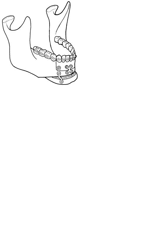
FIGURE 50.10 Miniplate fixation of anterior mandibular segmental and genioplasty osteotomies. (From Shafer and Assael60)
into a surgical stent and after MMF has been attained, rigid fixation may be performed with a variety of miniplates, microplates, or monocortical screws. If an anterior mandibular horizontal osteotomy for genioplasty is simultaneously performed, fixation of all the segments may be performed. The dentoalveolar segment should be repositioned and rigidly fixated first. Then, the chin bony segment may be repositioned and secured either separately with special preformed chin plates or with the same miniplates used to secure the dentoalveolar segment. Additional holes may be left in the plates for fixation of the chin (Figure 50.10). After fixation, bone
grafts may be wedged into the interdental osteotomy sites to aid in the stability of the dentoalveolar segments and reduce the incidence of periodontal defects. The osteotomy is made with a reciprocating saw. The length and angle of the horizontal cut can have profound effects on the resultant position of the segment (see Figure 50.11). It is recommended60 that this osteotomy be done with a power saw because extensive use of chisels in interdental areas can result in mucosal lacerations that adversely affect blood supply and periodontal health. Care should be taken to avoid root apices and lateral root surfaces. Midline anterior osteotomies (Figure 50.12) through the inferior border may also be performed in conjunction with anterior mandibular osteotomies. Their fixation should occur after the anterior dentoalveolar segmental osteotomy has been completed. Of major concern with subapical osteotomies is the preservation of the inferior alveolar nerves and their mental branches. Vitality of the teeth and the whole bony segment will be affected by the level of the horizontal cut, which should be at least 5 mm below the teeth apices. Soft tissue pedicles are important for blood supply, and the lingual mucosa should be preserved. If ramus osteotomies are performed they should be done following the anterior mandibular procedures.
Wound closure is accomplished in two layers—muscle and mucosa—with resorbable sutures. Care should be taken to reapproximate the mentalis muscles. Tape is placed on the lip and chin for 24 to 48 hours to minimize hematoma formation and to aid in supporting the suture lines.29
Anterior Mandibular Midline Osteotomy
In addition to subapical osteotomies, a useful procedure is the anterior midline (Figure 50.12) or paramidline osteotomy used to correct arch width discrepancies. Surgical technique
a |
b |
c |
d |
FIGURE 50.11 (a) Preoperative. (b) Long obliquely angled segment. (c) Short obliquely angled segment. (d) Horizontally angled segment. Effects of angle and lenth of mandibular osteotomy. (From Bloomquist29)
612 |
V. Escobar, A.M. Greenberg, and A. Schwimmer |
FIGURE 50.12 Rigid fixation of midline mandibular osteotomy with reconstruction plate. Lingual splint in place for alignment of segments. (From Shafer and Assael60)
includes anterior mandible infiltration with local anesthetic and epinephrine. The incision is created in the labial vestibular mucosa as in the subapical osteotomy described earlier. The periosteum is elevated preserving the mucoperiosteal attachment of the anterior mandibular segment. Smaller incisions including vertical types are sometimes preferred by surgeons. A fissure bur is then used to create a vertical osteotomy line between the roots of the mandibular central incisors and carried through to the inferior border of the mandible. The osteotomy is completed with the help of an osteotome. The mandibular segments are then mobilized into a stent, and the mandible can be either narrowed or widened. For this type of osteotomy, paired mandibular compression plates or a 2.4 mm standard reconstruction plate should be used (Figure 50.12). In addition to the rigid internal fixation, an acrylic lingual splint is prepared for alignment of the segments into the planned positions and to provide additional stability, especially if fixation is done with less rigid plates than a standard reconstruction 2.4 mm plate60 (see Figure 50.12).
Horizontal Osteotomy of the Symphysis
The surgical approach to a horizontal osteotomy of the symphysis (sliding genioplasty) is similar to the approach used for the subapical osteotomy, in which the incision through the buccal mucosa is placed at the depth of the vestibular sulcus, but the subperiosteal dissection extends down to the inferior border of the mandible from first molar to first molar. Again, the mental nerves are isolated as they exit their foramina. The incision should be angled toward bone and should include mentalis muscle in both flaps to aid in closure of the wound and suspension of the musculature to avoid lip ptosis.60 It is desirable to maintain the periosteal attachment at the anterior inferior border to preserve the soft tissue contours of the chin.
The osteotomy is made with a reciprocating saw. The length and angle of the horizontal cut can have profound effects on the resultant position of the segment (see Figure 50.11). For routine advancements using nonrigid fixation, the amount of advancement is limited by the width of the mobilized segment. With rigid fixation, this is not critical because bone grafts can be used to close the gap between bony segments. Initial stability is provided by rigid internal fixation (RIF) rather than bone-to-bone contact. The plates should sit passively with at least two screws placed in each bony segment (Figure 50.13).
The skin is then taped across the lip and chin and the wound closed in two layers, muscle and mucosa, as described for the other anterior mandibular osteotomies.
Mandibular Sagittal Split Ramus Osteotomy
Bilateral sagittal split ramus osteotomies (SSRO) are used for most types of mandibular movements that involve the entire mandibular body. The surgical procedure is technique sensitive, especially when accompanied by rigid internal fixation.
The sagittal split ramus osteotomy now widely performed is the original osteotomy described by Obwegeser36 with modifications by Hunsuck,55 Dal Pont,54 and Epker57 (see Figures 50.9e,f). As with other osteotomies, local anesthetic with epinephrine is infiltrated before the incision. A 3-cm curvilinear incision is made medial to the fat pad and along the external oblique ridge (Figure 50.14). Following a fullthickness mucoperiosteal flap dissection, the lateral aspect of the angle and anterior ramus of the mandible are stripped to the inferior and posterior borders. With a J-shaped stripper, the dissection is extended to the inferior border of the mandible and forward to the second molar. This allows for optimal positioning of screws and does not increase the risk of devitalizing the proximal bone segment. A V-notched retracter is then placed inferiorly on the anterior aspect of the ascending ramus, which is then elevated above the mandibular foramen, and the ramus is stripped of the temporalis muscle attachments up to the sigmoid notch. Once the medial fullthickness mucoperiosteal flap has been developed and the lingula identified, the location of the sigmoid notch may be checked with a suitable instrument.
The medial cortex horizontal osteotomy is made with a Lindemann bur or a reciprocating saw about 1.5 cm inferior to the sigmoid notch but superior to the mandibular foramen. Soft tissues are retracted with a channel retractor while protecting the inferior alveolar neurovascular bundle. The osteotomy is created through the cancellous space down to “bleeding bone” until approximately 50% of the thickness of the ramus is cut. It is then continued along the natural concave boundary between the medial aspect of the ramus and the internal oblique ridge. This creates an osteotomy between the medial and lateral cortices. The vertical cut is continued to the level of the distal aspect of the second molar. A vertical osteotomy is then created perpendicular to the inferior
50. Mandibular Osteotomies and Considerations for Rigid Internal Fixation |
613 |
a |
b |
c |
d |
FIGURE 50.13 Examples of miniplate fixation for anterior horizontal mandibular osteotomies (genioplasties): (a) bilateral Y plates; (b) X plate; (c) offset double-bend plate for preset 6-mm advancement; (d) bilateral 2.0-mm adaption plates.
FIGURE 50.14 Intraoral buccal mucosa incision along the external oblique ridge, positioned to allow for the cuff of medial mucosa pedicle needed for closure.

614
FIGURE 50.15 Bivector positioning of the proximal segment with digital pressure along the angle of the mandible for superior seating of the condyle, and downward pressure with an instrument such as a
border of the mandible along the buccal aspect of the second molar. It should only be through the buccal cortex and inferior border of the mandible to prevent damage to the inferior alveolar nerve. This vertical osteotomy connects the inferior border of the mandible with the sagittal ramus osteotomy along the internal oblique ridge. Thin narrow osteotomes may be used to begin the sagittal split osteotomy, slowly advancing to larger osteotomes until the sagittal splitting is completed. Great care should be undertaken to avoid fracturing the buccal plate or the proximal extension of the distal segment, especially if the Smith spreader instrument is used. Either of those fractures may preclude the use of rigid internal fixation. Any prying or torquing of these segments should be minimized.
As splitting of the segments is performed, attention must be directed to the mandibular nerve, which usually remains along the lateral aspect of the distal segment. If it appears that the nerve is still attached to the medial aspect of the proximal segment, it needs to be released to avoid its laceration or transection. Once the segments are fully mobilized, if the mandible is being advanced, maxillomandibular fixation is attained and bivector positioning (see Figure 50.15) of the proximal segments is performed in association with measurements of the planned movement. Anterior positioning of the condyle in the fossa reduces condylar resorption.61 Transoral or transcutaneous rigid fixation of the segments is then performed with either screws or miniplates depending on the surgeon’s choice. For large forward movements, the proximal aspect of the distal segment needs to be stripped from medial pterygoid attachments. If the mandible is to be repositioned posteriorly, trimming of the distal aspect of the proximal segment needs to be performed to avoid interference between the segments (Figure 50.16). When the distal portion of the proximal segment fractures, it may be repositoned and rigidly fixated with miniplates.
V. Escobar, A.M. Greenberg, and A. Schwimmer
gauze directer for anterior rotation of the condyle. Positioning of the condyle in this manner decreases the risk of condylar resorption.
Vertical Ramus Osteotomy
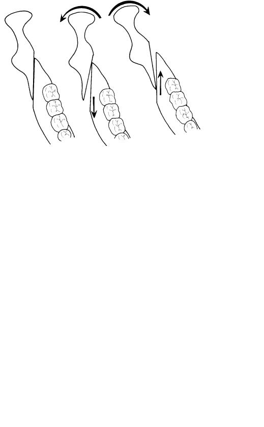
Osteotomies of the vertical ramus (intraoral vertical ramus osteotomy, IVRO) may be considered when the dental arch has to be moved as a unit into its new position, or if the patient prefers to avoid the increased risk of paresthesia associated with SSRO. Most commonly it is used to rotate or reposition the mandible posteriorly in patients with mandibular horizontal excess or asymmetry. It is also used for secondary correction of prior SSRO failures. Robinson44 has suggested its use for mandibular advancements, but because the segment lacks perioperative stability, IVRO is normally not recommended for this type of movement.
These osteotomies extend from the sigmoid notch vertically
FIGURE 50.16 For large setbacks the distal portion of proximal segment needs to be trimmed to avoid interferences. (From Bloomquist29)
50. Mandibular Osteotomies and Considerations for Rigid Internal Fixation |
615 |
behind the inferior alveolar nerve foramen to the inferior border or angle of the mandible (see Figure 50.8h). Originally the procedure was done transcutaneously but with the development of oscillating saws and blades a transoral approach became the standard surgical approach.
The surgical incision is similar to the full-thickness mucoperiosteal flap of the sagittal split ramus osteotomy (Figure 50.14). The incision is curvilinear and medial to the buccal fat pad, along the external oblique ridge, and approximately 3 cm in length. The periosteum is reflected laterally to expose the entire ramus from the sigmoid notch to the angle of the mandible. The soft tissues are then retracted and the antelingula is identified as an anatomic reference mark of the inferior alveolar nerve position. The osteotomy can now be safely completed about 7 mm anterior to the posterior border of the ramus but posterior to the mandibular foramen.29 The osteotomy extends from the sigmoid notch down to the angle of the mandible and involves both medial and lateral cortices (see Figure 50.8h). After completion of the IVRO, the proximal segment is placed lateral to the distal segment and the patient is placed in maxillomandibular fixation. Medial pterygoid attachments are preserved to maintain superior seating of the condyles.
The main fixation method for this type of osteotomy is 6 to 8 weeks of MMF, which is necessary to allow bony union. In order to ensure seating of the condyle in the fossa,29 minimal medial pterygoid stripping is performed.
When using rigid fixation, a percutaneous trocar is used for appropriate screw placement. The easiest method is to apply two to three screws in a linear fashion securing the proximal to the distal segment.37 Application of miniplates provides the most rigid fixation, but their placement must be passive to avoid condylar displacement.
Rigid Internal Fixation
Early reports of mandibular osteotomies described fixation of segments via direct wire osteosynthesis and maxillomandibular fixation. With the advent of internal rigid fixation techniques and the development of hardware for maxillofacial applications, orthognathic surgery is now possible without prolonged MMF fixation. This advance has reduced the discomfort associated with weight loss, perioperative airway management difficulties, extended disability, and poor control of segment position during the immediate healing phase.
The best osteosynthesis method for sagittal split ramus osteotomies is still a matter of controversy. Many techniques however have been described for fixation of the proximal seg- ment—wire,62 pins,63 bone plates,64 lag screws or positional screws,3,58,65–68 and recently resorbable PLLA screws.69 Spiessl’s technique58 utilized decortication of the external oblique ridge to avoid injury to the mandibular nerve. He recognized the importance of flat sectioning of the mandible to avoid the Hunsuck effect (Figure 50.17) and the role that dif-
ferences in cross-sectional anatomy between the angular and supraangular regions play in achieving this goal. In this way, he was able to achieve interfragmentary compression with 2.7-mm lag screws65 applied transcutaneously at a right angle to the osteotomy. Jeter et al.66 advocated using 0.062-in. threaded Kirschner wire to prepare holes in both distal and proximal segments. Then, 2-mm screws were placed transcutaneously perpendicular to the osteotomy. The pattern of placement depended on location of the mandibular canal and varied from three screws on the superior border to two above and one below the canal and vice versa (Figure 50.18). Based on their modification, they claimed that the frequency of neurologic deficiencies decreased from 22% to 4%.66 Since then, several patterns of screw positioning have been proposed: two tapped lag screws above and one below the mandibular canal67; two lag screws below the canal and one above3; two self-tapping screws at the superior border of the ramus to avoid neurovascular bundle compression59,70; three self-tap- ping screws placed obliquely at the superior aspect of the ramus71,72; etc. Lindorf68 proposed the use of tandem screw fixation with tapping of the medial and lateral cortex to provide a passive fit and noncompressive positioning of the segments. Recently, Schwimmer et al.73 studied the effect of screw pattern position and found that the position of the lag screws did not significantly affect the stability of mandibular osteotomies. They also found that a larger 2.7-mm screw provided the same degree of stability as a 2.0-mm screw for stabilization of mandibular osteotomies and supported the already widespread concept that 2.0-mm screws provide adequate stability of mandibular sagittal split osteotomies.74
The effect of screw angle on the stability of the mandibular sagittal split ramus osteotomy has been investigated.71,72,75 It appears that screws oriented at an oblique angle across the osteotomy behave no differently than those placed transcutaneously and perpendicular to the osteotomy.75 No statistically significant difference in stability is gained by the transoral approach.75
Other reported methods for internal rigid fixation of mandibular sagittal split osteotomies have included the use of singleand double-plate systems.76,77 Single plates are considered to be semirigid fixation and may require the use of skeletal wires or elastic traction to complete immobilization (Figure 50.19). Comparisons of nonrigid wire fixation with rigid fixation osteosynthesis78,79 have shown that rigid fixation with lag screws results in fewer skeletal changes and improved stability as compared to wire fixation.
An increased risk of mandibular nerve damage has been reported to occur when rigid internal fixation80 is accomplished with bicortical screws. This may result from compression, direct laceration, or even transection of the nerve during screw placement. The use of miniplates with monocortical screws offers an advantage because compression and direct nerve injury may be avoided.80 If nerve repairs are necessary, plating also prevents tension on the repaired nerve and facilitates its recovery.

616 |
V. Escobar, A.M. Greenberg, and A. Schwimmer |
a
b
c
FIGURE 50.17 Spiessl’s technique for mandibular sagittal split ramus osteotomies without Hunsuck effect to facilitate lag screw fixation.
(a) External oblique ridge corticotomy to allow mandibular ramus sagittal splitting. (b) Cross-sectional view demonstrating complete
outer cortex splitting to avoid Hunsuck effect and nerve injury. (c) Complete anteroposterior and superoinferior mandibular sagittal ramus splitting without Hunsuck effect. (From Spiessl,98 with permission)
50. Mandibular Osteotomies and Considerations for Rigid Internal Fixation |
617 |
FIGURE 50.18 Screw positioning for maximun bony interface. (From Swift50)
There has been concern regarding the use of rigid internal fixation and its effects on intercondylar width, condylar torquing, and the condyle-to-fossa relationship.17 The changes observed are different in advancement versus setback procedures (Figure 50.20). When advancement is performed, there is agreatertendencyfortheintercondylardistancetoincrease(Figure 50.20b). When the distal segment is retropositioned, the intercondylar distal is narrowed (Figure 50.20c). This has longterm effects on TMJ remodeling, osteoarthrosis, and degenerative changes resulting in progressive condylar resorption (PCR).
Other mandibular osteotomies are now also stabilized with rigid internal fixation, some to a greater degree than others depending on the orientation and shape of the segments. Because of the need for the placement of screws in a linear vertical fashion has been advocated,37 the intraoral vertical ramus osteotomy (IVRO) is usually nonrigidly fixated. The perisurgical stability depends on 6 to 8 weeks of MMF fixation. RIF is limited in most cases by the small amount of bone overlap that occurs after the osteotomy. Application of miniplates37 has also been suggested, but an extraoral approach has been used and although this provides the most rigid fix-
FIGURE 50.19 Bone plate for rigid fixation of sagittal split ramus osteotomy (SSRO). (From Swift50)
ation, it increases the number of screws that must be placed passively to avoid condylar displacement. The inverted “L” osteotomy lends itself to RIF with application of miniplates and bone grafts. In areas of small bone gaps, position screws must be used to maintain the interbony gap and again prevent condylar displacement.37
Horizontal osteotomies of the symphysis (sliding genioplasty) lend themselves to rigid fixation. The flexibility of the various configurations of rigid internal fixation allows a great range of choices. The preferable method, however, is the selection of X-shaped plates (Figure 50.21) bent accordingly to the movement or the use of special genioplasty prebent, preshaped, premeasured X-shaped plates (Figure 50.13c). These plates allow for unequal movements to be accomplished. If an X-shaped plate is not available, paired straight plates should be used to minimize rotation and help distribute forces (Figure 50.13d). It is recommended60 that two screws be placed in each segment and that these be bicortical if possible. Rigid internal fixation also allows for improvement of stability if a bone gap occurs because the graft may be rigidly fixated into position.
For mandibular subapical osteotomies, rigid internal fixation and an acrylic splint are necessary. Again, X-shaped plates are preferable (Figure 50.10) and should be bent into a passive position to avoid occlusal displacement. Although monocortical screws can be used in the toothbearing segment, bicortical screws are preferable.60 The X-shaped plates are preferred because their geometry prevents rotation and distributes the forces with minimum hardware. It should be of concern that the osteotomy be accomplished in such a way that good bone buttressing and contact exist after repositioning of the segment. Bone buttressing makes less hardware for fixation necessary.
For the paramidline or anterior midline osteotomy, the use of rigid fixation is more than appropriate. Here, paired plates, mandibular compression plates, or a standard reconstruction plate is indicated together with a lingual acrylic splint to reposition the segments and provide additional stability (see Figure 50.11).
618 |
V. Escobar, A.M. Greenberg, and A. Schwimmer |
c
a |
b |
FIGURE 50.20 (a) Position of osteotomy segments before movement. (b) Lateral condylar torquing from mandibular distal segment advancement. (c) Medial condylar torquing from mandibular distal segment setback.
Complications of Rigid Internal Fixation
The primary reported advantages of rigid internal fixation are increased stability, rapid bone healing, and avoidance of longterm MMF. Uneventful bone healing is usually observed in most patients and makes RIF an effective and predictable way of treating mandibular osteotomies37,50,60,81 and frac-
tures.8,82–84
When compared to MMF,50 the risk of temporomandibular joint arthrosis secondary to immobility is minimized with rigid internal fixation when compared to maxillomandibular fixation.50 However, 9 months after SSROs these patients are
FIGURE 50.21 Double Y miniplate fixation of genioplasty. (From Shafer and Assael25)
still 2.5 mm short of their preoperative maximal interincisal opening (MIO)85 regardless of whether rigid fixation was used.7,86,87 This reduction in MIO does not occur in patients undergoing simultaneous maxillary and vertical ramus osteotomies who undergo 6 to 8 weeks of MMF,81 suggesting that this phenomenon may be a complication of the osteotomy itself, rather than the MMF. The reason patients with rigid fixation seem to return to their preoperative function more rapidly is that rapid fixation allows early physiotherapy and opening exercises, rather than actually reducing the risk of iatrogenic temporomandibular joint arthrosis. There is no doubt that changes in intercondylar width,88–90 axial inclination of the condylar heads,91 and horizontal vertical positioning of the condylar heads in the glenoid fossa50 occur with application of rigid internal fixation. Tuinzing and Swart92 found that when compression and bicortical screws are used, intercondylar width increases with mandibular advancement and decreases with mandibular retropositioning (Figure 50.20).
In most patients, condylar heads undergo remodeling, resorption, and other adaptive changes with little or no temporomandibular joint symptoms. In addition, during sagittal split ramus osteotomy surgery condylar torquing occurs, which causes edema in and around the joint. This results in immediate occlusal discrepancies and “condylar sagging”.50 Within a few days, this edema decreases and the inferior condylar position resolves with no adverse effects, malocclusion, or TMJ symptoms.50 It is during this period (7–10 days) that postoperative maxillomandibular training elastics are encouraged93 to help retrain the patient’s jaw movements and allow for condylar adaptation to the new occlusion. It also compensates for the muscle spasms that usually accompany extensive surgical movements.
50. Mandibular Osteotomies and Considerations for Rigid Internal Fixation |
619 |
Fibrous ankylosis of the temporomandibular joint following rigid fixation has been reported.94 Ordinarily, when rigid fixation is used, intermaxillary fixation is not necessary. In the patients described, MMF was also used for 6 to 8 weeks in addition to rigid fixation. It has been speculated that in those patients long-term compression of the cartilage by the condylar head resulted in its disintegration and the formation of fibrous adhesions.94
Postoperative infection is a rare occurrence, but it has been reported95 to be three times more frequent among patients with rigid internal fixation than among those with wire osteosynthesis. This may be related to increased soft tissue stripping, associated dead space, and length of operation. It is now routine for surgeons performing orthognathic surgery to use a bolus of antibiotics at the inception of the surgical procedure and to continue their IV use in the immediate postoperative period.
In addition to the trauma caused by the osteotomy procedure, rigid internal fixation has also been reported to increase trauma to the alveolar nerve.3,81 The literature is replete with reports describing an increased incidence of neurosensory disturbances. Neurosensory disturbances may be as high as 60%3 and as low as 1 in 25 patients.96 Experimental studies97 suggest that rigid internal fixation should only be done with bicortical screws, rather than with lag screws. This allows for a constant space between the osteotomy segments and prevents undesirable compression of the inferior alveolar nerve. Placement of the screws themselves may compress, lacerate, or transect the inferior alveolar nerve. Avoidance of screw placement at the inferior border and good radiographic evaluation are necessary to prevent this complication.
The goal of rigid fixation is to achieve stability of segments for predictable and uneventful healing. The fact that rigid fixation permits bone healing by primary intention speaks for a compression technique. Yet, when two segments of bone are held together with compressive force, union will occur without callus formation, but this union will not be stronger. Spiessl98,99 emphasizes this point: “From a clinical standpoint, primary bone union is not the true therapeutic goal” (of rigid fixation). “It is immaterial whether the bone consolidation is accomplished indirectly, through differentiation of fibrous tissue, or by direct regeneration of osteons.” The aim of rigid fixation is to provide a functionally stable fixation of bone segments until bone healing is achieved. One should keep in mind that because the condyle is seated in the fossa and the occlusion is fixed, any gaps between the osteotomy segments should not be rigidly fixated using a compression technique because this would draw the segments together, unseat the condyles, or disturb the occlusion. Conventional bicortical screws will then suffice because stability of the segments is accomplished with avoidance of nerve entrapment by compression. Miniplates and elastic traction MMF can also achieve this goal.
Removal of bone plates and screws has received much attention81 for several reasons. It is argued that such plates act
as stress protectors, not allowing bone to be exposed to the full range of physiologic stresses and increasing the risk of fractures at the osteosynthesis site.81 An increase of metal alloy content has also been found in soft tissues, serum, and
urine.100,101
The basic argument against removal of these implants is that although corrosion does occur, the incidence of infections and other complications is relatively low.3 Also, a surgical procedure to remove the implants carries a morbidity risk for infection, nerve damage, and anesthesia complications.81 With the change to titanium implants, the question of hardware removal is no longer important because titanium, at present, does not appear to have any of the clinical characteristics and properties of the aluminum alloys.100–104 Implant removal should be based on the presence of infection, patient discomfort, or exposure and loosening of the implant. Resorbable plates and screws when used in selected cases avoid the need for later hardware removal.69
Addendum: Distraction Osteogenesis
Distraction osteogenesis has become a new method for mandibular lengthening and widening.105–107 New techniques utilizing extraoral and intraoral devices permit lengthening of the ramus and body.108–110 Lengthening of the ramus and body can be performed as a simultaneous procedure using multivector distraction devices.111 Mandibular widening has also been reported with satisfactory periodontal health main- tained.112–113 Mandibular distraction is particularly advantageous for its use in pediatric patients with microgenia, allowing earlier tracheostomy cannulation and in some cases the avoidance of tracheostomy.114 Mandibular ramus lengthening for hemifacial microsomia can also avoid the multiple combined osteotomy and bone grafting procedures that have been traditionally used.115 Continued evolution of these techniques, such as manual molding of the regenerate callus116 will influence the indications and methods used for traditional mandibular osteotomies.
Conclusions
Mandibular osteotomies have undergone dramatic evolution over the past 150 years with modern refinements of both bonecutting techniques and fixation methods. The future may bring new developments in the form of lighter semirigid resorbable implants. Distraction osteogenesis is a promising new methodology that will undoubtedly provide additional treatment modalities for mandibular osteotomies in the future.
References
1.Hullihen SP. A case of elongation of the under jaw and distortion of the face and neck, caused by a burn: successfully treated. Am J Dent Sci. 1849;9:157–165.
620
2.McDonald WR. Champy bone plate fixation in sagittal split osteotomy for mandibular advancement. Int J Adult Orthod Orthognath Surg. 1987;2:89–97.
3.Steinhauser EW. Bone screws and plates in orthognathic surgery. Int J Oral Surg. 1982;11:209–216.
4.Krekmanov L, Lilja J. Orthognathic surgery with no post-op- erative intermaxillary fixation. Scand J Plast Reconstr Surg. 1986;21:189–197.
5.Cawood J. Small plate osteosynthesis of mandibular fractures.
Br J Oral Maxillofac Surg 1985;23:77–91.
6.Leonard MS, Ziman P, Bevis R, Cavanaugh G, Speidel MT, Worms F. The sagittal split osteotomy of the mandible. Oral Surg Oral Med Oral Pathol. 1985;60:459–466.
7.Aragon SB, Van Sickels JE, Dolwick F, Flanary CM. The effects of orthognathic surgery on mandibular range of motion.
J Oral Maxillofac Surg. 1985;43:938–943.
8.Storum KA, Bell WH. Hypomobility after maxillary and mandibular osteotomies. Oral Surg Oral Med Oral Pathol. 1984;57:7–12.
9.Aragon SB, Van Sickels JE. Mandibular range of motion with rigid/non-rigid fixation. Oral Surg Oral Med Oral Pathol. 1987;63:408–411.
10.Lydiatt DD, Davis LF. The effects of immobilization on the rabbit temporomandibular joint. J Oral Maxillofac Surg. 1985; 43:188–293.
11.Van Sickels JE, Flanary CM. Stability associated with
mandibular advancement treated by rigid osseous fixation.
J Oral Maxillofac Surg. 1985;43:338–341.
12.Kirkpatrick TB, Woods MG, Swift JQ, Markowitz NR. Skeletal stability following mandibular advancement and rigid fixation. J Oral Maxillofac Surg 1987;45:572–576.
13.Van Sickels JE. Relapse after rigid fixation of mandibular advancement. J Oral Maxillofac Surg. 1986;44:698–702.
14.Storum KA, Bell WH. The effects of physical rehabilitation on mandibular function after ramus osteotomies. J Oral Maxillofac Surg. 1986;44:94–99.
15.Epker BN, Turvey T, Fish LC. Indications for simultaneous mobilization of the maxilla and mandible for the correction of dentofacial deformities. Oral Surg Oral Med Oral Pathol. 1982;54:369–381.
16.Turvey T, Hale DJ, Fish LC. Surgical-orthodontic treatment planning for simultaneous mobilization of the maxilla and mandible in the correction of dentofacial deformities. Oral Surg Oral Med Oral Pathol. 1982;54:491–498.
17.Kraut RA. Simultaneous maxillary and mandibular orthognathic surgery stabilized by rigid internal fixation. Oral Surg Oral Med Oral Pathol. 1990;69:427–430.
18.Spitzer W, Rettinger G, Sitzmann F. Computerized tomographic examination for the detection of positional changes in the temporomandibular joint after ramus osteotomies with screw fixation. J Oral Maxillofac Surg 1984;12:139–242.
19.Persson G, Hellem S, Nord PG. Bone plates for stabilizing of the LeFort I osteotomy. J Oral Maxillofac Surg. 1986;14:69–73.
20.Dingman RO. Surgical correction of mandibular prognathism.
Am J Orthod Oral Surg. 1944;30:683–692.
21.Blair VP. Report of a case of double resection for the correction of protrusion of the mandible. Dent Cosmos. 1906;48: 817–820.
22.Harsha WM. Bilateral resection of the jaw for prognathism: report of a case. Surg Gynecol Obstet. 1912;15:51–53.
V.Escobar, A.M. Greenberg, and A. Schwimmer
23.Hofer O. Die Operative behandlung der alveolären Retraktion des Unterkiefers u ihre Anwendungsmöglichkeit für Prognathie und Mikrogenie. Dtsch-Zahn-Mund Kieferheilkd. 1942;9(3): 121–132.
24.Köle H. Indikation und Plauong von kieferorthopädischen Operationen (Distalbiss, alveolare protursion und retrusion).
Oesterr Z Stomatol. 1960;57:212.
25.Shafer DM, Assael LA. Rigid internal fixation of mandibular segmental osteotomies. Atlas Oral Maxillofac Surg Clin North Am. 1993;1:41–51.
26.Kent JN, Hinds E. Management of dental facial deformities by anterior alveolar surgery. J Oral Surg. 1971;29:13–26.
27.MacIntosch RB. Total mandibular alveolar osteotomy: encouraging experiences with an infrequently indicated procedure. J Maxillofac Surg. 1974;2:210–218.
28.Angle EH. Double resection of the lower maxilla. Dent Cosmos. 1889;40:635–638.
29.Bloomquist DS. Principles of mandibular orthognathic surgery. In: Peterson LJ, Andresano AT, Marciani RD, Roser SM, eds.
Principles of Oral and Maxillofacial Surgery. Philadelphia: JB Lippincott; 1992:1415–1463.
30.Von Eiselberg J. Ueber plastik bei Ektropium des Unterkiefers (progenie). Wien Klin Wochenschr. 1906;19:1505–1508.
31.Blair VP. Operations of the jaw bone and face. Surg Gynecol Obstet. 1907;4:67–78.
32.Limberg A. Treatment of open-bite by means of plastic oblique osteotomy of the ascending rami of the mandible. Dent Cosmos. 1925;67:1191–1200.
33.Moose SM. Surgical correction of mandibular prognathism by intraoral subcondylar osteotomy. J Oral Surg Anesth Hosp Dent Serv. 1964;22:197–202.
34.Kazanjian V, Converse J. The Surgical Treatment of Facial Injuries. 2nd ed. Baltimore: Williams & Wilkins; 1959.
35.Ernst F. Uber die chirurgische beseitigung der prognathie des unterkiefers. Zentralbl Chir. 1938;65:179.
36.Obwegeser H, Trauner R. Zur Operationstechnik bei der Progenie und anderen Unterkieferanomalien. Dtsch Zahn Kieferheilkd. 1955;23:H1–H2.
37.Tucker MR. Surgical correction of mandibular excess. Atlas Oral Maxillofac Surg Clin North Am. 1993;1:29–39.
38.Kostec˘ka F. A contribution to the surgical treatment of open bite. Int J Orthod. 1934;28:1082–1092.
39.Thoma KH. Surgical treatment of deformities of the jaw. Am J Orthod. 1946;32:333–339.
40.Robinson M. Prognathism corrected by open vertical subcondylotomy. J Oral Surg. 1958;16:215–219.
41.Caldwell JB, Letterman GS. Vertical osteotomy in the mandibular rami for correction of prognathism. J Oral Surg. 1954;12:185–202.
42.Robinson M. Micrognathism corrected by vertical osteotomy of ascending ramus and iliac bone graft: a new technique. Oral Surg Oral Med Oral Pathol. 1957;10:1125–1130.
43.Caldwell JB, Amaral WJ. Mandibular micrognathism corrected by vertical osteotomy in the rami and iliac bone graft. J Oral Surg. 1960;18:3–15.
44.Robinson M, Lytle JJ. Micrognathism corrected by vertical osteotomies of the rami without bone grafts. Oral Surg Oral Med Oral Pathol. 1962;15:641–645.
45.Wassmund M. Fracturen und Luxationen des Gesichtschadels unter Beruksichtigung der Komplikationen des Hirnschadels.
50. Mandibular Osteotomies and Considerations for Rigid Internal Fixation |
621 |
In Klinik und Therapie. Praktischen Lehrbuch, Vol. 20 Berlin: Hermann Meusser; 1927.
46.Hawkinson RT. Retrognathia correction by means of an arcing osteotomy in the ascending ramus. J Prosthet Dent. 1968; 20:77–86.
47.Hayes PA. Correction of retrognathia by modified “C” osteotomy of the ramus sagittal osteotomy of the mandibular body. J Oral Surg. 1973;31:682–686.
48.Fox GL, Tilson HB. Mandibular retrognathia: a review of the literature and selected cases. J Oral Surg. 1976;34:53–61.
49.Caldwell JB, Hayward JR. Lister RL. Correction of mandibu-
lar retrognathia by vertical-L osteotomy: a new technique. J Oral Surg. 1968;26:259–264.
50.Swift JQ. Mandibular advancement. Atlas Oral Maxillofac Surg Clin North Am. 1993;1:17–27.
51.Trauner R, Obwegeser H. The surgical correction of mandibular prognathism and retrognathia with considerations of genioplasty. I. Surgical procedures to conect mandibular prognathism and reshaping the chin. Oral Surg Oral Med Oral Pathol. 1957;10:677–689.
52.Neuner O. Circa un nuovo metodo per la cura chirugica della progenia. Riv Ital Stomatol. 1958;13:1573.
53.Neuner O. Beitrag zur progenie operationen am aufsteigenden unterkieferast. Dtsch Zahnaerztl Z. 1958;13:1416.
54.Dal Pont G. Retromolar osteotomy for the correction of prognathism. J Oral Surg. 1961;19:42–47.
55.Hunsuck EE. A modified intraoral sagittal splitting technic for correction of mandibular prognathism. J Oral Surg. 1968;26: 250–253.
56.Wolford LM, Davis WM. The mandibular inferior border split: a modification in the sagittal split osteotomy. J Oral Maxillofac Surg. 1990;48:92–94.
57.Epker BN. Modification in the sagittal osteotomy of the mandible. J Oral Surg. 1977;35:157–159.
58.Spiessl B. Osteosynthese bei sagittaler Osteotomie nach Obwegeser-Dal Pont. Fortschr Kieferheilkd Gesichtschir. 1974;18:145–148.
59.Shufford EL, Kraut RA. Passive rigid fixation of sagittal split osteotomy. Oral Surg Oral Med Oral Pathol. 1989;68:150–153.
60.Shafer DM, Assael LA. Rigid internal fixation of mandibular segmental osteotomies. Atlas Oral Maxillofac Surg Clin North Am. 1993;1:41–51.
61.Ritzau M, Wenzel A, Williams S. Changes in condyle position after bilateral vertical ramus osteotomy with and without osteosynthesis. Am J Orthod Dentofacial Orthop. 1989;96:507–513.
62.Booth DF. Control of the proximal segment by lower border wiring in the sagittal split osteotomy. J Oral Maxillofac Surg. 1981;9:126–128.
63.Gingrass DJ, Messer EJ. Rigid non-compressive pin fixation of the mandibular sagittal split osteotomy. J Oral Maxillofac Surg. 1986;44:413–416.
64.McDonald WR, Stoelinga PJ, Blijdorp PA, Schoenaers JA. Champy bone plate fixation in sagittal split osteotomies for mandibular advancement. Int J Adult Orthod Orthognath Surg. 1987;2:89–97.
65.Spiessl B. The sagittal splitting osteotomy for correction of mandibular prognathism. Clin Plast Surg. 1982;9:491–507.
66.Jeter TS, Van Sickels JE, Solwick MF. Modified techniques for internal fixation of sagittal ramus osteotomies. J Oral Maxillofac Surg. 1984;42:270–272.
67.Spiessl B. Rigid internal fixation after sagittal split osteotomy of the ascending ramus. In: Spiessl B. New Concepts in Maxillofacial Bone Surgery. New York: Springer-Verlag; 1976:21.
68.Lindorf HH. Sagittal split osteotomy with tandem screw fixation: techniques and results. J Oral Maxillofac Surg. 1986;14: 311–316.
69.Suuronen R, Laine P, Sarkiala E, Pohjnen T, Lindqvist C. Sagittal split ramus osteotomy fixed with biodegradable selfreinforced poly-L-lactide screws. A pilot study in sheep. Int J Oral Maxillofac Surg. 1992;21:303–308.
70.Shepherd JP, Dohvoma CN, Harradine NW. Screw fixation after mandibular sagittal split osteotomy: an intraoral approach.
Br J Oral Maxillofac Surg. 1991;29:325–329.
71.Turvey T, Hall D. Intraoral self-threading screw fixation for sagittal osteotomies: early experiences. Int J Adult Orthod Orthognath Surg. 1986;1:243–250.
72.Kempf KK. Transoral technique for rigid fixation of sagittal ramus osteotomies. J Oral Maxillofac Surg. 1987;45:1077– 1079.
73.Schwimmer A, Greenberg AM, Kummer G, Kaynar A. The effect of screw size and insertion technique on the stability of the mandibular sagittal split osteotomy. J Oral Maxillofac Surg. 1994;52:45–48.
74.Van Sickels JE, Richardson DA. Stability of orthognathic surgery: a review of rigid fixation. Br J Oral Maxillofac Surg. 1996;34:279–285.
75.Foley WL, Frost DE, Paulin WB, et al. Internal screw fixation: Comparison of placement pattern and rigidity. J Oral Maxillofac Surg. 1989;47:720.
76.Luhr HG. Compression plate osteosynthesis through the Luhr system. In: Kruger E, Schilli S, eds. Oral and Maxillofacial Traumatology. Vol. 1. Chicago: Quintessence; 1982:319.
77.Champy M, Pape HD, Gerlach KL. The Strasbourg miniplate osteosynthesis. In: Kruger E, Schilli, W, eds. Oral Maxillofacial Traumatology. Vol. 2. Chicago: Quintessence; 1986:19.
78.Thomas PM, Tucker MR, Prewitt JR, Proffit WR. Early skeletal and dental changes following mandibular advancement and rigid internal fixation. Int J Adult Orthod Orthognath Surg. 1986;3:171–178.
79.Van Sickels JE, Flanary CM. Stability associated with
mandibular advancement treated by rigid osseous fixation.
J Oral Maxillofac Surg. 1985;43:338–341.
80.Nishioka G, Zysset M, Van Sickels J. Neurosensory disturbance with rigid internal fixation of the bilateral sagittal split osteotomy. J Oral Maxillofac Surg. 1987;45:20–26.
81.Leonard MS. Rigid internal fixation: facts vs. fallacies. Atlas Oral Maxillofac Clin North Am. 1990;2:737–743.
82.Strelzow VV, Strelzow AG. Osteosynthesis of mandibular fractures of the angle region. Arch Otolaryngol. 1983;109:403– 406.
83.Tu HK, Tenhulzen D. Compression osteosynthesis of mandibular fractures: a retrospective study. J Oral Maxillofac Surg. 1985;43:585–589.
84.Iizuka T, Lindqvist C. Rigid internal fixation of fractures in the angular region of the mandible: an analysis of factors contributing to different complications. Plast Reconstr Surg. 1993; 91:265–271.
85.Stacy GC. Recovery of oral opening following sagittal ramus osteotomy for mandibular. J Oral Maxillofac Surg. 1987;45: 487–492.
622
86.Storum KA, Bell WH. Hypomobility after maxillary and mandibular osteotomies. Oral Surg Oral Med Oral Pathol. 1984;57:7–12.
87.Storum KA, Bell WH. The effect of physical rehabilitation on mandibular function after ramus osteotomies. J Oral Maxillofac Surg. 1986;44:94–99.
88.Freihofer HP Jr. Modellversuch zur Lageveranderung des kieferkopfchens nach sagittaler Spaltung des Unterkiefers. SSO Schweiz Monatsschr Zahnheilkd. 1977;87:12–22.
89.Kundert M, Hadjianghelou O. Condylar displacement after sagittal splitting of the mandibular rami. A short term radiographic study. J Maxillofac Surg. 1980;8:278–287.
90.Spitzer W, Rettinger G, Sitzmann F. Computerized tomography examination for the detection of positional changes in the temporomandibular joint after ramus osteotomies with screw fixation. J Oral Maxillofac Surg. 1984;12:139–142.
91.Hackney FL, Van Sickels JE, Nummikoski PV. Condylar displacement and temporomandibular joint dysfunction following bilateral sagittal split osteotomies and rigid fixation. J Oral Maxillofac Surg. 1989;47:223–227.
92.Tuinzing DB, Swart JGN. Lageveranderungen des caput mandibular bei verwendung von zugschrauben nach sagittaler osteotomie des unterkiefers. Dtsch Zahn Mund Kieferheilkd Gesichts Chir. 1978;2:94.
93.Bock JT. Horizontal condylar angulation changes following forward and backward sagittal split ramus osteotomy with rigid screw fixation. Dissertation. Minneapolis: University of Minnesota; 1986.
94.Nitzan DW, Dolwick MF. Temporomandibular ankylosis following orthognathic surgery: report of eight cases. Int J Adult Orthodon Orthognath Surg. 1989;4:7–11.
95.Buckley MJ, Tullock JF, White RP Jr, Tucker MR. Complications of orthognathic surgery: a comparison between wire fixation and rigid internal fixation. Int J Adult Orthodon Orthognath Surg. 1989;2:69–74.
96.Souryis F. Sagittal splitting and bicortical screw fixation of the ascending ramus. J Maxillofac Surg. 1978;6:198–203.
97.Ikemura K, Hidaka H, Etoh T, Kabata K. Osteosynthesis in facial bone fractures using miniplates: clinical and experimental studies. J Oral Maxillofac Surg. 1988;46:10–14.
98.Spiessl B. The stability principle. In: Internal mandible. New York: Springer-Verlag; 1988:16.
99.Spiessl B. Biomechanics. In: Internal fixation of the Mandible. New York: Springer-Verlag; 1988:28.
100.Balck J. Does corrosion matter? J Bone Joint Surg. 1988;70: 517–520.
101.Gillespie WJ, Frampton CMA, Henderson PJ. The incidence of cancer following total hip replacement [published erratum
V. Escobar, A.M. Greenberg, and A. Schwimmer
appears in J Bone Joint Surg [Br] 1996 Jul;78(4):680]. J Bone Joint Surg. 1988;70:539–542.
102.Bell WH. Modern Practice in Orthognathic and Reconstructive Surgery. Vol. III. Philadelphia: WB Saunders; 1992.
103.Powell NB, Riley RW. Obstructive sleep apnea. Atlas Oral Maxillofac Surg Clin North Am. 1990;2(4):843–853.
104.Edwards RC, Kiely KD, Eppley BL. Fixation of bimaxillary osteotomies with resorbable plates and screws: experience in 20 consecutive cases. J Oral Maxillofac Surg. 2001;59:271–276.
105.McCarthy JG, Stelnicki EJ, Grayson BH. Distraction osteogenesis of the mandible: a ten year experience. Semin Orthod. 1999;5:3–8.
106.van Strijen PJ, Perdijk FB, Becking AG, et al. Distraction osteogenesis for mandibular advancement. Int J Oral Maxillofac Surg. 2000;29:81–85.
107.Carls FR, Sailer HF. Seven years clinical experience with mandibular distraction in children. J Craniomaxillofac Surg. 1998;26:197–208.
108.Dessner S, Razdolsky Y, El-Bialy T, et al. Mandibular lengthening using programmed intraoral tooth-borne distraction devises. J Oral Maxillofac Surg. 1999;57:1318–1322.
109.McCarthy JG, Williams JK, Grayson BH, et al. Controlled multiplanar distraction of the mandible: device development and clinical application. J Craniofac Surg. 1998;9:322–329.
110.Haug RH, Nuveen EJ, Barber JE, et al. An in vitro evaluation of distractors used for osteogenesis. Oral Surg Oral Pathol Oral Radiol Endod. 1998;86:648–659.
111.Gateno J, Teichgraeber JF, Aguilar E. Distraction osteogenesis: a new surgical technique for use with the multiplanar mandibular distractor. Plast Reconstr Surg. 2000;105:883–888.
112.Guerero CA, Bell WH, Costasti GI, et al. Mandibular widening by intraoral distraction osteogenesis. Br J Oral Maxillofac Surg. 1997;35:383–392.
113.Kewitt GF, Van Sickels JE. Long term effect of mandibular midline distraction osteogenesis on the status of the temporomandibular joint, teeth, peridontal structures, and neurosensory function. J Oral Maxillofac Surg. 1999;57:1419–1425.
114.Morovic CG, Monasterio L. Distraction osteogenesis for obstructive apneas in patients with congenital craniofacial malformations. Plast Reconstr Surg. 2000;105:2324–2330.
115.Kusnoto B, Figueroa AA, Polley JW. A longitudinal threedimensional evaluation of the growth pattern in hemifacial microsomia treated by mandibular distraction osteogenesis: a preliminary report. J Craniofac Surg. 1999;10:480–486.
116.Kunz C, Hammer B, Prein J. Manipulation of the callus after linear distraction: a “lifeboat” or an alternative to multivectorial distraction osteogenesis of the mandible? Plast Reconstr Surg. 2000;10:674–679.