

23
Aesthetic Considerations in Reconstructive and Corrective Craniomaxillofacial Bone Surgery
R. Gregory Smith and Luc M. Cesteleyn
Although the bony architecture of the face is a major component in the perception of facial harmony, it must not be viewed as the most important. The truly aesthetic face is defined by the abstract interplay of symmetry, balance, projection, and animation created within the soft tissue envelope overlying its bony foundation.
Concepts of Facial Harmony
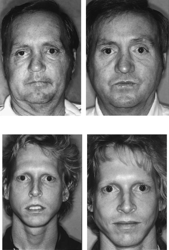
Many attempts have been made to quantify facial beauty dating back to early times. Da Vinci was a student of body proportion and symmetry. His studies furnished important data upon which many modern concepts of facial beauty are based.1 In the study of facial harmony, symmetry is one of the most obvious yet most critical concepts to appreciate, especially when dealing with the facial bony base. The aesthetic face requires reasonable symmetry. Any disturbance of equality of the facial halves is usually quite obvious to even the most casual observer. When planning a surgical procedure (either reconstructive or cosmetic), the surgeon must have symmetry as a primary goal.2 Even with the use of state-of- the-art rigid internal fixation to align bony fragments, this is not always possible, especially in the trauma patient. The following two examples will illustrate this concept. The patient in Figure 23.1a,b is shown preoperatively after multiple midface and mandible fractures and postoperatively after multiple reconstructive procedures. Final symmetry was achieved after realignment of bony segments using rigid internal fixation, and later insertion of a right malar implant to compensate for overlying soft tissue atrophy on the right and previous overprojection of the zygoma on the left. The patient in Figure 23.2a,b is shown preoperatively status post open reduction with rigid internal fixation of a displaced left zygomaticomaxillary complex fracture, operated elsewhere. The patient felt this fracture was still minimally displaced and requested reoperation. The postoperative view shows nearperfect reduction of this fracture with the use of microplates via a bicoronal approach. At a separate operation, the patient’s preexisting congenital facial asymmetry of the lower half of
the face was corrected. Improved facial symmetry resulted with improvement in overall aesthetic balance.
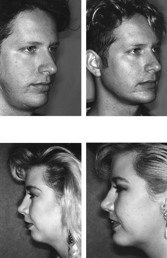
Balance among the aesthetic units of the face plays a very important role in creating facial harmony. The facial units that shouldbesystematicallystudiedpreoperativelyincludetheforehead, nose, eyes, malar prominences, lips, chin, and mandibular angles.3 No one feature should overpower the interrelationship between units. Features that are out of proportion should be considered for change during the planned cosmetic or reconstructive procedure. Once again, several examples will serve to illustrate the concept of balance. Figure 23.3a,b demonstrate a patient, preoperatively and postoperatively, who was treated for multiple complaints of facial imbalance in the areas of the malar prominences, chin, and mandibular angles. His facial balance was dramatically improved by placement of malar and mandibular angle implants and performance of a rigidly fixated bony genioplasty. The patient in Figure 23.4a,b shows preoperative and postoperative results after a rigidly fixated bony genioplasty and full face/neck liposuction. Again, remarkable improvement was achieved by reestablishing more favorable facial balance of the aesthetic units.
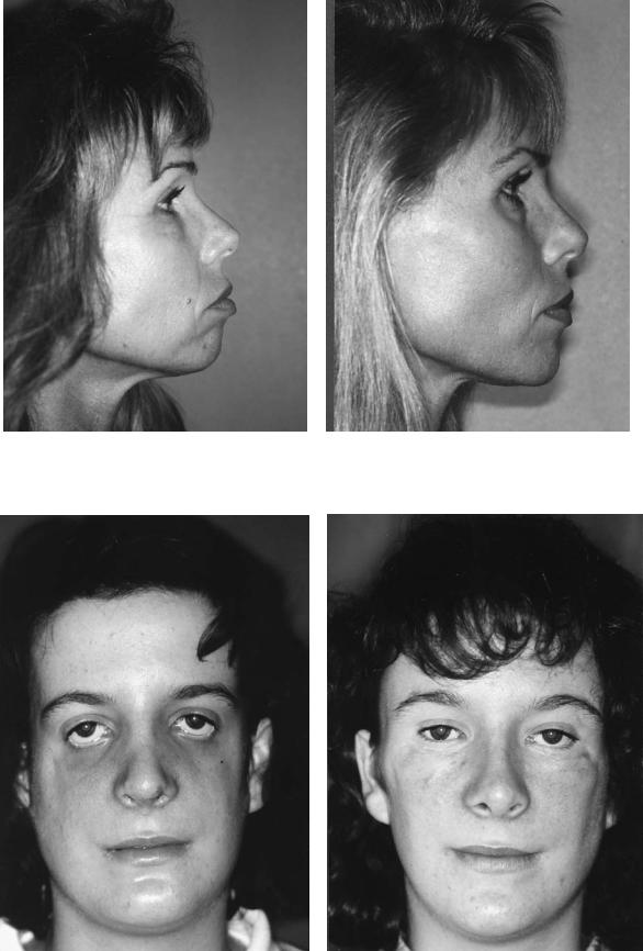
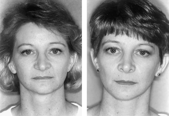
The projection of bony aesthetic units provides contours over which the facial soft tissue may drape. Lack of appropriate projection of the underlying facial skeleton leads to an amorphous facial appearance, which often appears more aged or plain.4,5 The facial units appear to blend together and inelastic soft tissue may sag in the older patient, adversely impacting facial aesthetics as seen in Figure 23.5a,b.
Maintenance of jaw and bony landmark projection is imperative to accentuate the transitions between the aesthetic units of the face. This concept is especially important in craniomaxillofacial and orthognathic surgery.
Two patients who lack important areas of facial projection are illustrated by the following examples. Figure 23.6a,b show preoperative and postoperative views of a TreacherCollins syndrome patient treated by cranial bone reconstruction of the zygomas, orthognathic surgical correction of malocclusion with rigid fixation, lower lid-switch blepharoplasties, malar implant insertion, and conservative rhinoplasty. In this case, both hard and soft tissues required
280
a |
|
|
|
|
|
|
|
b |
|
|
|
|
|
|
|
|
|
|
|
|
|
|
|
|
|
|
|
|
|
|
|
|
|
|
|
|
|
|
|
|
|
|
|
|
FIGURE 23.1 (a) Patient shown preoperatively status post multiple midface and mandibule fractures. (b) Patient shown postoperatively status post reduction of multiple midface and mandible fractures and placement of right malar implant.
a |
|
|
|
b |
||||
|
|
|
|
|
|
|
|
|
|
|
|
|
|
|
|
|
|
|
|
|
|
|
|
|
|
|
|
|
|
|
|
|
|
|
|
|
|
|
|
|
|
|
|
|
FIGURE 23.2 (a) The patient shown preoperatively status post open reduction of left zygomatic maxillary complex (ZMC) fracture operated elsewhere. (b) The patient shown postoperatively status post reoperation of left ZMC fracture and reduction via micro-miniplates
and status post Le Fort I maxillary osteotomy and bilateral sagittal split mandibular osteotomies with rigid internal fixation. Greater facial balance has been achieved.
a |
|
|
|
|
|
|
|
b |
|
|
|
|
|
|
|
|
|
|
|
|
|
|
|
|
|
|
|
|
|
|
|
|
|
|
|
|
|
|
|
|
|
|
|
|
FIGURE 23.3 (a) The patient shown preoperatively with facial imbalance secondary to malar deficiency, mandibular angle deficiency, and genial deficiency. (b) The patient shown postoperatively after
placement of bilateral malar implants, mandibular angle implants, and rigidly fixated advancement bony genioplasty.
a |
|
|
|
b |
|
|
|
|
|
|
|
|
|
|
|
|
|
|
|
|
|
|
|
|
FIGURE 23.4 (a) Patient shown preoperatively with genial deficiency and increased facial liposity. (b) Patient shown postoperatively status post rigid fixated bony genioplasty and full face/neck liposuction.
a |
|
|
|
b |
|
|
|
|
|
|
|
|
|
|
|
|
|
|
|
|
|
|
|
|
FIGURE 23.5 (a) Patient shown preoperatively with genial and malar deficiency. (b) Patient shown postoperatively status post placement of bilateral malar implants and rigidly fixated advancement bony genioplasty.
a |
|
|
|
|
|
|
|
b |
|
|
|
|
|
|
|
|
|
|
|
|
|
|
|
|
|
|
|
|
|
|
|
|
|
|
|
|
|
|
|
|
|
|
|
|
FIGURE 23.6 (a) Patient shown preoperatively with Treacher-Collins syndrome with absent zygomas. The patient is also status postorthognathic surgical correction of her malocclusion and has had con-
servative rhinoplasty. (b) Patient shown 1 year postoperatively. Patient shown status post cranial bone graft reconstruction of zygomas, lower lid-switch blepharoplasties, and placement of malar implants.
284 |
|
|
|
|
|
R.G. Smith and L.M. Cesteleyn |
||
a |
|
|
|
|
|
|
|
b |
|
|
|
|
|
|
|
|
|
|
|
|
|
|
|
|
|
|
|
|
|
|
|
|
|
|
|
FIGURE 23.7 (a) Patient with bimaxillary retrusion status post previous orthodontic correction of Class II malocclusion and extraction of four bicuspid teeth. (b) Patient shown status postmaxillary ad-
augmentation to achieve the appearance of adequate projection. The patient in Figure 23.7a,b had undergone four previous bicuspid extractions and orthodontic treatment of a class II malocclusion, which left her with a sunken-in appearance secondary to maxillomandibular deprojection. The postoperative views show the correction achieved with application of internal rigid fixation to allow downgraft advancement of the maxilla and advancement of the mandible, which was combined with simultaneous conservative rhinoplasty. Reestablishment of proper facial projection has achieved dramatic improvement.
Finally, facial animation plays a paramount role in the aesthetic appearance of the face. In short, if the soft tissues do not move, no alteration of the amount of symmetry, balance, and projection will make it aesthetic. Often, major soft tissue injuries to muscle, skin, and nerves leave little chance of normal animation, even if the bony framework is restored to a normal position. Knowledge and skill in soft tissue repair is mandatory for the surgeon. However, coverage of these areas is beyond the scope and mission of this text.
Quantifying Facial Harmony
To this point, the authors have dealt only with the basic abstract concepts that they believe define the aesthetic face. However, numerous works have been completed that objec-
vancement and down grafting with rigid internal fixation, bilateral sagittal split advancement osteotomies, with rigid internal fixation, and conservative rhinoplasty.
tively measure both hard and soft tissue aesthetic characteristics, and these must not go unnoticed. Although numbers cannot completely describe the aesthetic face, they provide useful references when attempting to quantify relationships. This is especially useful to the surgeon who has not yet developed an “aesthetic sense.”
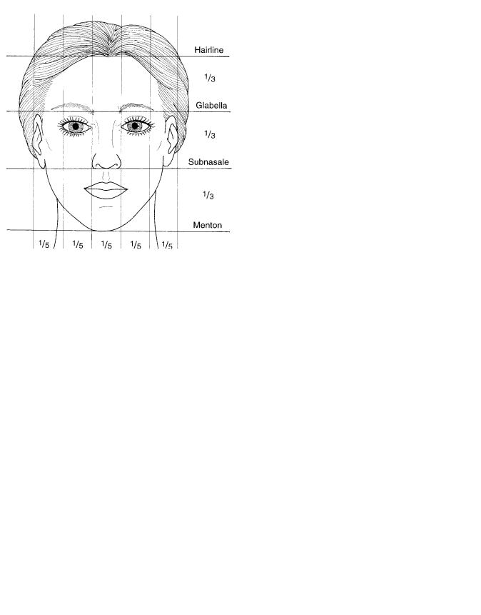
In general, when viewed from the front, the face is divided by the midline vertically, and similar structures in the respective halves are symmetrical. The face is normally broken up into “fifths,” being five average “eye widths” wide (Figure 23.8).2 Facial height is proportionally divided into equal thirds by lines drawn horizontally through the junction of the hairline and forehead skin, subnasale, and menton (Figure 23.8). Trauma victims frequently increase or decrease the various facial thirds owing to the displacement or impaction of facial bones. This is also common in congenital maxillomandibular deformities expressed as too much or too little jaw growth.
In profile, the projection and interrelationships of facial aesthetic units such as the forehead, nose, dental structures, and jaws are extremely important. Their “normalization” can greatly enhance facial aesthetics as previously shown.
Beginning with lateral cephalometric analysis of hard tissue structures, the two most important landmarks are the Frankfort horizontal line, defined as a line drawn from the upper part of the external auditory meatus to the infraorbital
23. Aesthetic Considerations |
285 |
|
|
TABLE 23.1 Cephalometric analysis. |
|
|
|
|
|
|
Range |
|
|
|
|
Mandibular plane angle (FH-MP) |
26° 4.5 |
|
Maxillary depth (FH-NaA) |
90° 3 |
|
Facial depth (FH-NaPO) |
87° 3 |
|
Point A to nasion perpendicular |
1 mm |
|
PO to nasion perpendicular |
2 to 4 mm |
|
Overbite |
3 1.5 mm |
|
Overjet |
1–2 mm |
|
Upper incisor—NaA(mm) |
0–8 mm |
|
Lower incisor—NaB(mm) |
2–10 mm |
|
Upper incisor to lower incisor |
120°–140° |
|
Lower anterior dental height |
40 mmF/44 mmM |
|
|
|
FIGURE 23.8 Facial dimensions divided vertically into fifths and horizontally into thirds.
rim, and MacNamara’s line, which is a line that begins at nasion and is dropped perpendicular to the Frankfort horizontal (Figure 23.9).6–8 Using only these two reference lines, the surgeon may identify anomalies of jaw and teeth position using the normal values listed in Table 23.1. For surgeons who are
unfamiliar with dental structures and occlusion, we recommend review of texts dedicated to orthognathic surgery of the jaws.
Soft tissue profile aesthetics have been studied extensively, but several numbers bear remembering. Again, we feel that the two most useful landmarks are the Frankfort horizontal line and the Smith nasion perpendicular (SNP) (Figure 23.10). SNP is defined as a line perpendicular to the Frankfort horizontal line and tangent to the depth of soft tissue nasion and extending through soft tissue pogonion. The aesthetic nasal dorsum takes off from SNP at approximately 35°.3,9 The height of the nasal dorsum measured from the medial canthus area is approximately 15 mm according to Goldman.10 The
FIGURE 23.9 Lateral cephalometric radiographic references for Frankfort horizontal and MacNamara’s line.
FIGURE 23.10 Smith nasion perpendicular (SNP) and Frankfort Horizontal SNP extends through soft tissue nasion and pogoion which differs by these defined landmarks from the zero meriden of Gon- zalez-Ulloa.3
286
nasolabial angle should be in the range of 90° for men and up to 110° for women.3,9,11
The aesthetic forehead slopes away from the SNP at an angle of 20°.12 The glabella is slightly rounded in the midline, not flat, and it projects 2 to 3 mm anterior to SNP.13 This point must be considered during craniofacial reconstruction.
Summary
Although the list of “numbers” presented here is by no means exhaustive, it represents a starting point for an objective assessment of the face as it relates to craniomaxillofacial surgery. When it is used in conjunction with the abstract concepts of beauty previously presented, the surgeon should be able to effectively analyze the individual patient’s face and formulate a surgical plan that will maximize the aesthetic outcome.
References
1.Beeson WH. Facial analysis in aesthetic surgery of the aging face. Beeson WH, McCollough EG, eds. St. Louis: C.V. Mosby Co.; 1980.
2.Bell WH, Proffitt WB, White RP. Surgical Correction of Dentofacial Deformities. Philadelphia: WB Saunders Co.; 1980:115–123.
R.G. Smith and L.M. Cesteleyn
3.Powell N, Humphreys B. Proportions of the Aesthetic Face.
New York: Thieme-Stratton Inc.; 1980:1–9;1984:15–39.
4.Binder WJ, Schoenrock LD, Terino EOL. Augmentation of the malar-submalar/midface. Facial Plastic Surg Clin. 1994;2(3):265– 284.
5.Mittleman H. The anatomy of the aging mandible and its importance to facelift surgery. Facial Plastic Surg Clin. 1994;2(3): 301–310.
6.Tweed CA. The Frankfort-mandibular plane angle in orthognathic diagnosis, classification, treatment planning and prognosis. Am J Orthod Oral Surg. 1946;32:175–230.
7.Mourvees CFA, Kean MR. Natural head position: a basic consideration for analysis of cephalometric radiographs. Am J Phys Anthropol. 1958;16:213–234.
8.McNamara JA Jr. A method of cephalometric analysis. In: McNamara JA Jr, Ribbens KA, Howe RP, eds. Clinical Alteration of the Growing Face. Monograph No. 14, Craniofacial Growth Series. The Center for Human Growth and Development, The University of Michigan, Ann Arbor, MI; 1983.
9.Sheen JH, Sheen AP. Aesthetic Rhinoplasty, Vol 1, 2nd ed. St Louis, MO: C.V. Mosby; 1987:67–127.
10.Goldman IB. Rhinoplasty Manual. NY Restricted Publication, 1968.
11.Aiach G, Levignac J. Aesthetic Rhinoplasty. Edinburgh: Churchill Livingstone; 1991:21–23.
12.Bon M. L’Esthetic Face a la Chirurgie Esthetique. Private Publication. Paris 1977.
13.Tessier P. Personal Communication, 1989.
Section IV
Craniomaxillofacial Reconstructive
Bone Surgery
This page intentionally left blank