
- •Preface
- •Acknowledgments
- •Contents
- •Contributors
- •1. Introduction
- •2. Evaluation of the Craniomaxillofacial Deformity Patient
- •3. Craniofacial Deformities: Review of Etiologies, Distribution, and Their Classification
- •4. Etiology of Skeletal Malocclusion
- •5. Etiology, Distribution, and Classification of Craniomaxillofacial Deformities: Traumatic Defects
- •6. Etiology, Distribution, and Classification of Craniomaxillofacial Deformities: Review of Nasal Deformities
- •7. Review of Benign Tumors of the Maxillofacial Region and Considerations for Bone Invasion
- •8. Oral Malignancies: Etiology, Distribution, and Basic Treatment Considerations
- •9. Craniomaxillofacial Bone Infections: Etiologies, Distributions, and Associated Defects
- •11. Craniomaxillofacial Bone Healing, Biomechanics, and Rigid Internal Fixation
- •12. Metal for Craniomaxillofacial Internal Fixation Implants and Its Physiological Implications
- •13. Bioresorbable Materials for Bone Fixation: Review of Biological Concepts and Mechanical Aspects
- •14. Advanced Bone Healing Concepts in Craniomaxillofacial Reconstructive and Corrective Bone Surgery
- •15. The ITI Dental Implant System
- •16. Localized Ridge Augmentation Using Guided Bone Regeneration in Deficient Implant Sites
- •17. The ITI Dental Implant System in Maxillofacial Applications
- •18. Maxillary Sinus Grafting and Osseointegration Surgery
- •19. Computerized Tomography and Its Use for Craniomaxillofacial Dental Implantology
- •20B. Atlas of Cases
- •21A. Prosthodontic Considerations in Dental Implant Restoration
- •21B. Overdenture Case Reports
- •22. AO/ASIF Mandibular Hardware
- •23. Aesthetic Considerations in Reconstructive and Corrective Craniomaxillofacial Bone Surgery
- •24. Considerations for Reconstruction of the Head and Neck Oncologic Patient
- •25. Autogenous Bone Grafts in Maxillofacial Reconstruction
- •26. Current Practice and Future Trends in Craniomaxillofacial Reconstructive and Corrective Microvascular Bone Surgery
- •27. Considerations in the Fixation of Bone Grafts for the Reconstruction of Mandibular Continuity Defects
- •28. Indications and Technical Considerations of Different Fibula Grafts
- •29. Soft Tissue Flaps for Coverage of Craniomaxillofacial Osseous Continuity Defects with or Without Bone Graft and Rigid Fixation
- •30. Mandibular Condyle Reconstruction with Free Costochondral Grafting
- •31. Microsurgical Reconstruction of Large Defects of the Maxilla, Midface, and Cranial Base
- •32. Condylar Prosthesis for the Replacement of the Mandibular Condyle
- •33. Problems Related to Mandibular Condylar Prosthesis
- •34. Reconstruction of Defects of the Mandibular Angle
- •35. Mandibular Body Reconstruction
- •36. Marginal Mandibulectomy
- •37. Reconstruction of Extensive Anterior Defects of the Mandible
- •38. Radiation Therapy and Considerations for Internal Fixation Devices
- •39. Management of Posttraumatic Osteomyelitis of the Mandible
- •40. Bilateral Maxillary Defects: THORP Plate Reconstruction with Removable Prosthesis
- •41. AO/ASIF Craniofacial Fixation System Hardware
- •43. Orbital Reconstruction
- •44. Nasal Reconstruction Using Bone Grafts and Rigid Internal Fixation
- •46. Orthognathic Examination
- •47. Considerations in Planning for Bimaxillary Surgery and the Implications of Rigid Internal Fixation
- •48. Reconstruction of Cleft Lip and Palate Osseous Defects and Deformities
- •49. Maxillary Osteotomies and Considerations for Rigid Internal Fixation
- •50. Mandibular Osteotomies and Considerations for Rigid Internal Fixation
- •51. Genioplasty Techniques and Considerations for Rigid Internal Fixation
- •52. Long-Term Stability of Maxillary and Mandibular Osteotomies with Rigid Internal Fixation
- •53. Le Fort II and Le Fort III Osteotomies for Midface Reconstruction and Considerations for Internal Fixation
- •54. Craniofacial Deformities: Introduction and Principles of Management
- •55. The Effects of Plate and Screw Fixation on the Growing Craniofacial Skeleton
- •56. Calvarial Bone Graft Harvesting Techniques: Considerations for Their Use with Rigid Fixation Techniques in the Craniomaxillofacial Region
- •57. Crouzon Syndrome: Basic Dysmorphology and Staging of Reconstruction
- •58. Hemifacial Microsomia
- •59. Orbital Hypertelorism: Surgical Management
- •60. Surgical Correction of the Apert Craniofacial Deformities
- •Index
18
Maxillary Sinus Grafting and Osseointegration Surgery
Jeffrey I. Stein and Alex M. Greenberg
Posterior maxillary dental implant reconstruction for advanced alveolar ridge atrophy has become possible through bone grafting procedures involving the maxillary sinus. This procedure involves augmentation of either the internal or the external aspects of the sinus, or both. Bone grafting of the external aspect is usually performed with guided bone regeneration using allogeneic, autogenous cortical, or corticocancellous grafts as onlays with immediate or delayed implant placement.1 Sandwich techniques of bone grafting both the external ridge and the internal sinus with simultaneous dental implant placement has also been reported.2 Sailer and Keller et al. have reported the use of iliac crest bone grafts with the immediate placement of dental implants with Le Fort I osteotomies.3,4 However, this is a more extensive technique with increased possibilities for morbidity in this older patient population.
What has become the most common procedure for this region of reconstruction, however, is the sinus lift graft procedure, which was first reported in 1976 at the Alabama Implant Congress by Tatum et al.5 This procedure is further supported by Jensen et al., who prefer sinus grafting as opposed to onlay grafts, which tend to have greater resorption.6 In this technique, a bony window in the lateral sinus wall is infractured, and the sinus membrane is preserved intact and elevated superiorly. Initially, Tatum’s technique involved the use of autologous bone. A bone graft of various reported compositions is placed, and immediate or delayed dental implant placement is performed. A review of the literature reveals that this new procedure has various reports and that several methods of bone grafts have been proposed. The reports of sinus lifting technique are basically the same with regard to the type of incisions, lateral sinus wall osteotomy, sinus membrane elevation, and use of root form implants. It is with regard to various types of bone grafts that numerous authors report differences in their methods.
Tatum et al. reported more than 1500 cases using either 100% autogenous bone of iliac crest origin, demineralized freeze-dried bone, or irradiated bone and some experience with mixtures of these types with various forms of hydroxyapatite.5 Jensen et al. reported only the use of 100% autoge-
nous iliac crest cancellous bone grafts.6 Raghoebar et al. reported 25 patients who had various autogenous bone grafts of iliac crest (22), symphysis (2), and maxillary tuberosity (1) origin.7 Block and Kent advocate the use of a 1:1 mixture of autogenous bone with demineralized bone.8
Vlassis et al. reported using a mixture of demineralized allogeneic bone gel (Grafton; Musculoskeletal Transplant Foundation, Little Silver, NJ, USA) and resorbable hydroxyapatite (Osteogen; Stryker, Kalamazoo, MI, USA).9 Fugazzotto reported the use of a 1:1 mixture of demineralized bone and resorbable tricalcium phosphate (Augmen; Miter and Co.).10 Moy et al. have performed a comprehensive histomorphometric investigation regarding four different maxillary sinus grafting materials, which consisted of autogenous symphysis bone (44.4% bone), hydroxyapatite (20.3% bone), hydroxyapatite and demineralized bone, 7:1 (4.6% bone), and hydroxyapatite and autogenous symphysis bone, 1:1 (59.4% bone).11
Treatment Planning
A multitude of prosthetic and surgical alternatives exist for treatment of the partially or completely edentulous posterior maxilla. It must first be determined if conventional nonimplant dentistry is a viable alternative. Numerous questions need to be addressed. In the partially edentulous patients, are the remaining teeth in sufficient number, periodontally fit, and strategically located to serve as abutments for a traditional fixed bridge or removable partial denture? In complete edentulism, a determination must be made whether a denture can be fabricated with satisfactory retention. If not, would an implant supported overdenture be acceptable to the patient? In the presence of advanced atrophy with altered ridge relationships, will esthetic and occlusal demands be reasonably met without skeletal correction?
If implant treatment is to be considered, initial factors to be evaluated are the patient’s age, medical history, and psychologic status. The patient’s tolerance for surgery and a prolonged treatment time with the possibility of an extended pe-
174
18. Maxillary Sinus Grafting and Osseointegration Surgery |
175 |
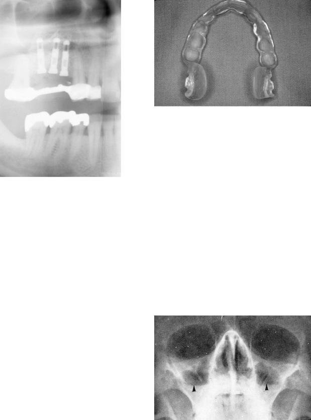
FIGURE 18.1 Maxillary sinus bone graft and placement of three dental implants to prevent fracture of a long span fixed bridge and failure of natural tooth abutments.
riod without a prosthesis needs to be assessed. The surgicalrestorative team must have a clear comprehension of the patient’s complaints and desires and of each other’s vision of the final restoration. Many individuals may not wish to further compromise remaining teeth to serve as abutments (Figure 18.1). Frequently, the only desire stated may be for a plan that would allow for a fixed rather than a removable prosthesis.
Load requirements must be delineated. Nonaxial loading from malaligned abutments/fixtures as well as long edentulous spans, more posterior locations, parafunctional habits, increase in crown/root (implant) ratio, and an opposing natural dentition all place a greater strain on the restoration. This problem is further compounded because the posterior maxillary bone density, especially in long-standing edentulism, is spongy with fewer trabeculae. Compromised stress-bearing capacity is inherent. Modification of the treatment plan needs to account for these biomechanical demands. If inadequate natural abutments are present, the use of fixture(s) generally restored as a free-standing unit should be considered (Figure 18.1). Fixture load tolerances are elevated by positioning implants along the long axis of force vectors as well as by increasing fixture number, length, diameter, and available surface area for osseointegration. This plan may be accomplished by altering the implant surface coating via plasma spraying.
These and future determinations are made on the basis of the initial patient discussion, examination, mounted cast evaluation, and diagnositc setup. These casts can later be utilized
FIGURE 18.2 Surgical guide stent for bilateral maxillary sinus bone graft and dental implant placement.
for fabrication of a surgical guide stent (Figure 18.2). Radiographic assessment is an essential element of the workup. Minimally, a panoramic radiograph supplemented by periapical films is necessary. The magnification factors must be known either by comparison to a standard measure placed in the region under examination or by use of a film with a measured grid. From these radiographs, the periodontalendodontic status of the remaining teeth are first considerations. Shortand long-term prognostic determinations as to the viability of teeth individually and whether they may serve as abutments needs to be judged. Edentulous areas as well as the tuberosity-pterygoid plate region should be surveyed for pathology and quantitatively assessed for vertical bone heights. A qualitative assessment of bone type may also be approximated. If no panoramic machine is available, the maxillary sinus may be screened with a Waters’ view (Figure 18.3).
Should further radiographic data be necessary, or to delineate sinus pathology or septae detected on plain films, a
FIGURE 18.3 Waters’ view demonstrating bilateral air/fluid levels indicating sinus infection (black arrows).
176 |
J.I. Stein and A.M. Greenberg |
a
b
c
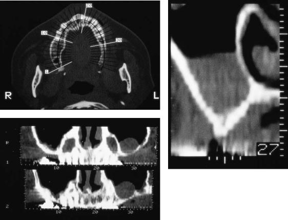
FIGURE 18.4 Computerized tomography scan reformatted with dental software. (a) Axial plane with numerically identified 3-mm sectional cuts correlated to each cross-sectional view. (b) Chronic left
computed tomographic (CT) study should be ordered.12,13 Patients with a significant history of sinus disease or smoking considering a sinus lift procedure also are required to obtain CT scanning. Reformatted CT utilizing specialized dental software will clearly demonstrate the residual bone length, width, and angulation. Qualitative bone assessment is also enhanced. Stents with radiopaque markers may be used to delineate prosthetically desirable implant locations in all views. Study and cross-referencing of the various dimensions will demystify and facilitate treatment planning (Figures 18.4a–c). Availability, cost, and additional radiation exposure dictate the need for CT scanning are made on an individual case-by- case basis.
Once it is evident that the residual dentition cannot serve as abutments and a fixed or removable restoration is desired, an implant treatment plan must be developed. Assessment of bone density and volume of individualized implant sites in light of biomechanical demands is critical. Generally, bone rising vertically 10 mm or more with adequate width to encase the fixture circumferentially permits utilization of the standard surgical approach. Minor vertical deficiencies may
maxillary sinus membrane thickening and subantral bone atrophy.
(c) Cross-sectional view also demonstrating left maxillary sinus membrane thickening and advanced subantral alveolar bone atrophy.
be compensated for by placing the fixture apex slightly beyond the sinus floor and allowing for secondary bony doming. This can also be accomplished by localized apical sinus lifts which enhance length by imploding the fixture site sinus floor via an osteotome technique described later.14,15 Localized width deficiencies may be augmented via a guided tissue regeneration procedure with simultaneous fixture placement or as a staged procedure. Fixture sites approximating 7 mm of height, particularly if poor bone quality is evident, require alternative planning. In this intermediary range, if biomechanical forces are not excessive, bony ridge inadequacies may be compensated by increasing the number or the diameter of fixtures utilized.
Unfortunately, minimum bony thresholds are frequently not met. Posterior maxillary atrophy inferiorly (crestally) and laterally results from periodontal disease and postextraction disuse resorption. Pneumatization of the maxillary sinus further compromises the residual alveolus. This expansion of the maxillary antrum results from a slight increase in intrasinus pressure during expiration, inducing osteoclastic activity in the periosteal layer of the Schneiderian membrane. The resid-
18. Maxillary Sinus Grafting and Osseointegration Surgery |
177 |
a |
b |
c
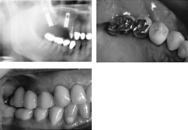
FIGURE 18.5 Posterior maxillary implant reconstruction without sinus grafting. (a) Panoramic radiograph with dental implants placed in the maxillary tuberosity and second premolar site in order to avoid sinus bone grafting. (b) Occlusal clinical view of fixed implant prosthesis. (c) Right buccal clinical view.
ual ridge may be narrow with a medial inclination and of minimal osseous volume.
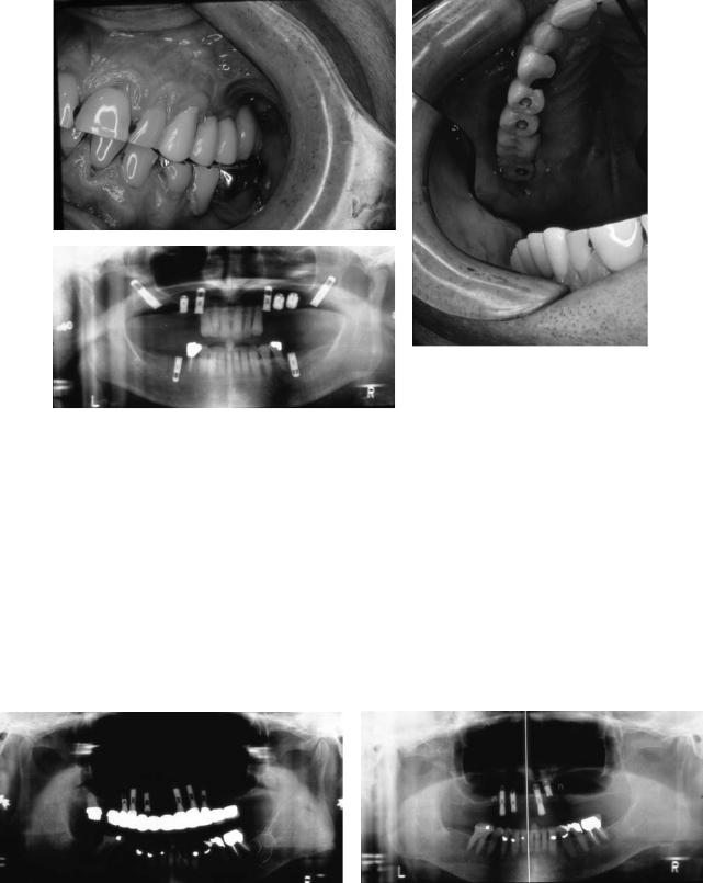
If inadequate bone is present for traditional fixture placement, an alternative to subantral grafting to be considered is to bypass this region and place a fixture within the maxillary tuberosity, possibly extending into the pterygoid plates16–18 (Figures 18.5a–c and 18.6a–c). This will provide a distal abutment for bridge fabrication. This option is only viable for short spans and if mesial and distal fixtures are of sufficient length and diameter. Excessive angulation of the distal fixture as well as other unfavorable biomechanical factors will commonly preclude implementation of this procedure.
The use of a distal cantilevered pontic is often entertained at this junction. Commonly, this is at the misguided prodding of the patient in their desire “to keep it simple” and avoid a graft procedure. The strain placed on the distal fixture in a cantilevered situation, especially if it is short or in poor quality bone, may lead to fixture failure or fracture (Figures 18.7a,b), or possible frequent abutment–prosthetic screw loosening or fracture. Similar considerations contraindicate the use of excess anterior lever arms when only posterior fixtures are available in the totally edentulous situation.
Restoration of the severely atrophic posterior maxilla re-
quires graft augmentation by either Le Fort I osteotomy with an autogenous interpositional corticocancellous graft,3,4,19 an onlay corticocancellous graft, or a maxillary antrostomy with membrane elevation and a subantral graft, commonly referred to as a sinus lift or sinus inlay graft procedure. Limited indication exists for the Le Fort I approach. Because of the procedure’s complexity and its lowest success rate of osseointegration at 68%,20 the Le Fort I approach should be reserved for those patients who require a major correction of a class III ridge relationship. The maxillary onlay graft is a procedure of potentially greater complexity and postoperative complications, with a lower osseointegration success rate than the sinus inlay graft. Inital onlay results reported range from 50% to 90% but appear to approach 80% to 90% with experi- ence.2,19–24 An onlay graft procedure is indicated in lieu of the sinus inlay approach in the following circumstances: when severe buccal alveolar resorption would necessitate palatal implant placement or angulation, excessive interocclusal space would result in an unfavorable prosthesis/implant ratio, maxillary anterior augmentation is required, or when the patient has significant sinus disease or smoking addiction. It should be noted that anterior atrophy or posterior buccal atrophy may be alternatively addressed with adjunctive graft-
178 |
J.I. Stein and A.M. Greenberg |
a
b
c
FIGURE 18.6 Bilateral posterior maxillary implant prosthesis supported with tuberosity and Subantral implants. (a) Left buccal clinical view.
(b) Panoramic radiograph. (c) Left occlusal clinical view.
ing procedures (i.e., nasal inlay graft, localized guided tissue regeneration) in combination with the sinus lift technique.
The sinus lift procedure has, comparatively, the highest success rate and may be of less complexity and morbidity. Contraindications to sinus elevation and augmentation include sinusitis, presence of a cyst, tumor, or displaced root tip. These conditions may interfere with normal sinus drainage through the ostium, leading to a mucopurulent accumulation within the antrum. Sinus pathology must be resolved before grafting
to permit a well-ventilated, draining, aseptic antrum. Sinus inlay grafting alone will not address excess intermaxillary space or correct major skeletal discrepancies in the transverse or anteroposterior dimension. Alternative or adjunctive procedures should be considered. Smoking may be a relative to absolute contraindication depending on severity. Bain and Moy25 reported smokers have more than twice the failure rate, 11.3% versus 4.8%, compared to nonsmokers in standard implant cases. Impaired polymorphonuclear neutrophil (PMN) func-
a |
b |
FIGURE 18.7 Failure of bilateral cantilevered prosthesis with resultant implant fractures. (a) Panoramic radiograph demonstrates right single cantilever and left double cantilever. (b) Panoramic radiograph
following removal of fixed bridge. Right distal implant removed with retained implant apex, and left two distal implants with retained implant apices.
18. Maxillary Sinus Grafting and Osseointegration Surgery
tion as well as local and systemic vasoconstrictive effects are believed responsible. Increased complications and failure have been evident in heavy smokers undergoing the sinus elevation procedure.26–28 It is recommended that smoking be discontinued 2 months preoperatively and throughout the healing period.29 Clinical data have yet to clearly define necessary time parameters. Patient compliance with such requirements is frequently difficult to monitor and achieve, despite new spoking cessation treatments.
Anatomy
The maxillary sinus is a paired, pyramidally shaped, pneumatic cavity occupying the body of the maxilla. The base of the pyramid lies medially, serving as the lateral nasal wall. This medial sinus wall–lateral nasal wall merges with the anterior wall to form the tallest vertical strut of the maxilla. Posteriorly, the maxillary antrum abuts the pterygoid plates. The lateral sinus wall is contiguous with the buccal plate of the alveolar bone. Superiorly, the sinus roof helps form the orbital floor. Inferiorly, the floor of the maxillary sinus extends below the level of the nasal cavity. Bony septae (buttress) frequently join the medial or the lateral walls to reinforce the sinus cavity. These septae may divide the sinus into two or more cavities that may or may not communicate.
The dimensions and capacity of the sinus demonstrate marked variability, dependent on sinus expansion. It is the largest of the four paranasal sinuses, with an approximate height at the base of 35 mm. The mediolateral base width is 35 mm, which tapers to 25 mm in the first molar regions. Its anteroposterior depth is generally in excess of 30 mm. The maxillary antrum has an average volume capacity of 15 ml.
The sinus is lined with a thin delicate pseudostratified ciliated cuboidal to columnar epithelium tightly bound to and indistinguishable from the underlying periosteum. The periosteum has few elastic fibers and is loosely bound to bone, facilitating surgical elevation. In the lining there are three glandular cell types—goblet, mucous, and serous—with the latter two concentrated near the ostium. Approximately 2 liters of fluid is produced per day. The fluid layer is divided into an outer gel-like mucous layer for transport that overlies a less viscous serous fluid layer surrounding the cilia. The cilia beat in a coordinated undulating manner, propelling a blanket of mucus and intrasinus debris toward the ostium for drainage into the nose.
The ostium is located along the superior aspect of the medial wall, 25 to 35 mm above the antral floor, and drains through the anterior aspect of the middle meatus. The ostium is a ductlike orifice 3.5 6 mm in cross section, extending superomedially 3.5 to 10 mm in length. The ostium position, high on the medial wall, is further hindered by the ductlike configuration, making passive (gravitational) drainage ineffective.
The sinus functions to lighten the weight of the skull and to
179
warm and debride inhaled air, as well as acting as a resonance chamber for voice modulation. Normal sinus function requires the patency of the ostium, a functioning ciliary apparatus, and secretions qualitatively and quantitatively appropriate.
The arterial vasculature to the maxillary antrum derives from branches of the internal maxillary artery (ethmoidal, infraorbital, facial, and palatine). Venous drainage medially is into the sphenopalatine vein while the remaining walls drain through the pterygomaxillary plexus. Innervation is provided by branches of the trigeminal nerve, second division (lateral posterior superior nasal, superior alveolar, and infraorbital nerves).
Sinus inlay grafting in reports discussed previously has demonstrated success rates generally well in excess of 90%. In managing the severely atrophic posterior maxilla, this procedure most frequently fulfills a cardinal goal of surgery, which is to obtain a successful if not optimal long-term outcome with the least amount of intervention, complications, and risks. This statement is particularly valid if the procedure morbidity can be further decreased by performing it in one stage (simultaneous graft and implant insertion) and if the graft material utilized does not necessitate an iliac crest or mandibular symphyseal donor site. Various graft materials that have been successfully used independently or in combination are autogenous, allogeneic, alloplastic, and xenogeneic.
Selection of Graft Material
The graft material selected must be able to provide long-term support for an implant-borne prosthesis. Present materials utilized alone or in combination include autologous, allogeneic (demineralized freeze-dried bone), alloplastic (synthetic hydroxyapatite), and xenogeneic grafts. A potential additional class of bone substitutes likely to be available in the near future are genetically engineered osteoinductive bone morphogenic proteins. The characteristics of the ideal subantral graft material are that it be nontoxic, nonantigenic, nonmigratory, infection resistant, readily available, easily fabricated, inexpensive, strong, resilient, capable of functional remodeling, provide ease of manipulation, minimize surgical time, eliminate donor morbidity, eliminate need for general anesthesia, enhance early stability of implants, and permit long-term osseointegration.
Bone Healing
Autologous bone grafts may be cortical, cancellous, or corticocancellous in composition. These type of grafts contain many live cells capable of osteogenesis. Success of any graft is dependent on a variety of host and surgical factors. In selecting which graft material to utilize, whether autologous in nature or a substitute, the healing process of autogenous bone grafts must be clearly comprehended.30
180
Axhausen28 delineated a two-phase healing model of bone grafts. During phase 1, cortical and cancellous grafts heal similarly, with blood coagulated around transplanted bone and an acute inflammatory reaction evident. Initial grafted cell survival is through nutrient diffusion followed by angiogensis from the graft bed. Transplanted cells proliferate and differentiate to form osteoid surrounding avascular grafted trabeculae. With progressive osteoid deposition, the graft becomes joined with new bone. The quantity of bone regenerated is directly proportional to the amount of cellular density of transplanted bone cells that survived. By the end of week two, inflammation has decreased, and fibrous granulation tissue around the graft and increased osteoclastic activity are present. Osteocytes die, as evidenced by vacant lacunae. Necrotic tissue in the Haversian system is removed by macrophages.
Cancellous grafts are rapidly revascularized by host ingrowth and end-to-end anastomosis by the end of week two. Primitive mesenchymal cells differentiate into osteogenic cells. These cells together with transplanted osteogenic cells differentiate into osteoblasts that envelop cores of necrotic bone with osteoid, which is then replaced completely by new bone strengthening the graft.
Cortical grafts behave differently and are revascularized slowly, taking approximately 1 to 2 months. Remodeling differs in that repair is driven by osteoclastic, not osteoblastic, resorption. Neovascularization is facilitated by osteoclastic bone resorption through and following along preexisting Haversian and Volkmann’s canals. This initial resorptive component, which begins within the first 4 weeks, is followed by appositional bone deposition sealing off bone from further osteoclastic activity. This process is termed creeping substitution. Areas of necrotic bone may persist and are histologically unique to cortical bone healing. New bone continues to form via creeping substitution until the graft is remodeled. The early osteoclastic component with delayed osteoblastic activity leads to early mechanical weakness, which may last 6 weeks to 6 months. Maturation to normal bone strength may require 1 to 2 years.
These differing processes between cortical and cancellous graft behavior occur predominately during Axhausen’s phase 2. This phase begins during week two and becomes critical in weeks four and five. Fibroblasts and other mesenchymal cells from the host bed differentiate into osteoblasts and begin to produce new matrix. This programming of cells is termed osteoinduction and is believed to be regulated by bone matrix proteins. BMP (bone morphogenic protein), the best known, is an acid-insoluble, oligosaccharide glycoprotein of low molecular weight (15,000–18,000). Additionally, passive ingrowth of osteogenic cells occurs from the surrounding bone with the grafted bone acting as a scaffold or new bone formation, a process designated as osteoconduction. Neovascularization, osteogenesis, osteoinduction, and osteoconduction combined with osteoclastic activity derived from circulating monocytes allow for continued resorption, remodeling, and replacement, eventually leading to an incorporated bone graft.
J.I. Stein and A.M. Greenberg
Autogenous Grafts
Autologous bone is the gold standard by which other graft materials are judged. Sinus inlay grafting utilizing an iliac crest particulate cancellous graft was first performed by Tatum5,29 in the 1970s and later published by Boyne and James.30 Jensen et al.6 similarly described the use of a particulate cancellous graft into which implants were placed 4 to 5 months later. Simultaneous one-stage implant and graft insertion was reported by Tatum5,29 and by Block and Kent8,26 utilizing cancellous chips, and by Keller et al.4,19,31 and oth- ers7,32–34 using a corticocancellous block.
Autogenous bone has been a reliable source of immunocompatible viable bone cells to allow for osteogenesis. Via transplanted osteoprogenitor cells and their osteoconductive and osteoinductive properties, bone is produced and maintained to stabilize fixtures. Graft healing is generally accelerated in comparison to other material, allowing for implant insertion at 4 to 6 months in a staged procedure, or facilitates earlier fixture uncovering at 6 to 8 months for single-stage implant graft procedures. The major disadvantages of autogenous bone grafts relates to the donor site morbidity, additional surgical time, anesthesia, and cost. The potential for graft resorption in cases of delayed implant placement may also exist.
A cancellous graft contains particulate medullary bone and hematopoietic marrow with the highest concentration of osteogenic cells. This graft type allows for rapid revascularization and bone production. Available intraoral sources of cancellous bone may be within the surgical field from fixture preparation sites or the maxillary tuberosity. Additional volume can be obtained from the mandibular symphysis, extraction sites, or the retromolar pad–ramus region. Should large quantities be necessary, the iliac crest is an abundant source. Autologous grafts should be stored in saline, not hypotonic or hypertonic solutions, which may cause osteogenic cell death before transplant. Unlike intraoral sources, which are membranous in nature, the iliac crest provides endochondral bone and may be more prone to resorption. This seems to be of minimal if any clinical significance in terms of fixture integration and long-term stability. Particulate cancellous bone is readily combined and osteogenically enhances allogeneic, alloplastic, and xenogeneic material that may serve as a volume expander or the bulk of the graft. The major drawbacks of a cancellous graft to those previously stated for autogenous bone in general are associated with excess resorption and an inability, as all particulate grafts, to rigidly stabilize and maintain implant position and angulation where minimal alveolar bone exists.
The corticocancellous block graft is indicated in such situations to allow for rigid fixture stabilization. Immediate fixture placement, particularly of threaded types, allows for precise implant positioning as well as rigid primary stabilization of the graft, thereby minimizing mobility and resultant resorption. Corticocancellous grafts provide greater initial
18. Maxillary Sinus Grafting and Osseointegration Surgery
strength and possibly less postremodeling resorption than cancellous grafts. However, because of slow revascularization block grafts are more prone to infection and weakness during early remodeling. When the graft is subjected to force transmission by direct loading through the implants, it is not known whether a corticocancellous or a particulate cancellous graft will behave differently. Shaping, placement, and stabilization of a corticocancellous block is more technically demanding. Sinus septae must be removed to optimize the interface between the donor block and its bed. Residual voids between the block and the sinus walls are packed with particulate bone. In cases in which the sinus membrane has been perforated, there is less risk of fragment migration and dissemination than when only a particulate cancellous graft is utilized.
Corticocancellous blocks may be harvested from the symphysis. The iliac crest is used if a large graft is necessary and commonly when a bilateral augmentation is planned. Iliac crest bone harvesting increases the relative magnitude of the overall procedure in comparison to utilizing other graft materials to be discussed. Faltering patient motivation and acceptance, if not frank objection, may be encountered. Graft procurement from the iliac crest usually entails hospitalization, general anesthesia, increased blood loss, surgical time, cost, convalescence, and possibly the need for a second surgeon. Donor site morbidity may include gait disturbances (related to disruption of the tensor fascia latae and psoas major muscles), neurosensory deficits (lateral femoral cutaneous nerve), scarring, and abdominal and urologic complications (hernia, meralgia parathetica, adynamic ileus, hematoma, seroma, pain, and infections). Tibial plateau bone grafts may also be a consideration, especially for outpatient or in office ambulatory procedures.
Intraoral graft procurement from the symphysis, ramus, extraction sites, tuberosity, or sinus retromolar pad eliminates many of these disadvantages. Problems of donor site morbidity, neurosensory complication (inferior alveolar, mental, and lingual nerves), increased surgical time, inadequate graft volume, and cost, however, still persist and dictate consideration of alternative graft materials for sinus lift procedures. Particular care is needed in harvesting bone from the tuberosity. One must avoid invasion into the sinus. Utilizing the tuberosity may compromise the primary procedure, eliminate a potential implant recipient site, and make retention of a transitional prosthesis during the healing period difficult. However, harvesting bone from regions other than the surgical field once again may raise patient objections.
Allogeneic Grafts
Allogeneic grafts have been quite successful as a volume expander in combination with autologous grafts or for providing osteoconductive properties with hydroxyapatite of various forms. Allografts are materials taken from another individual within the same species. Mineralized allografts possess
181
osteoconductive properties, but are unacceptable because of slower revascularization and resorption with increased infection liability. Demineralized allografts are capable of more predictable repair. Minerals may be removed by means of an acid treatment, then washed and freeze-dried (lyophilized). This demineralized freeze-dried bone (DFDB) retains bone morphogenic protein (BMP) and thus osteoinductive activity.35 DFDB is available as cancellous or cortical chips (1–5 mm) or powder (250–1000 m). Demineralized freeze-dried cortical powder of 250–500 m particle size provides enhanced osteoinductivity and surface area compared to the other forms. A 20-min saline reconstitution period is minimally necessary.
DFDB should be obtained from a reputable, accredited bank that adheres to the guidelines and standards of the American Association of Tissue Banks (AATB). The AATB criteria delineate protocols for donor selection and screening, recovery techniques, testing, processing, storage, distribution, and record keeping. All donors are screened and tested to exclude transmissible diseases, infections, malignancy, toxic exposures, parenteral drug use, immunosuppression, and other disease states, Donor tissue undergoes extensive serologic assays with aerobic-anaerobic microbiologic monitoring from recovery throughout processing, including sampling at final packaging.36,37 Buck et al.38 in 1989 estimated the risk of HIV transmission as 1 in 1.2 million allograft procedures. Introduction in 1991 of the polymerase chain reaction test has proven to be an extremely sensitive screen for HIV and further diminishes the risk. Patient education is critical in minimizing anxiety and objection to allografts. Clinicians should utilize only those tissue banks that abide by AATB guidelines and employ advanced tissue testing and processing technique.
Urist,35 Reddi and Hascall,39 Glowacki et al.,40 and many others27,41,42 over the past 30 years have delineated the osteoinductive healing process of DFDB. DFDB has no osteoprogenitor cells and serves to enhance phase 2 bone healing. The graft is initially bound by fibrin and fibronectin, followed by mesenchymal cell proliferation and chemotaxis to the graft. These pluripotential cells differentiate into chrondroblasts, hematopoietic cells, and osteoblasts, eventually producing bone. Adequate oxygenation as determined by local vascularity is critical to this process.
DFDB as a lone subantral graft material has been somewhat unpredictable.8,43–45 Extended healing periods of 12 to 16 months, prolonged rubbery consistency, and comparatively sparser bone formation contraindicate its routine use alone as an independent subantral graft material. However, success is evident in the high 90th percentile since the midto late 1980s with DFDB as part of a composite graft with autologous bone or hydroxyapatite porous46 or resorbable.10,43,47–49 In a 2.5-year period, Smiler et al.43 reported 95% success using DFDB with a xenogeneic material, Bio-Oss (Osteohealth, Shirley, NY, USA) in a 1:3 proportion in 21 graft sites with 56 implants.
DFDB composite grafts may enhance bone formation compared to either component used independently.50 This allo-
182
graft does not demonstrate antigenicity and provides predictable results. As an autograft expander, it may be synergistic for bone formation while diminishing donor site morbidity and other unfavorable consequences when larger autologous grafts would be otherwise necessary. If autogenous bone is not utilized, the osteoinductive features of DFDB appear to complement the osteoconductive properties of alloplastic materials. This provides an excellent subantral graft option with a healing period of 9 to 12 months.
Alloplastic Grafts
The use of synthetic hydroxyapatite as the sole graft material or in combination with autografts or allografts has been expanding during the past 10 years. Hydroxyapatite (HA) is a natural mineral component of hard tissue composing 97% of enamel, 70% to 80% of dentin, 50% to 60% of cementum, and 60% to 70% of bone. HA is biocompatible, nontoxic, nonantigenic, readily available, inexpensive, and a time-effi- cient material. Synthetic HA is derived from calcium phosphate crystals compacted under high pressure (10,000– 20,000) and temperature (1100°–1300°C). This fusion process is termed sintering. Porosity size approximating 100 m is necessary for effective bony ingrowth. Somewhat larger porosity size allows for more rapid bone infiltration.51
A frequently utilized form of HA is derived by a hydrothermal chemical exchange reaction from coral (family Portidae) commercially available as Interpore 200 (Interpore, Irvine, CA, USA). This HA is a highly organized material with a pore average of 230 m and a labyrinth of continuous uniform interconnected porosities averaging 190 m.50 The pore percentage is uniform with a solid-to-void ratio of 1. Porous forms of HA provide a passive scaffold for bony infiltration. It is an osteophilic and osteoconductive material encouraging bony ingrowth from the surrounding graft bed into areas where bone would not otherwise form. It does not possess any intrinsic osteogenic potential and lacks the bone matrix proteins required for osteoinduction. Bony infiltration is enhanced by increasing the exposed surface area of surrounding bone. Bone formation is slower and less predictable in regions further distant from the recipient bone bed. Creation of a three-wall defect by adequately reflecting the medial sinus wall mucoperiosteum is critical for graft maturation with this material as well as others discussed. The contribution to bone formation from the imploded lateral cortical sinus wall is somewhat speculative.
Natural occurring porous HA structure mimics the macrostructure of bone, optimizing fibrovascular invasion and new bone incorporation. Following osteogenic cell ingrowth, osteoblasts organize on the HA surface with apposition of lamellar bone until the final regeneration of cortical bone in the form of osteons. Normal bone healing is evident on the surface of both porous and nonporous HA. The bond between HA and bone consists of an amorphous zone rich in mu-
J.I. Stein and A.M. Greenberg
copolysaccharides with calcification evident. Cells directly attach to the HA by collagen fibers.
Dense, sintered HA has low microporosity. Resorbability of HA is dependent on density, crystal size, and porosity. High-density HA of relatively large particle size as well as HA derived from coral are slowly resorbable or essentially nonresorbable in vivo.50 A nonsintered (nonceramic) graft material composed of small crystal clusters is a nonporous resorbable form of HA marketed as Osteogen (Stryker, Kalamazoo, MI, USA). As osteoclasts resorb this form of HA, new bone formation is facilitated through osteoconduction with HA acting as a mineral reservoir.
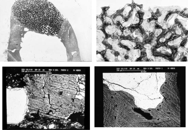
Tatum,29 Misch,47,48 Smiler et al.,43 and Smiler and Holmes,52 followed by numerous clinicians,46,49,53,54 began reporting the use of HA as an independent or combination subantral graft material in the midto late 1980s. Predictable successful outcomes generally approaching 100% were evident with HA or HA in combination with autologous grafts or allogeneic grafts. Smiler et al.43 in 1992 published a 100% success rate on the use of Interpore in 66 sinus lifts on 36 patients with 198 implants supporting a prosthesis. Histomorphometic examination confirmed bony ingrowth into the porous HA granules. Core biopsies on specimens demonstrated bone present in as much as 10 mm of the 12-mm core length, with a mean amount of HA covered by bone ingrowth of 40.9% and a mean bony ingrowth of 23.10%. Small and Zinner46 reported on a 6-year experience using Interpore 200 in a 1:1 ratio with demineralized freeze-dried cortical bone. To date (in yet unpublished data) in 68 sinus lifts only 4 of 211 implants have failed, for a success rate of 98%. Limited histomorphometric and volume fraction studies were consistent with Smiler’s results (Figures 18.8a–d) (Ralph E. Holmes, Department of Plastic Surgery, University of California, San Diego, CA). Approximately equal thirds of HA, bone, and soft tissue were noted in the consolidated subantral augmentation. Resorbable HA has also demonstrated predictable successful outcomes as an independent or as a composite graft in multiple reports.9,43,53,55
A multitude of varying proportions in composite grafts have been suggested. Many clinicians have utilized an alloplast as a minor component serving as an autograft extender when less than optimal autologous bone was available. Others, wishing to limit donor site morbidity, used only readily available autogenous bone in whatever limited quantities from intraoral sites, generally harvested from the tuberosity or fixture osteotomies as a supplement to the predominant HA component. Autologous bone serves to enhance the osteogenic, osteoinductive, and osteoconductive potential of the graft. Similarly, DFDB providing osteoinductive properties is added by some with this mix or with HA only. On the basis of core biopsies studying bone volume and clinical consistency, it appears that DFDB should be limited maximally to 50% or less of the composite graft.43–46 Further bone volume fraction studies considering healing time variabilities appear necessary to determine the optimal graft material or combination proportions.
18. Maxillary Sinus Grafting and Osseointegration Surgery |
183 |
a |
b |
c |
d |
FIGURE 18.8 Histomorphometic and volume fraction studies of core biopsies from human sinus bone grafts with Interpore 200 hydroxyapatite and demineralized freeze dried bone in a 1:1 ratio (courtesy of Ralph Holmes, MD, Department of Plastic Surgery, University of California San Diego, San Diego, California). (a) Routine stain
Predominantly anecdotal evidence suggests the minimal healing period for autogenous grafts is 4 to 6 months for fixture placement in a two-stage procedure, and 6 to 9 months for a one-stage protocol. DFDB alone, which is not recommended, takes approximately 12 to 16 months to mature. HA alone necessitates a wait of 9 to 12 months until abutment surgery in simultaneous graft/implant insertion or 6 months until fixture placement if staged with an additional 9 months to allow for osseointegration. HA-DFDB combinations require 9 months minimally in a one-stage procedure or fixture placement no earlier than 6 months post grafting in a twostage procedure.
Autologous grafts or autologous-dominated combinations demonstrate the fastest healing and the highest bone volume fractions. Despite these results, similar high levels of clinical success in the development and maintenance of osseointegration is apparent with HA as a solitary graft or in combination as an equal or dominant component mixed with allogeneic grafts. It must be kept in mind that the ultimate goal
low power reveals mixture of bone regeneration and ingrowth into Interpore 200 pores. (b) High power light photomicrograph. (c) Scanning electon micrograph of mature new bone growth with fibrous tissue. (d) Scanning electron micrograph of bone growth in relation to hydroxyapatite Interpore 200 granules.
of the sinus lift procedure is stable long-term osseointegration. Pragmatically, porous nonresorbable HA combined with readily available autologous bone or possibly DFDB ( 50%) would seem a prudent choice.
HA grafts or combinations, owing to their particular nature, may not provide adequate fixture positional stability. Cases of extreme osseous atrophy leaving an eggshell residual ridge may dictate a staged procedure or use of a corticocancellous graft to allow for reliable implant positioning. Additional disadvantages of HA to consider are increased healing time and an increase in difficulty in implant site preparation because of the HA hardness compared to autogenous grafts. Although HA independently or as part of a composite graft does not satisfy all the criteria for an ideal graft material, it has demonstrated clinical success equal if not superior to other graft options. It does so with the advantage of eliminating or limiting donor site morbidity, hospitalization, general anesthesia, cost, operative time, and potential disease transmission. HA lacks toxicity and antigenicity and permits a direct
184
bone bond facilitated via osteoconduction. In our experience of 68 sinus lifts with 211 implants, we have had a success rate of 98% utilizing a composite graft consisting of 50% Interpore 200 with 50% DFDB, and locally available intraoral bone from the tuberosity or implant preparation sites is substituted on a volume basis for the DFDB when available. The autologous bone fraction constituted 0% to 25% of the final composite graft.
Xenogeneic Grafts
Xenografts (heterografts) are grafts taken from a genetically different species. BioOss (Osteohealth, Shirley, NY) and Osteograf/N (Ceramed, Lakewood, CO, USA) are calcium-de- ficient carbonate apatite crystals derived from a bovine source. Osteograf/N (Ceramed) has a smaller particle size than BioOss. This natural bone mineral is chemically and physically identical to bone. It is deorganified and deproteinated to render it nonantigenic. As a result of deproteination it does not possess osteoinductive capacity. Because of its natural structure and network of macropores, micropores, and small crystal formations, it provides considerably greater surface area than synthetic HA.27 This natural porous HA undergoes a three-phase healing process. Initially particles are surrounded by host bone, then particles are resorbed by osteoclastic activity, and finally new bone is formed by osteoblasts replacing the particles with dense lamellar bone. The conversion rate is dependent on cellularity and other local and systemic factors.
Initial short-term reports of less than 3 years of these materials combined with autogenous bone in varying proportions are encouraging.43,56 Utilization in composite grafts with either autogenous bone or DFDB seems promising but optimal fractions need to be defined. Further long-term evaluation of these xenografts as an independent antral graft material or as a composite graft is necessary.
Selection of Endosseous Implant
Endosseous fixtures approximately 15 mm in length and 4 mm in diameter are generally utilized to optimize the bone–implant surface area interface. Narrower diameter implants may be necessary when only a thin crestal ridge remains. Use of a wider diameter fixture in such circumstances may lead to residual ridge fracture. Shorter fixtures are indicated for patients with skeletal vertical deficiency with decreased sinus height or when the level of the superior horizontal osteotomy is inappropriately placed. The maximal number of implants allowing 2 mm of spacing between fixtures should be placed. At 2 to 3 months preoperatively, any tooth with a poor long-term prognosis is extracted to permit bone healing for fixture placement and sufficient time for soft tissue closure.
J.I. Stein and A.M. Greenberg
Varying combinations of implant types and graft materials have been successful. Uncoated screw-type fixtures require adequate bone quantitatively and qualitatively for initial stabilization and success. It is well documented that implant success rates are lower in type IV bone, particularly for screwtype implants.57–59 This most likely results from an inability to obtain initial (bi)cortical stabilization. In subantral grafting procedures, threaded implants require a minimal amount of residual bone for stabilization, of approximately 5 mm. Qualitatively, the bone may still be unfavorable, presenting with a thin cortex and low density of trabeculae. Use of screw-type implants more frequently may therefore require the implementation of a two-stage procedure (subantral grafting followed by implant insertion) or consideration of using a corticocancellous block to enhance stabilization. Threaded fixtures and corticocancellous blocks stabilize each other reciprocally. Screw-type fixtures are very successful when employed for subantral grafting when a sufficient residual alveolus is present. These are most commonly used with a predominantly autogenous graft material. However, when minimal bone volume or density is available, use of threadedtype implants would mandate a more complex procedure. Complexity may be in the form of an additional surgical step, increased treatment time, or the necessity of using an autogenous corticocancellous block from the mandibular symphysis or iliac crest. This increase in complexity seemingly derives from some clinicians’ interest in utilizing one type of implant universally and procedures are modified as necessary around the “chosen” implant. As there are well-documented simplified alternatives for extremely deficient bone status that use coated cylinders, frequently with nonautogenous graft material, and are equally or more successful, logic would dictate reassessment of such planning.
Plasma-sprayed cylinders do not require as rigid initial stability as screw-type implants and possess a significally higher surface area for integration. HA-coated fixtures, owing to their osteoconductive surface, stabilize earlier than non-HA-coated implants. In subantral grafting procedures, unlike those in other regions, problems relating to HA coating dissolution, bacterial colonization of the roughened surface with resultant bone loss, and fixture failure have not been encountered. This may be because the basilar bone of the sinus floor does not resorb as easily, and therefore the HA surface is not exposed to the oral environment. A fixture with a polished titanium collar should be used. Coating variabilities among manufacturers must also be considered in selecting fixtures. HA coatings with higher crystallinity percentages have lower dissolution rates and are therefore desirable. Inadequate data are currently available for the use of coated screw-type implants. HA-coated implants have been successfully implemented in one-stage simutaneous graftng and insertion procedures in extremely poor residual bone situations.46 Cylinders do not provide as satisfactory rigid stabilization as threaded fixtures when a corticocancellous graft is used.

18. Maxillary Sinus Grafting and Osseointegration Surgery
In summary, titanium threaded fixtures, HA, or titanium plasma-sprayed cylinders have all been successfully utilized in the sinus lift procedure. However, in situations of diminished bone volume and density, the plasma-sprayed fixtures, particularly those that are HA coated, appear useful if a onestage procedure is desired.
185
to 6 months later. For HA grafts and combinations with autologous or DFDB, the waiting period is 6 to 9 months, depending on proportions. A longer bone maturation phase is necessary to account for a graft subjected to osteoconductive revitalization.
One-Stage Versus Two-Stage Procedure
The advantages of simultaneous subantral grafting and implant insertion are the avoidance of the trauma of a second surgery, reduction in total treatment time, and the possibility of providing a stimulus to the graft for consolidation around the fixtures. For press-fit implants, 3 mm or possibly even less of residual bone may be adequate for proper immediate positioning and stabilization.8,46 Fixture displacement may be further avoided by minimal countersinking of the preparation site. This will necessitate corresponding relief to a removable prosthesis or use of a fixed temporary prosthesis to avoid premature loading. Utilizing HA-coated implants will facilitate early stabilization because bonding with bone may begin to occur as early as 4 weeks. Care in packing the particulate graft so as to avoid deflection of the implant body is necessary. Corticocancellous blocks if utilized are best stabilized rigidly by screw-type fixtures. Healing periods required are 6 to 8 months for autogenous grafts, 9 to 12 months for HA grafts, and 9 months for HA combined with autogenous bone or DFDB.
The advantages of a two-stage approach are possibly better control of implant alignment as well as less risk of fixture displacement and better tolerance to iatrogenic premature loading. Aside from the obvious disadvantage of additional surgery and treatment time, there are concerns about autogenous graft disuse resorption or pneumatization should significant delays in implant insertion occur. Fixtures may be placed 4 to 6 months following autogenous grafts and uncovered 5
a
Surgical Technique
Preoperatively, the patient rinses with chlorhexidine gluconate, and ingests an appropriate antibiotic (discussed later), glucocorticoid, and NSAID are administered. The procedure may be performed in the office or hospital under local anesthesia, with sedation or general anesthesia. The patient is prepped, draped, and then infiltrated with a local anesthetic containing a vasoconstrictor. The sinus boundaries are indentified by radiographic evaluation and fiberoptic transillumination.
Incision and Reflection
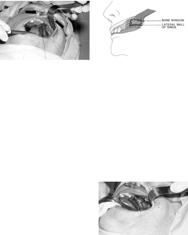
In the totally edentulous posterior maxilla, a horizontal anteroposterior incision is made slightly palatal to the crest from the region of the hamular notch to the canine region (Figures 18.9a,b and 18.10). Anteriorly and posteriorly, vertical releasing incisions are placed approximately 1 cm beyond the vertical walls of the antrum. The relief incisions are brought from the palate horizontal and laterally over the crest and extended superiorly toward the vestibule anteriorly. The posterior vertical release should remain conservative so as to contain the buccal fat pad. A full-thickness mucoperiosteal flap is reflected superiorly to 5 mm beyond the proposed superior horizontal osteotomy or the malar buttress if encountered first. Once adequate exposure to the residual alveolar crest and the lateral wall of the maxilla has been attained, multiple 3-0 silk sutures are used to suture the flap laterally to the cheek in a self-retentive fashion.
b
FIGURE 18.9 Design of incision for sinus bone grafting. (a) Incision at crest of alveolar ridge. (b) Reflected flap and planned position of implant.
186
FIGURE 18.10 Clinical view of incision at alveolar ridge with buccally retracted flap.
J.I. Stein and A.M. Greenberg
FIGURE 18.11 Diagram of buccally retracted flap and creation of bone window in the lateral wall of the maxillary sinus.
Osteotomy
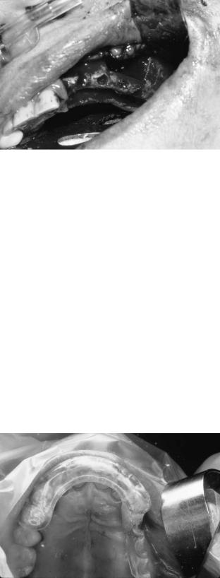
Once again, on the basis of radiographs and fiberoptic illumination, the sinus boundaries are defined. A surgical marking pencil may be used to delineate the location and extent of the antrostomy. Using a high-speed drill with a small round diamond bur under saline irrigation, a rectangular box with rounded corners or an ovoid osteotomy is scored just within the anterior, posterior, and inferior extent of the sinus. The inferior horizonal cut is minimally 2 mm above the alveolar crest to facilitate Schneiderian membrane reflection. This cut may need to be placed slightly superiorly to leave sufficient buccal bone to place fixtures simultaneously while avoiding a ridge fracture. It is also important to make the inferior horizontal osteotomy within the antrum and above the thicker alveolar bone. The further superior the cut is made, however, the more difficulty is encountered in raising the membrane inferiorly. This horizontal osteotomy extends approximately 25 mm from the anterior sinus border to the molar region posteriorly as necessary. Full anterior extension is necessary to avoid blind spots during sinus membrane elevation and grafting. The superior horizontal osteotomy parallels the inferior at a level to permit placement of 15-mm-long implants. The horizontal osteotomies are then connected with parallel rounded vertical cuts. Once the bony window is accurately scored, the osteotomies are completed using the diamond bur in a delicate paintbrush stroke until the bluish hue of the membrane is apparent (Figures 18.11 and 18.12). When mobility of the lateral wall of the window is noted, the osteotomy is complete circumferentially. Should these cuts be complete and immobility persist, a sinus septum may be suspected. Radiographic review and transillumination will likely reveal the presence of septa preoperatively. These sinus septa may be negotiated in one of two methods. A thin osteotome or specialized curette can be used to section the septa inferiorly. Alternatively, two smaller bone windows may be created on either side of the septae partition.
Membrane Reflection
A curved side of a dull surgical curette displaces the bony window slightly inward. The concave portion is positioned between the membrane and the inferior margin of the residual alveolus. The inferolateral membrane is reflected with a sliding motion anteroposteriorly. The Schneiderian membrane has few elastic fibers and is easily separated from its underlying bone. Maintaining contact with bone throughout, the mucoperiosteal lining is released circumferentially around the outer window margin. With increasing medial and superior mobility of the bony window, the membrane can next be raised to the medial sinus wall. Vertically, the mucoperiosteal lining is reflected anteriorly, posteriorly, and medially to accomodate placement of 15-mm fixtures. The attainment of adequate height should be measured. The bony window has simultaneously been infractured and hinged superiorly (Figure 18.13). Membrane reflection without perforation is facilitated by utilizing specialized sinus membrane elevators of appro-
FIGURE 18.12 Clinical view of buccal window in the lateral wall of the maxillary sinus.
18. Maxillary Sinus Grafting and Osseointegration Surgery
FIGURE 18.13 Clinical view of infractured buccal window of lateral wall of the maxillary sinus with osteotomy sites prepared for dental implants.
priate curvature and size. These curettes should always lie subperiosteally against bone. Under direct visualization the rounded back is used to elevate the soft tissue. With an intact sinus membrane, a bellows effect will be observed during breathing, with the hinged lateral sinus wall rising and falling.
Graft and Fixture Insertion
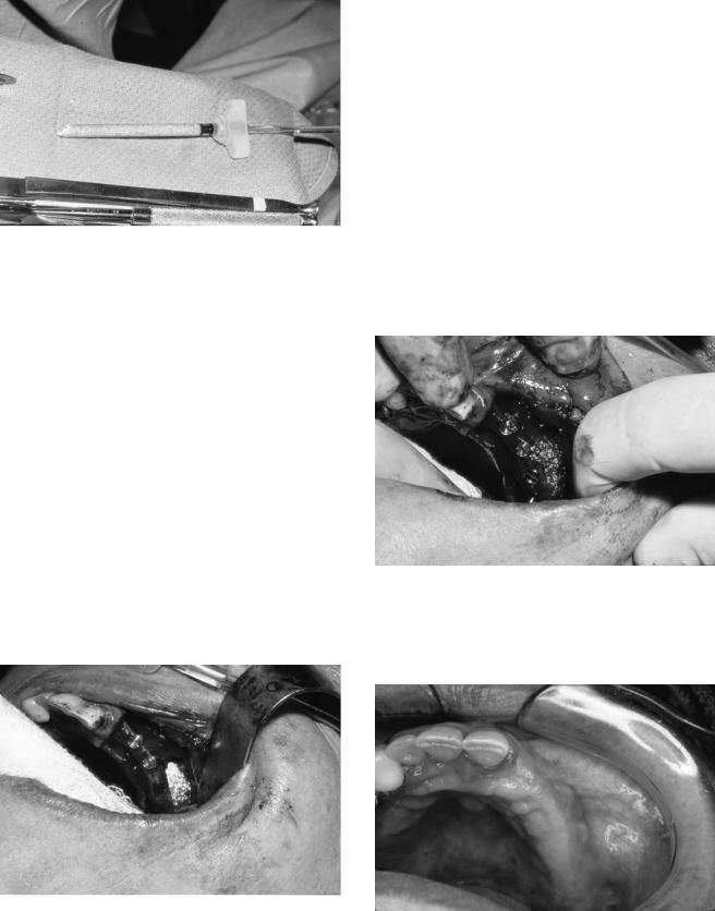
In a two-stage procedure, the antral void is obturated with a graft material. For one-stage procedures, after completion of the sinus membrane elevation a surgical guide stent is placed and stabilized (Figure 18.14). Receptor sites for implants are prepared. Minimal if any countersinking may assist in stabilizing fixtures, if necessary. Placement of a particulate graft is facilitated by loading the graft into a small-diameter glass syringe, which will allow for easier manipulation, control, and access to the medial aspects of the recipient bed (Figure
FIGURE 18.14 Example of surgical guide stent for exact placement of osteotomies for dental implants.
187
18.15). Alternatively, a 3-cc plastic syringe with the tip cut off may be used. The graft material is compacted initially in the medial half and extreme anterior and posterior aspects of the antral void (Figures 18.16 and 18.17). Press-fit fixtures are subsequently inserted, and the residual lateral aspect and the regions between implants are condensed with graft material (Figures 18.18 and 18.19). Observation and care in placement of the lateral graft is necessary to prevent any deflection of the fixtures in cases of severe atrophy. A plastic or titanium instrument can be used to reorient the displaced implant if necessary.
If the screw-type fixtures are utilized with a particulate graft, the fixtures may need to be placed before graft insertion. This will prevent washout of the graft material from the cooling irrigant but complicates medial graft placement. If an autogenous corticocancellous block is chosen as the graft material, it must be custom shaped to the bed. With the cortical surface lying superiorly, the graft is rigidly stabilized with fixtures. Residual voids should be packed with particulate graft material.
The lateral wall of the maxilla is restored to normal contour with particulate graft material. A resorbable membrane such as a collagen sheet or laminar bone is custom fitted to prevent extravasation of material and to delay fibrous tissue invasion. The flap is repositioned and sutured closed with 3-0 slowly resorbable sutures (Figure 18.20). Rarely, a periosteal releasing incision will be necessary to attain a tensionless closure.
Postoperative Management
Glucocorticoids and analgesics are prescribed in anticipation of mild to moderate edema and pain. Antibiotics are administered for a 7- to 10-day period. Amoxicillin clavulanate is commonly prescribed. This antibiotic will provide coverage against the typical oral organisms (anaerobic gram-negative rods, aerobic and anerobic streptococci) as well as common sinus pathogens (Streptococcus pneumoniae, Haemophilus influenzae, Staphylococcus aureus, Branhamella catarrhalis, and numerous anaerobes).60 Alternative antibiotics are either clindamycin or cefaclor, which have no or low cross-sensi- tivity, respectively. Chlorhexidine gluconate 0.12%, as an antiseptic rinse twice daily is additionally recommended. Sinus mucosa inflammation and edema may obstruct the ostium and lead to an infection. Short-term use of a systemic or topical decongestant, particularly if membrane perforation has occurred, may be prophylactically efficacious. These oral and nasal preparations containing sympathomimetric amines vasoconstrict the vascular bed and relieve congestion. Antihistamines are not routinely indicated.
Instructions are similar to other oral surgical procedures with sinus involvement. Temporization with an appropriately adjusted fixed toothborne prosthesis may be immediate. Retaining a nonrestorable, noninfected tooth may be worthy to act as
188
FIGURE 18.15 Glass syringe filled with particulate bone graft.
FIGURE 18.16 Cross-sectional diagram before insertion of dental implants; bone graft is placed along medial sinus wall.
J.I. Stein and A.M. Greenberg
FIGURE 18.18 Cross-sectional diagram demonstrating placement of dental implants and complete bone grafting beneath sinus membrane.
FIGURE 18.19 Clinical view of placement of dental implants and completion of bone grafting with complete filling of buccal window.
FIGURE 18.17 Clinical view before insertion of dental implants; bone graft is placed along medial sinus wall for medial placement of bone graft.
FIGURE 18.20 Clinical view of healed incision with closure of mucosa overlying bone graft and implants.
18. Maxillary Sinus Grafting and Osseointegration Surgery
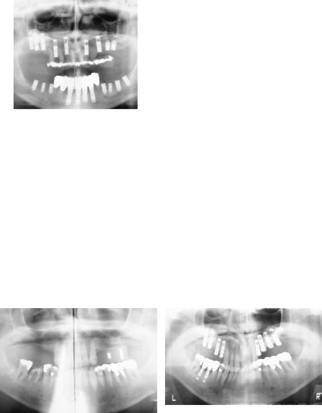
FIGURE 18.21 Panoramic radiograph of bilateral posterior maxillary partial edentulism with right maxillary sinus bone graft and implants, left subantral implant placement, retention of teeth for interim prosthesis, and total mandibular dental implant prosthesis reconstruction.
an interim abutment (Figure 18.21). More commonly, a removableprosthesismaybeinsertedafter2orpreferably4weeks (Figure 18.22a,b). The soft tissue-bearing prosthesis must then beappropriatelyrelievedtoavoidencroachmentontheimplants, particularly if not fully countersunk. Premature loading may cause micromovement and implant failure to osseointegrate.
189
23a,b). The incision should be midcrestal or slightly palatal to allow transposition of keratinized tissue buccally. Tissue height in excess of 3 mm will impair maintenance and should be trimmed.
Implants are most at risk during the first year of function as the result of stresses exceeding physiologic limits. The degree of bone contact and the density of the supporting bone will determine whether a load placed upon an implant is tolerable.48 The fabrication and insertion of an occlusally adjusted acrylic temporary with a metal substructure for 12 months allows additional time for bone maturation as well as incremental loading of the bone (Figures 18.24, 18.25, and 18.26). Progressive implant loading will enhance the amount of desirable mature lamellar bone to develop at the interface. The increase in trabeculae in contact with the implant and in the immediate surrounding bone will improve implant survival. Additionally, early temporization provides for immediate patient gratification (Figure 18.27a,b). It allows the restorative dentist time to perfect the final prosthesis while ameliorating the patient’s desire to “finish up” after a prolonged surgical period. Temporization can also permit the use of sinus bone grafted implants for use as posterior anchorage for fixed orthodontic treatment until idealized tooth positions allow final prosthesis fabrication or additonal implant placement (Figure 18.28a–d).
Abutment Surgery and
Progressive Bone Loading
Patients are followed clinically and when necessary radiographically during the healing period. At the appropriate time, depending on the residual bone and the graft material utilized, abutment placement surgery is performed (Figure
Complications
Sinus Membrane Perforation
Tearing of the Schneiderian membrane is the most common complication of the sinus lift procedure. These defects usually resultfromexcessdepthpenetrationduringtheinitialosteotomy, right-angle bone window corners, and presence of sinus septa,
b
a
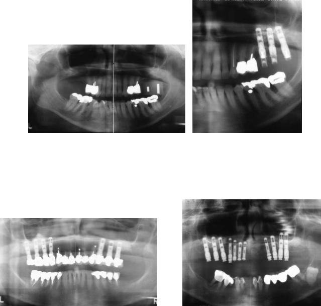
FIGURE 18.22 Bilateral posterior maxillary partial edentulism reconstructed with maxillary sinus bone grafts and placement of dental implants, with removable partial denture temporization possible ow-
ing to the presence of anterior dentition. (a) Preoperative panoramic radiograph. (b) Postoperative panoramic radiograph.
190 |
J.I. Stein and A.M. Greenberg |
b
a
FIGURE 18.23 Unilateral posterior maxillary partial edentulism reconstructed with maxillary sinus bone grafts and placement of dental implants and fixed prosthesis. Placement of cementable post type abutments after exposure of implants allows acrylic temporization
FIGURE 18.24 Postoperative panoramic radiograph with staged bilateral maxillary sinus bone grafts and placement of dental implants with fixed prosthesis. Left maxillary sinus bone graft and placement of dental implants with permanent fixed bridge, right maxillary sinus bone graft with cast metal temporary prosthesis for early loading.
and early loading prior to final prosthesis fabrication and insertion.
(a) Preoperative panoramic radiograph. (b) Postoperative panoramic radiograph.
FIGURE 18.25 Postoperative panoramic radiograph with total maxillary dental implant reconstruction with bilateral maxillary sinus bone grafts and placement of dental implants. Placement of temporary abutments and fabrication of temporary acrylic prosthesis following exposure of bilateral sinus bone grafts and dental implants and removal of remaining anterior maxillary teeth and placement of dental implants. Temporary fixed bridge allows early loading and transitional period until remaining maxillary implants can be utilized in the fabrication of a permanent fixed prosthesis.
a |
b |
c |
d |
e |
f |
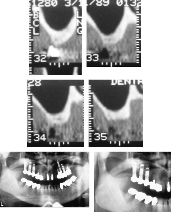
FIGURE 18.26 Right posterior maxillary sinus bone graft with dental implant reconstruction demonstrating adequate subantral bone to stabilize immediate placement of implants and bone graft. Early progressive loading is permitted with a temporary fixed bridge reinforced with a metal substructure. Preoperative CT scan reveals adequate subantral bone to stabilize immediate placement of dental
implants with sinus bone graft: (a) tooth #4 site, (b) tooth #3 site,
(c) tooth #2 site, (d) tooth #1/tuberosity site. (e) Postoperative panoramic radiograph of postoperative view with temporary cast metal reinforced prosthesis for early loading. (f) Postoperative panoramic radiograph close-up view.
Continued.
192 |
J.I. Stein and A.M. Greenberg |
g |
h |
i
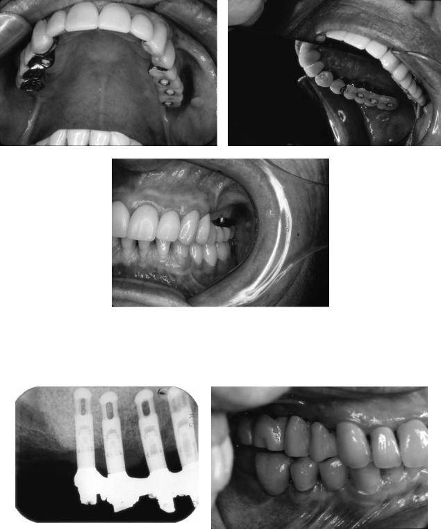
FIGURE 18.26 Continued. (g) Postoperative occlusal view with temporary prosthesis. (h) Postoperative lingual view with temporary prosthesis. Note the metal substructure. (i) Postoperative buccal view with “high water” design to permit easier hygiene.
a |
b |
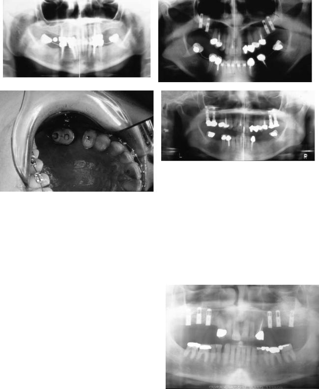
FIGURE 18.27 Right posterior maxillary sinus bone graft and implant placement with temporary metal substructure temporary prosthesis with natural crown emergence for better soft tissue site development
and esthetics, and early loading. (a) Postoperative panoramic radiograph. (b) Postoperative buccal view.
18. Maxillary Sinus Grafting and Osseointegration Surgery |
193 |
a |
b |
c |
d |
FIGURE 18.28 Bilateral posterior maxillary edentulous ridges with decreased vertical dimension and placement of bilateral maxillary sinus bone grafts and placement of dental implants. Sinus bone grafted dental implants with temporary bridges placed for early loading and use as posterior anchorage for fixed orthodontic treatment.
(a) Preoperative panoramic radiograph with fixed orthodontic me-
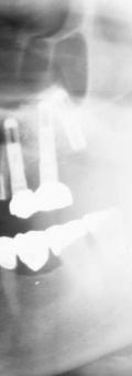
and most commonly occur inferiorly during the initiation of sinus membrane elevation and anteriorly if the osteotomy was inadequately extended and the reflection is being performed blindly. Earlier in the development of the procedure development, the osteotomy was designed so as to be incomplete superiorly. Subsequently, the superiorly hinged bony window was greenstick fractured inwardly. The modified osteotomy design and membrane reflection technique as described earlier will prevent and decrease perforation frequency. Membrane defects increase early and late complications. Tears in the membrane increase bacterial and mucus ingress into the graft, increasing the risk of infection and decreasing graft density, respectively. Egress of graft material through the defect into the residual sinus proper will decrease the graft volume as well as potentially block the ostium and impair drainage (Figure 18.29).
Appropriately managed sinus perforations are generally of minimal consequence. When a defect is noted, further membrane elevation should be performed distally, working toward the perforation circumferentially; this will prevent further enlargement of the tear. With enhanced membrane elevation, particularly along the medial wall, the membrane frequently
chanics for mandibular molar uprighting. (b) Postoperative panoramic radiograph. (c) Clinical occlusal view with temporary fixed prosthesis and fixed orthodontic appliances. (d) Post completion of fixed orthodontic treatment preparation for additional mandibular dental implant placement.
FIGURE 18.29 Postoperative panoramic radiograph with bilateral maxillary sinus bone grafts and dental implant placement. Left maxillary sinus extravasation of bone graft into the space above the sinus membrane perforation causing decreased bone graft volume, blockage of ostium, and impaired sinus drainage.
194
folds upon itself, sealing the opening. If small residual defects persist they are best occluded with a customized resorbable collagen sheet. Alternatively, laminar bone may be utilized as a patch for larger defects. The graft material, which normally should be compacted in all directions, is passively condensed toward this region. Extensive large defects may necessitate aborting the procedure. All internal septa should then be removed in anticipation of reentry months later.29 Ironically, patients with a history of sinus disease have a thicker membrane that is more resistant to perforation. Sinus membrane thickening in the absence of active sinus disease is usually not a contraindication to sinus inlay graft surgery.
Residual Ridge Fracture
In the presence of minimal crestal bone or an excessively inferior horizontal osteotomy, a ridge fracture may occur. This is an uncommon event and will occur during either fixture site preparation or insertion in eggshell ridges. A one-stage procedure must then be aborted and a two-stage procedure instituted. The antral void is grafted inclusive of the fractured region. Fixture insertion is performed following the period required for graft maturation.
Hemmorhage
Significant hemorrhage is rarely a problem. If the initial horizontal incision is placed too far palatal, the greater palatine artery may be lacerated. Increased bleeding from the lateral flap may occur if the periosteum is violated. Sinus membrane perforation may result in low-grade hemorrhage as well as postoperative epistaxis. If bleeding is not self-limiting, hemostasis is attained by conventional methods.
J.I. Stein and A.M. Greenberg
logic with an antibiotic protocol based on culture and sensitivity tests. Prolonged infections will significantly impair bone volume and maturation. Surgical management is with incision and drainage, and if refractory, open debridement and antrostomy or middle meatus widening may be necessary. Regrafting may be considered after a prolonged healing period. Chronic sinusitis is not evident and, anecdotally, patients have reported improvement over preoperative sinus states. It is conjectured that the subantral augmentation enhances drainage by elevation of the floor superiorly in closer proximity to the ostium.
Displaced or Malposed Implant
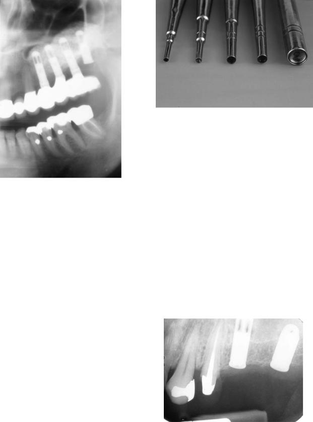
In one-stage procedures, inadequate crestal bone may lead to displaced or malposed fixtures. The crest will be thin and medial, resulting in bodily palatal implant placement or buccal emergence angulation. This will result in unfavorable biomechanics or prosthetic compromise requiring the burying of a fixture (Figure 18.30) or the use of an angled abutment (Figure 18.31) or an overdenture in lieu of a fixed restoration. Fixtures inadequately stabilized by crestal bone may be deflected during graft placement. Similarly, fixtures may be totally displaced into the subantral graft by a poorly adjusted prosthesis during the initial healing period. These complications may be avoided by having bone adequate for prosthetically desirable implant positioning and to provide sufficient stabilization. A two-stage procedure deferring fixture insertion is necessary if these criteria are not met.
Parathesia
Temporary neurosensory deficits of the infraorbital nerve may occur with excess superior flap reflection or improper retractor positioning. Greater palatine nerve distribution paresthesia may occur with an incision placed to medially or excessive medial flap reflection or traction.
Delayed Soft Tissue Healing
Superficial wound dehiscence and necrosis may occur if the horizontal incision is placed excessively palatal. This results in ischemia to the distal edges of the lateral flap as it crosses over the ridge. Prolonged elevated pain, infection, implant exposure, or loss of graft material may occur. Local wound care is supplemented with use of analgesics, chlorhexidine gluconate rinses, and possibly antibiotics until healing is completed by secondary intention.
Infection
Wound infection and acute sinusitis are infrequent and generally transient in nature. Initial care is predominately pharmaco-
FIGURE 18.30 Postoperative panoramic radiograph close-up view reveals left maxillary sinus bone graft with implant placement. Malposed most posterior implant that could not be utilized in the final prosthesis owing to poor position.
18. Maxillary Sinus Grafting and Osseointegration Surgery |
195 |
FIGURE 18.31 Postoperative panoramic radiograph left maxillary sinus bone graft with placement of dental implants. Poorly angled middle position fixtures requiring use of angulated abutments. The distal most implant could not be utilized in the final prosthesis.
Deficient Graft
Insufficient new bone volume or density will result in failure or loss of osseointegration. Supplementation with alternative materials should be considered. This regrafting may be performed with minimal morbidity in that the osteotomy, lateral access, and sinus elevation are preexisting.
Osteotome Sinus Floor Elevation
Summers14,15 has described a simpler, less invasive method of immediate implant insertion utilizing specialized serial osteotomes (Figure 18.32; Implant Innovations, West Palm Beach, FL, USA) in patients with a minimum of 5–6 mm of residual crestal bone. In this technique, bone of the implant recipient site is conserved and serially impacted upward. Bone graft material may be then added to the osteotomy site apically. Further malleting pressure from the osteotome and the graft causes infracturing of the sinus floor and elevation of the membrane. Additional graft material may then be incrementally added to gain additional height. With this technique, the sinus floor may be elevated and grafted, allowing for placement of a 10-mm-long fixture (Figure 18.33). Further multicenter studies are still necessary to determine the efficacy of this procedure. Extensive malleting necessary in the preparation of multiple sites may be disconcerting to the unsedated patient.
FIGURE 18.32 Summers Osteotomes of various sizes (Implant Innovations, Inc., Palm Beach Gardens, FL).
Conclusion
Osseous deficiencies quantitatively and qualitatively have made the posterior maxilla the least predictable region of endosseous implant placement. Sinus membrane elevation with subantral augmentation utilizing a variety of graft materials, including many that are nonautogenous in nature, has produced bone capable of responding to biomechanical demands. Endosseous implants may be placed simultaneously or as a staged procedure, which achieves and maintains long-stand- ing osseointegration. This provides a suitable foundation for an appropriately designed prosthesis following bone maturation. Techniques described here provide for low morbidity, few risks, and minimal and manageable complications, and may be performed in an office setting under local anesthesia. Introduced by Tatum in the 1970s and more widely performed for more than 15 years, sinus lift grafting has been highly successful in providing implant predictability equivalent to any intraoral region.
FIGURE 18.33 Summers technique for maxillary sinus bone grafting and placement of two dental implants.
196
References
1.Jensen J. Reconstruction of the atrophic alveolar ridge with mandibular bone grafts and implants (abstract). J Oral Maxillofac Surg. (special issue) 1990:125.
2.Brein V, Bránemark P-I. Reconstruction of the alveolar jaw bone. An experimental and clinical study of immediate and preformed autologous bone grafts in combination with osseointegrated implants. Scand J Plast Reconstr Surg. 1980;14:23–48.
3.Sailer HF. A new method of inserting endosseous implants in totally atrophic maxilla. J Craniomaxillofac Surg. 1989;17:299– 305.
4.Keller EE, VanReokel NB, Desjardins RP, Tolman DE. Pros- thetic-surgical reconstruction of the severely resorbed maxilla with iliac bone grafting and tissue integrated prosthesis. Int J Oral Maxillofac Implants. 1987;2:155–165.
5.Tatum OH, Leibowitz MS, Tatum CA, Borgner RA. Sinus augmentation rationale development, long-term results. NY State Dent J. 1993;5:43–48.
6.Jensen J, Simonson EK, Sindet-Pederson S. Reconstruction of the severely resorbed maxilla with bone grafting and osseointegrated implants: a preliminary report. J Oral Maxillofac Surg. 1990;48:27–34.
7.Raghoebar GM, Brouwer TJ, Reintsoma H, VanDort RP. Augmentation of the maxillary sinus floor with autogenous bone for the placement of endosseous implants: a preliminary report. J Oral Maxillofac Surg. 1993;51:1198–1203.
8.Block MS, Kent JN. Maxillary sinus grafting for totally and partially edentulous patients. J Am Dent Assoc. 1993;124:139–143.
9.Vlassis JM, Húrzeler MB, Quinones CR. Sinus lift augmentation to facilitate placement of nonsubmerged implants: a clinical and histologic report. Pract Periodontol Restor Dent. 1993;2:15–23.
10.Fugazzotto PA. Maxillary sinus grafting with and without simultaneous implant placement: technical considerations and case reports. Int Periodontics Restor Dent. 1994;14:544– 551.
11.Moy PK, Lundgren S, Holmes RE. Maxillary sinus augmentation: histomorphometric analysis of graft materials for maxillary sinus floor augmentation. J Oral Maxillofac Surg. 1993;51:857–862.
12.Williams MY, Mealey BL, Hallman WW. The role of computerized tomography in dental implantology. Int J Oral Maxillofac Implants. 1992;7:373–380.
13.Kraut R. Radiologic planning for dental implants. In: Block MS, Kent JN, eds. Endosseous Implants for Maxillofacial Reconstruction. Philadelphia: WB Saunders; 1995:113–133.
14.Summers RB. A new concept in maxillary implant surgery: the osteotome technique. Compend Contin Educ Dent. 1994;2:152– 160.
15.Summers RB. The osteotome technique: Part 3. Less invasive methods of elevating the sinus floor. Compend Contin Educ Dent. 1994;8:698–708.
16.Bahat O. Osseointegrated implants in the maxillary tuberosity: report on 45 consecutive patients. Int J Oral Maxillofac Implants. 1992;7:459–467.
17.Khayat P, Nader N. The use of osseointegrated implants in the maxillary tuberosity. Pract Periodontol Aesth Dent. 1994;4:53– 61.
18.Graves SL. The pterygoid plate implant: a solution for restor-
J.I. Stein and A.M. Greenberg
ing the posterior maxilla. Int J Periodontol Restor Dent. 1994; 14:513–523.
19.Keller EE. Composite graft reconstruction of advanced maxillary resorption. In: Block MS, Kent JN, eds. Endosseous Implants for Maxillofacial Reconstruction. Philadelphia: WB Saunders; 1995:504–536.
20.Isaksson S. Evaluation of three bone grafting techniques for severely resorbed maxilla in conjunction with immediate endosseous implants. Int J Oral Maxillofac Implants. 1994;9: 679–688.
21.Bránemark PI, Zarb GA, Albrekitsson T. Tissue-Integrated Prosthesis Osseointegration in Clinical Dentistry. Carol Stream, IL: Quintessence; 1985.
22.Adell R, Lekholm V, Grondahl K, Bránemark PI, Lindstrom J, Jacobson M. Reconstruction of the severely resorbed edentulous maxillae using osseointegrated fixtures in immediate autogenous bone grafts. Int J Oral Maxillofac Implants. 1990;5:233–246.
23.Jensen J, Sindet-Pederson S, Oliver AJ. Varying treatment strategies for reconstruction of maxillary atrophy with implants.
J Oral Maxillofac Surg. 1994;52:210–216.
24.Kahnberg KE, Nystrom E, Bartholdsson L. Combined use of bone grafts and Bránemark fixtures in the treatment of severely resorbed maxillae. Int J Maxillofac Implants. 1989;4:297–304.
25.Bain CA, Moy PK. The association between the failure of dental implants and cigarette smoking. Int J Oral Maxillofac Implants. 1993;8:609–615.
26.Block MS, Kent JN. Endosseous Implants for Maxillofacial Reconstruction. Philadelphia: WB Saunders; 1995.
27.Buckley MS. Bone substitutes. Sel Read Oral Maxillofac Surg. 1995;4:2.
28.Axhausen W. The osteogenetic phases of regeneration of bone, a historical and experimental study. J Bone Joint Surg. 1956; 38A:593–601.
29.Tatum OH. Maxillary and sinus implant reconstruction. Dent Clin North Am. 1986;30:207–229.
30.Boyne PJ, James RA. Grafting of the maxillary sinus floor with autogenous marrow bone. J Oral Surg. 1980;38:613–616.
31.Keller EE, Eckert SE, Tolman DE. Maxillary antral and nasal one stage inlay composite graft: preliminary report on 30 recipient sites. J Oral Maxillofac Surg. 1994;52:438–447.
32.Loukota RA, Isaksson SG, Linner ELJ, Blomquist JE. A technique for inserting endosseous implants in the atropic maxilla in a single stage procedure. Br J Oral Maxillofac Surg. 1992;30: 46–49.
33.Hall DH, McKenna SJ. Bone graft of the maxillary sinus floor for Branemark implants: a preliminary report. Oral Maxillofac Surg Clin North Am. 1991;3:869–875.
34.Hirsch JM, Erickson I. Maxillary sinus augmentations using mandibular bone grafts and simultaneous installation of implants. Clin Oral Implant Res. 1991;2:91–96.
35.Urist MR. Bone formation by autoinduction. Science 1965;150: 893.
36.Marx RE, Carlson ER. Tissue banking safety: caveats and precaution for the oral and maxillofacial surgeon. J Oral Maxillofac Surg. 1993;51:1372–1379.
37.Buck BE, Malinin TI. Human bone and tissue allografts: preparation and safety. Clin Orthop Relat Res. 1994;303:8–17.
38.Buck BE, Malinin TI, Brown MD. Bone transplantation and human immunodeficiency virus: an estimated risk of AIDS. Clin Orthop Relat Res. 1989;240:129–136.
18. Maxillary Sinus Grafting and Osseointegration Surgery
39.Reddi AH, Hascall VC. Changes in proteoglycan types during matrix reduced cartilage bone and marrow formation. Proc Natl Acad Sci USA. 1977;55:89.
40.Glowacki J, Altobelli D, Mulliken JB. Fate of mineralized and demineralized osseous implants in cranial defects. Calcif Tissue Int. 1981;33:71.
41.Lindholm TS, Nilsson OS, Lindholm TC. Extraskeletal and intraskeletal new bone formation induced by demineralized bone matrix combined with marrow cells. Clin Orthop Relat Res. 1982;171:251–255.
42.Wittbjer J, Palmer B, Robbin M, Thorngren KG. Osteogenetic activity in composite grafts of demineralized compact bone and marrow. Clin Orthop Relat Res. 1983;173:229–238.
43.Smiler DG, Johnson PW, Lozada JL, Misch C, Rosenlicht JL, Tatum OH, Wagner J. Sinus lift grafts and endosseous implants: treatment of the atropic posterior maxilla. Dent Clin North Am. 1992;36:151–188.
44.Collins T, Small S, Shepherd N, Buser D, Parel SM. Sinus floor elevations and the status of membranes. Panel discussion. Int J Oral Maxillofac Implants. 1994;9(suppl):85–96.
45.Becker W, Becker BE, Caffesse R. A comparison of demineralized freeze-dried bone and autologous bone to induce bone formation in human extraction sockets. J Periodontol. 1994;65: 1128–1133.
46.Small SA, Zinner ID, Panno FV, Shapiro HJ, Stein JI. Augmenting the maxillary sinus for implants: report of 27 patients.
Int J Oral Maxillofac Implants. 1993;8:523–528.
47.Misch CE. Maxillary sinus augmentaion for endosteal implants: organized alternative treatment plans. Int J Oral Implantol. 1987;4:49–58.
48.Misch CE. Contemporary Implant Dentistry. St. Louis: CV Mosby; 1993.
49.Whitaker JM, James RA, Lozada J, Cordova C, GaRey DJ. Histological response and clinical evaluation of autograft and allograft material in the elevation of maxillary sinus for the preparation of endosteal dental implant sites. Simultaneous sinus
197
elevation and root form implantation: an eight month autopsy report. J Oral Implantol. 1989;15:141–144.
50.Wittfbjer J, Palmer B. Osteogenetic activity in composite grafts of demineralized compact bone and marrow. Clin Orthop. 1988; 173:209.
51.Klinge B, Alberius P, Isaksson S, Jonsen J. Osseous response to implanted natural bone mineral and synthetic hydroxylapatite ceramic in the repair of experimental skull bone defects. J Oral Maxillofac Surg. 1992;50:241–249.
52.Smiler DG, Holmes RE. Sinus lift procedure using porous hydroxyapatite: a preliminary report. J Oral Implantol. 1987;13: 239–253.
53.Wagner J. A 31/2 year clinical evaluation of resorbable hydroxyapatite Osteogen (HA resorb) used for sinus lift augmentation in conjunction with the insertion of endosseous implants. J Oral Implantol. 1991;17:152–164.
54.Tidwell JK, Blijdorp PA, Stoelinga PJW, Brouns JB, Hinderks F. Composite grafting of the maxillary sinus for placement of endosteal implants. A preliminary report of 48 patients. Int J Oral Maxillofac Surg. 1992;21:204–209.
55.Vassos DM, Petrick DK. The sinus lift procedure: an alternative to the maxillary subperiosteal implant. Pract Periodontal Dent. 1992;9:14–19
56.Dario LJ, English R. Chin bone harvesting for autogenous grafting in the maxillary sinus: a clinical report. Pract Periodontol Restor Dent. 1994;9:87–91.
57.Jaffin RA, Berman CL. The excessive loss of Branemark fixtures in type IV bone: a 5-year analysis. J Periodontol. 1991;62:2–4.
58.Bahat O. Treatment planning and placement of implants in the posterior maxillae: report of 732 consecutive Nobelpharma implants. Int J Oral Maxillofac Implants. 1993;8:151–161.
59.Fugazzotto PA, Wheeler SL, Lindsay JA. Success and failure rates of cylinder implants in type IV bone. J Periodontol. 1993; 64:1085–1087.
60.Schow SR. Infections of the maxillary sinus. Oral Maxillofac Surg Clin North Am. 1991;32:343–353.
