


FACTS & HINTS
HIGH-YIELD FACTS
Clinical Points
page 261
page 262
Compartment Syndrome
Abnormallyhigh pressure within one of the leg's restricted compartments
Caused byhemorrhage, edema, or inflammation of the muscles of the leg, often secondaryto trauma or deep vein thrombosis Increased compartmental pressure causes ischemia and nerve damage
Maylead to permanent injuryof distal structures supplied byeffected arteries and veins Management is surgical: fasciotomyand debridement of anynecrotic tissue
Shin Splints (tibialis anterior strain)
Edema and pain over distal two thirds of the tibia
Caused byrepetitive microtrauma to the tibialis anterior muscle
Common in athletic overexertion
Often in persons who do not exercise, when theytrya long distance walk
Amild form of anterior compartment syndrome
Chronic condition can cause periostitis and bone remodeling, or lead to stress fractures
Ruptured Achilles' Tendon
Tendonitis of the calcaneal tendon
Acommon running injury
Rupture of the tendon usuallyoccurs in untrained middle-aged individuals with a historyof tendonitis
Presents as sudden calf pain with an audible snap and subsequent inabilityto plantarflexthe foot
Gap is palpable in complete rupture
MNEMONICS
Memory Aids
Deep posterior leg muscles (medial to lateral): |
Down The Hatch |
|
Flexor Digitorum longus |
|
Tibialis posterior |
|
Flexor Hallucis longus |
(structures behind medial malleolus-see Section 7-5: Ankle and Foot)
Symptoms of varicose veins: |
AEIOU |
|
Aching |
|
Eczema |
|
Itching |
|
Oedema |
|
Ulceration/Ugly(varicosities) |
409 / 425

53 Ankle and Foot
STUDYAIMS
At the end of your study, you should be able to:
Understand the structure of the ankle mortise and its supporting ligaments and appreciate its vulnerabilityto injury Know the bones of the foot and their organization
Understand the functional importance of the ligaments and fascia of the foot Describe the arches of the foot
Understand the arrangement of the intrinsic muscles of the foot Know the descriptive layers of the plantar surface of the foot
410 / 425
GUIDE
Lower Limb: Ankle and Foot
Ankle Joint (Talocrural Joint)
[Plate 514, Ankle: Radiographs]
Uniaxial, synovial, hinge type joint
Articulation between tibia (medial malleolus), fibula (lateral malleolus), and talus
Distal ends of tibia and fibula form a mortise
Trochlea of talus fits into mortise
Malleoli grip talus
Movements
Dorsiflexion
Produced bymuscles of anterior compartment of leg
Limited bytriceps surae
More stable when dorsiflexed
Plantarflexion-produced bymuscles of posterior compartment of leg
Some rotation, abduction, and adduction of joint possible in plantar flexion
Capsule
Thin
Supported bystrong collateral ligament
Attached to tibia and malleoli superiorlyand talus inferiorly
Ligaments
Lateral consists of three parts
Anterior talofibular-from lateral malleolus to neck of talus
Posterior talofibular-from malleolar fossa to lateral tubercle of talus
Calcaneofibular-from tip of lateral malleolus to lateral calcaneus
Medial or deltoid
Strong ligament
Originates on medial malleolus
Fibers can be identified as
411 / 425

Anterior and posterior tibiotalar
Tibionavicular
Tibiocalcaneal
Blood supply: malleolar branches of fibular, and anterior and posterior tibial arteries Nerve supply: Tibial nerve and deep fibular nerve (a branch of the common fibular)
Foot
[Plate 511, Bones of Foot]
page 263
page 264
412 / 425

[Plate 513, Calcaneus]
Bones
26 in number Tarsal bones (7)
Talus
Has head, neck, bodywith trochlea, posterior, and lateral processes
Articulates with fibula, calcaneus, and navicular
Has no muscular attachments
Head rests on lateral projection of calcaneus-sustentaculum tali
Wider anteriorlymaking the ankle more stable in dorsiflexion
Calcaneus
Largest and strongest bone
Posterior prominence-calcaneal tuberosity-with medial, lateral, and anterior tubercles
Articulates with the talus and cuboid
Transmits bodyweight from talus to ground
Lateral projection-sustentaculum tali-supports talar head
Navicular
Flattened bone with inferomedial tuberosity
Located between talar head and three cuneiform bones
Cuboid
Inferolateral groove for tendon of peroneus longus
Most lateral bone in distal row of tarsals
Cuneiform (3)
Medial, intermediate and lateral
Each articulates with navicular posteriorlyand base of related metatarsal anteriorly
 Lateral articulates with cuboid Metatarsals (5)
Have base (proximal), body, and head (distal)
Bases articulate with cuneiform and cuboid bones
Head articulates with proximal phalanges
 Medial and lateral sesamoid bones on plantar surface of first metatarsal in plantar ligament Phalanges (14)
Great toe (hallux) has two-proximal and distal
413 / 425

Other toes have three-proximal, middle and distal
Each consists of proximal base, body, and distal head
Joints
Important intertarsal joints
Where inversion and eversion occur
Subtalar
Synovial joint with where talus rests on calcaneus
Fibrous capsule supported bytalocalcaneal ligaments
Transverse tarsal, composed of
Calcaneocuboid joint
Talonavicular joint
Other joints where slight movement occurs
Talocalcaneal
Tarsometatarsal
Metatarsophalangeal (MTP)
Interphalangeal: proximal and distal (PIP and DIP)
page 264 page 265
Arches
Tarsal and metatarsal bones are arranged in longitudinal and transverse arches
Bonyarches maintained by
Interlocking bones
Plantar ligaments
Plantar aponeurosis
Action of plantar muscles
Functions
Shock absorbers for bodyweight
Distribute bodyweight
Make foot adaptable to changes in surface
Longitudinal arch composed of medial and lateral arches
Medial longitudinal arch
Higher arch than lateral
Composed of calcaneus, talus, navicular, three cuneiforms, three medial metatarsals
Talar head is keystone
Strengthened by
Tibialis anterior tendon and attachments
Fibularis longus tendon
Lateral longitudinal arch
Flatter than medial
Rests on the ground when standing
Composed of calcaneus, cuboid, and lateral two metatarsals
Transverse arch
Formed bycuboid, cuneiforms, bases of metatarsals
Has pillars formed bylateral and medial longitudinal arches
Maintained byfibularis longus tendon
Ligaments (major ligaments listed)
All foot bones are united byplantar and dorsal ligaments
Plantar calcaneonavicular ligament
Also called spring ligament
From sustentaculum tali to navicular
Maintains longitudinal arch of foot
Long plantar ligament
From plantar surface of calcaneus to cuboid
Maintains foot arches
Plantar calcaneocuboid ligament
Also called short plantar ligament
Deep to the long plantar ligament
From inferior surface of calcaneus to inferior surface of cuboid
Plantar Muscles
414 / 425
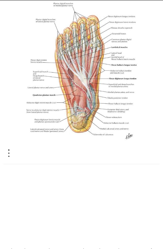
[Plate 522, Muscles of Sole of Foot: Second Layer]
Four layers in the sole of the foot
Individual muscles of little importance as fine control of the toes is not required
Aneurovascular plane exists between the first and second and third and fourth layers
page 265 page 266
Intrinsic Foot Muscles
|
Muscle |
Proximal Attachment |
Distal Attachment (Insertion) |
Layer of |
Innervation |
Action |
|
|
|
(Origin) |
|
plantar foot |
|
|
|
|
Abductor |
Medial process of |
Medial side, base of proximal |
First Layer: |
Medial plantar |
Abducts and flexes first toe |
|
|
hallucis |
calcaneal tuberosity |
phalanxof first toe |
deep to |
nerve (S2-S3) |
|
|
|
|
|
|
plantar |
|
|
|
|
|
|
|
aponeurosis |
|
|
|
|
Flexor |
Medial process of |
Four tendons split to allow |
First Layer: |
Medial plantar |
Flexes second through fifth |
|
|
digitorum |
calcaneal tuberosity |
passage of long flexor tendons, |
deep to |
nerve (S2-S3) |
toes |
|
|
brevis |
|
insert on middle phalanges |
plantar |
|
|
|
|
|
|
|
aponeurosis |
|
|
|
|
Abductor |
Calcaneus |
Lateral side, proximal phalanx |
First Layer: |
Lateral plantar |
Abducts and flexes fifth toe |
|
|
digiti |
|
of fifth toe |
deep to |
nerve (S2-S3) |
|
|
|
minimi |
|
|
plantar |
|
|
|
|
|
|
|
aponeurosis |
|
|
|
|
Quadratus |
Medial and lateral |
Lateral edge of flexor digitorum |
Second Layer |
Lateral plantar |
Corrects for oblique pull of |
|
|
plantae |
sides of plantar |
longus tendon |
|
nerve (S1-S3) |
FDL tendon; thus assists in |
|
|
|
surface of calcaneus |
|
|
|
flexion of toes |
|
|
Lumbricals |
First: Medial side of |
Medial side of dorsal digital |
Second Layer |
Medial one: |
Flexproximal phalanges at |
|
|
|
tendon to second toe |
expansions |
|
Medial plantar |
MP joint, extend distal |
|
|
|
Second through |
|
|
nerve |
phalanges at PIP and DIP |
|
|
|
fourth:Adjacent sides |
|
|
Lateral three: |
joints |
|
|
|
of contiguous |
|
|
Lateral plantar |
|
|
|
|
tendons |
|
|
nerve (S2-S3) |
|
|
|
|
|
|
|
|
|
|
415 / 425

|
Flexor |
Plantar surfaces of |
Divides in two, to each side of |
Third Layer |
Medial plantar |
Flexes proximal phalanxof |
|
hallucis |
cuboid and lateral |
base of proximal phalanxof |
|
nerve (S1-S2) |
first toe |
|
brevis |
cuneiform |
first toe |
|
|
|
|
Adductor |
Oblique head: bases |
Lateral side, base of proximal |
Third Layer |
Deep branch of |
Adducts first toe, maintains |
|
hallucis |
of second through |
phalanxof first toe (both heads) |
|
lateral plantar |
transverse arch |
|
|
fourth metatarsals |
|
|
nerve (S2-S3) |
|
|
|
Transverse head: |
|
|
|
|
|
|
Ligaments of |
|
|
|
|
|
|
metatarsophalangeal |
|
|
|
|
|
|
joints |
|
|
|
|
|
Flexor digiti |
Base of fifth |
Base of proximal phalanxof |
Third Layer |
Superficial |
Flexes proximal phalanxof |
|
minimi |
metatarsal |
fifth toe |
|
branch, lateral |
fifth toe |
|
brevis |
|
|
|
plantar nerve |
|
|
|
|
|
|
(S2-S3) |
|
|
Plantar |
Bases and medial |
Medial sides of bases of |
Fourth Layer |
Lateral plantar |
Adduct second through fourth |
|
interossei |
sides of third through |
proximal phalanges of third |
|
nerve (S2-S3) |
toes and flexMP joint |
|
(3) |
fifth metatarsals |
through fifth toes |
|
|
|
|
Dorsal |
Adjacent sides of first |
First: medial side of proximal |
Fourth Layer |
Lateral plantar |
Abduct second through fourth |
|
interossei |
through fifth |
phalanxof second toe |
|
nerve (S2-S3) |
toes and flexMP joints |
|
(4) |
metatarsals |
Second through fourth: lateral |
|
|
|
|
|
|
sides of second through fourth |
|
|
|
|
|
|
toes |
|
|
|
Dorsal Muscles of Foot |
|
|
|
|
||
|
Form bulge on dorsolateral surface of foot, anterior to lateral malleolus |
|
|
|||
|
Two muscles blend together |
|
|
|
|
|
|
Extensor digitorum brevis |
|
|
|
|
|
|
Proximal attachment: superior surface, anterolateral calcaneus |
|
|
|||
|
Splits into four muscles, each with a tendon that blends with that of long extensor |
|
||||
|
One tendon to great toe |
|
|
|
|
|
|
Other tendons to second to fourth toes |
|
|
|
||
|
Extensor hallucis brevis |
|
|
|
|
|
|
Largest and most medial bellyof extensor digitorum brevis |
|
|
|
||
|
Inserts on proximal phalanxof great toe |
|
|
|
||
|
Supplied bydeep fibular nerve |
|
|
|
|
|
|
Assist extensor digitorum longus in extending toes |
|
|
|
||
Fascia |
|
|
|
|
|
|
|
Deep fascia on dorsum of foot |
|
|
|
|
|
|
Thin on dorsum |
|
|
|
|
|
|
Continuous with inferior extensor retinaculum |
|
|
|
||
|
Over lateral and posterior aspects it is continuous with plantar fascia |
|
|
|||
|
Deep fascia of plantar surface of foot |
|
|
|
||
|
Central condensation of plantar fascia forms plantar aponeurosis |
|
|
|||
|
Arises from calcaneus |
|
|
|
|
|
|
Divides into five fibrous bands that split to enclose digital tendons |
|
|
|||
|
Vertical septa from deep surface divide the foot into medial, central, and lateral compartments |
|
||||
|
Functions of plantar fascia |
|
|
|
|
|
|
Holds foot together |
|
|
|
|
|
|
Protects sole of foot from injury |
|
|
|
||
|
Supports longitudinal arches |
|
|
|
||
416 / 425