

25 Body Wall
STUDYAIMS
At the end of your study, you should be able to:
Know the layers of the anterior abdominal wall from superficial to deep
Understand the orientation and functions of the muscles of the anterolateral abdominal wall
Describe the structure of the rectus sheath and its contents
Describe the neurovascular supplyof the abdominal wall
Understand the boundaries of the inguinal canal and layers of the spermatic cord
Describe the boundaries of Hesselbach's triangle
Know the etiologyand location of the umbilical peritoneal folds
Understand the structure and organization of the posterior abdominal wall and its neurovascular relations
191 / 425
GUIDE
Abdomen: Body Wall
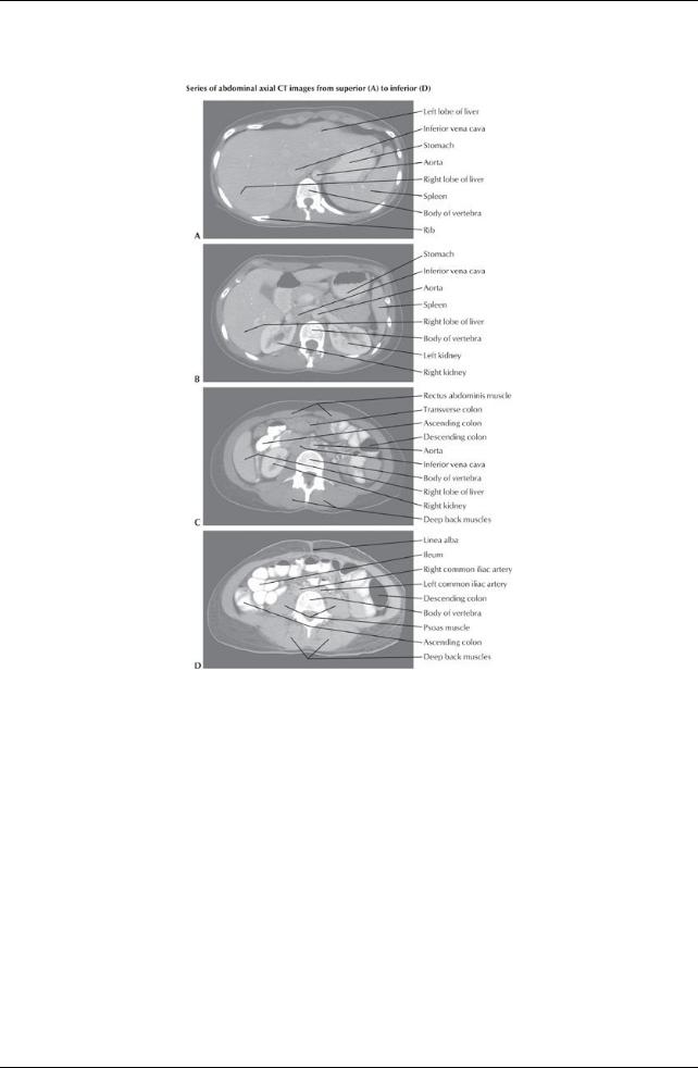
[Plate 324, Abdominal Scans: Axial CT Images]
192 / 425
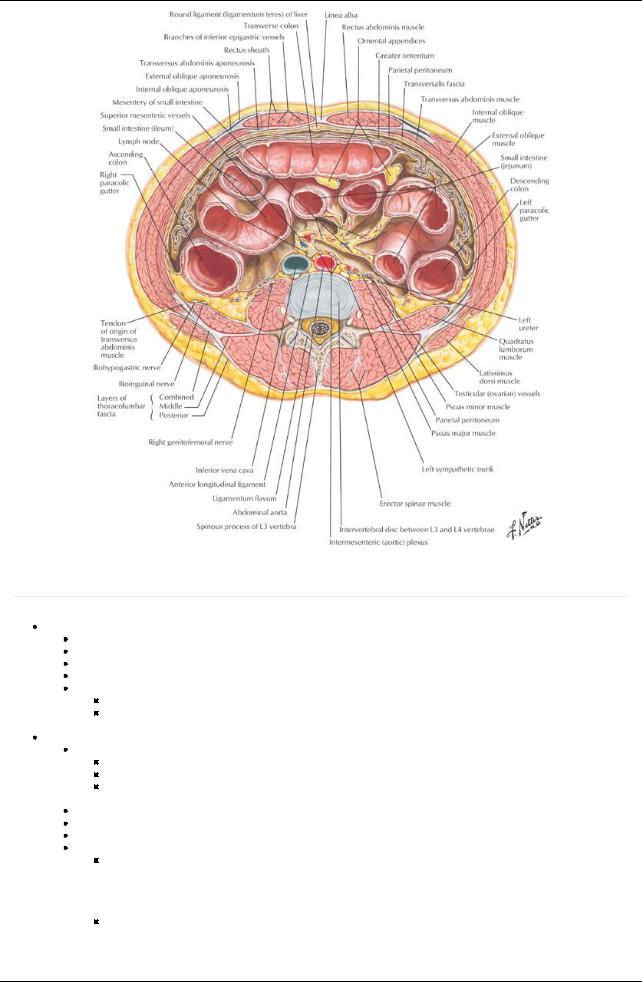
[Plate 330, Schematic Cross Section of Abdomen at L3, 4]
Anterolateral Abdominal Wall
page 129 page 130
Fascial layers
Superficial fascia: two layers in abdomen
Fattysuperficial layer (Camper's fascia)
Deeper membranous layer (Scarper's fascia)
Deep fascia-a verythin layer investing the most superficial muscles.
Transversalis fascia (endoabdominal fascia)
Athin membranous sheet lining most of the abdominal wall
Lies deep to the transversus muscles and the linea alba
 Endoabdominal fat separates the transversalis fascia from the parietal peritoneum Muscles
Functions
Protect the viscera
Help maintain posture
Can compress the abdominal contents, thus raising intra-abdominal pressure, such as in sneezing, coughing, defecating, micturating, lifting, and childbirth
Four paired muscles make up the anterolateral abdominal wall
Three flat muscles
Asingle vertical muscle.
Three flat muscles include
The external abdominal oblique
a.Largest and most superficial
b.Fibers run inferiorlyand mediallyand end in aponeurosis that contributes to the rectus sheath.
c.Inferior border of its aponeurosis forms the inguinal ligament, where it thickens and folds back on itself
d.Innervated segmentallybyT6-T12 spinal nerves and subcostal nerve
The internal abdominal oblique
a.Athin muscular layer
b.Fibers run inferiorlyand laterallyand end in an aponeurosis that contributes to the rectus sheath
c.Inferior aponeurotic fibers join with those of the rectus abdominis to form the conjoint tendon, inserting onto the pubic crest
193 / 425
d. Innervated segmentallybythe ventral rami of T6-T12 spinal nerves The transversus abdominis
a.Innermost of the three flat muscles
b.Fibers run transverselyand mediallyand end in an aponeurosis that contributes to the rectus sheath.
c.Innervated segmentallybythe ventral rami of T6-T12 spinal nerves
Linea alba
a.Tendinous raphe running verticallyin the midline
b.Formed bythe union of the aponeuroses of the flat muscles on either side
c.Largelyavascular
d.Umbilicus found in midline
Vertical muscle = rectus abdominis
Paired
Separated bythe linea alba in the midline
Wider superiorlythan inferiorly
Typicallycomposed of four segments connected bytendinous intersections that attach anteriorlyto the sheath of this muscle
Innervated segmentallybythe ventral rami of T6-T12 spinal nerves
Have the superior epigastric and the inferior epigastric arteries running inferiorlyand superiorly, respectively, on their deep surfaces.
Pyramidalis muscle
Small, insignificant, triangular muscles arising from the bodyof the pubis inferiorly
Inserts into the linea alba medially
 Absent in 20% of people Rectus sheath
Atough, fibrous sheath composed of the aponeuroses of the three flat muscles
Extends from the xiphoid process and fifth through seventh costal cartilages to pubic symphysis and crests
Contains the superior and inferior epigastric vessels, lymphatics and branches of the ventral primaryrami of T7-T12
Encloses the rectus abdominis and the pyramidalis muscle
Semilunar line marks lateral border
Has a crescent-shaped line-the arcuate line-on its posterior wall approximatelythree fourths of the waydown the wall
Above the arcuate line:
Anterior wall composed of the aponeurosis of the external abdominal oblique and the anterior layer of the aponeurosis of the internal abdominal oblique
Posterior wall composed of the posterior layer of the aponeurosis of the internal abdominal oblique, the aponeurosis of the transversus abdominis, the transversalis fascia of the abdomen, and parietal peritoneum
Below arcuate line
Aponeuroses of all three flat muscles pass anterior to the rectus muscle, reinforcing the anterior wall
Posterior wall composed of just transversalis fascia and parietal peritoneum
Has vessels and nerves entering the sheath at its lateral edge, the semilunar line, to supplythe rectus muscle.
page 130 page 131
Muscle |
Proximal Attachment (Origin) |
Distal Attachment |
Innervation |
Blood Supply |
Main Actions |
|
|
(Insertion) |
|
|
|
External |
External surfaces of 5th to 12th |
Linea alba, pubic tubercle, |
Inferior seven |
Superior and |
Compresses and |
oblique |
ribs |
and anterior half of iliac |
thoracic nerves |
inferior epigastric |
supports |
|
|
crest |
|
arteries |
abdominal viscera; |
|
|
|
|
|
flexes and rotates |
|
|
|
|
|
trunk |
Internal |
Thoracolumbar fascia, anterior |
Inferior borders of 10th to |
Ventral rami of |
Superior and |
Compresses and |
oblique |
two thirds of iliac crest, and |
12th ribs, linea alba, and |
inferior six |
inferior epigastric |
supports |
|
lateral half of inguinal ligament |
pubis via conjoint tendon |
thoracic and |
and deep |
abdominal viscera; |
|
|
|
1st lumbar |
circumflexiliac |
flexes and rotates |
|
|
|
nerves |
arteries |
trunk |
Transversus |
Internal surfaces of 7-12 costal |
Linea alba with |
Ventral rami of |
Deep circumflex |
Compresses and |
abdominis |
cartilages, thoracolumbar |
aponeurosis of internal |
inferior six |
iliac, and inferior |
supports |
|
fascia, iliac crest, and lateral |
oblique, pubic crest, and |
thoracic and |
epigastric arteries |
abdominal viscera |
|
third of inguinal ligament |
pecten pubis via conjoint |
1st lumbar |
|
|
|
|
tendon |
nerves |
|
|
Rectus |
Pubic symphysis and pubic |
Xiphoid process and costal |
Ventral rami of |
Superior and |
Flexes trunk and |
abdominis |
crest |
cartilages 5-7 |
inferior six |
interior epigastric |
compresses |
|
|
|
thoracic nerves |
arteries |
abdominal viscera |
Pyramidalis |
Bodyof pubis, anterior to rectus |
Linea alba |
|
Inferior epigastric |
Tenses the linea |
|
abdominis |
|
|
artery |
alba |
page 131 page 132
Nerve supply
Neurovascular plane
Found between the internal abdominal oblique and the transversus abdominis
Contains the vessels and nerves supplying the skin and muscles of the anterior and lateral abdominal wall.
Nerves and vessels are transverselyoriented and segmental
Nerves
Thoracoabdominal nerves
Anterior cutaneous branches of the ventral primaryrami of T7-T11
a.T7-T9 supplyskin above the umbilicus
b.T10 supplies skin around the umbilicus
c.T11 (plus subcostal and ilioinguinal and iliohypogastric nerves) supplies skin below umbilicus
194 / 425
d.Subcostal nerves (T12) supplyskin below umbilicus
e.Iliohypogastric and ilioinguinal nerves (terminal branches of L1) supplies skin below umbilicus
Vascular supply
Arteries
Anterior and collateral branches of posterior intercostal arteries
Branches of the internal thoracic arteries
a.Superior epigastric
b.Musculophrenic
Inferior epigastric (from external iliac)
Branches of the femoral artery
a.Superficial epigastric
b.Superficial circumflexiliac
Veins
Venous drainage is via venae comitantes (veins corresponding to the arteries listed)
Blood drains awayfrom the umbilicus
Venous drainage to the caval system
Lymphatics
Superficial lymphatics above the umbilicus lymph drains to the axillarynodes
Superficial lymphatics below the umbilicus drain to the superficial inguinal nodes
Deep lymphatics
a.Accompanydeep veins
b.Drain to external iliac, common iliac, and lumbar nodes
Posterior abdominal wall
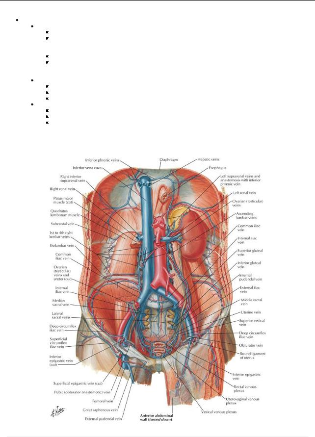
[Plate 258, Veins of Posterior Abdominal Wall]
page 132 page 133
|
Muscle |
Superior Attachment |
Inferior |
Innervation |
Blood Supply |
Actions |
|
|
|
(Origin) |
Attachment |
|
|
|
|
|
|
|
(Insertion) |
|
|
|
|
|
Psoas |
Transverse processes of |
Lesser trochanter |
Lumbar |
Lumbar arteries |
Acting superiorlywith iliacus, flexes |
|
|
major |
lumbar vertebrae; sides of |
of femur |
plexus via |
|
hip; acting inferiorly, flexes vertebral |
|
|
|
bodies of T12-L5 |
|
ventral |
|
column laterally; used to balance |
|
195 / 425
|
vertebrae, and intervening |
|
branches |
|
trunk in sitting position; acting |
|
intervertebral discs |
|
of L1-L4 |
|
inferiorlywith iliacus, flexes trunk |
|
|
|
nerves |
|
|
Iliacus |
Superior two thirds of iliac |
Lesser trochanter |
Femoral |
Iliolumbar artery |
Flexes hip and stabilizes hip joint; |
|
fossa, ala of sacrum, and |
of femur and |
nerve (L2- |
|
acts with psoas major |
|
anterior sacroiliac |
shaft inferior to it, |
L4) |
|
|
|
ligaments |
and to psoas |
|
|
|
|
|
major tendon |
|
|
|
Quadratus |
Medial half of inferior |
Iliolumbar |
Ventral |
Iliolumbar artery |
Extends and laterallyflexes vertebral |
lumborum |
border of 12th rib and tips |
ligament and |
branches |
|
column; fixes 12th rib during |
|
of lumbar transverse |
internal lip of iliac |
of T12 and |
|
inspiration |
|
processes |
crest |
L1-L4 |
|
|
|
|
|
nerves |
|
|
Diaphragm |
Xiphoid process, lower six |
Converge into |
Phrenic |
Pericardiacophrenic |
Draws central tendon down and |
|
costal cartilages, L1-L3 |
central tendon |
nerve (C3- |
musculophrenic, |
forward during inspiration |
|
vertebrae |
|
C5) |
superior and |
|
|
|
|
|
inferior phrenic |
|
|
|
|
|
arteries |
|
page 133 page 134 page 134 page 135
Fascia
Superficial fascia: single layer
Deep fascia-a verythin layer investing the most superficial muscles.
Transversalis fascia (endoabdominal fascia)
Endoabdominal fat separates the transversalis fascia from the parietal peritoneum
Psoas sheath
Fascia covering the psoas muscle
Attaches to lumbar vertebrae and pelvic brim
Thickened superiorlyto form the medial arcuate ligament-a site of origin of the muscle of the diaphragm
Fascia of quadratus lumborum
Fuses mediallywith psoas fascia
Thickened superiorlyto form the lateral arcuate ligament-a site of origin of the muscle of the diaphragm
Thoracolumbar fascia (Section 2: Back and Spinal Cord)
Composed of anterior and posterior layers
Encloses the deep muscles of the back
Thick and strong in the lumbar region
Extends from 12th rib to iliac crest and continuous laterallywith deep fascia of internal oblique and transversus abdominus muscles
Muscles
Psoas major
Long and thick
Lies lateral, and is attached to, the lumbar vertebrae
Tendon passes deep to inguinal ligament to lesser trochanter of femur
Together with iliacus forms iliopsoas muscle, which flexes the hip, helps maintain erect posture
Lumbar plexus of nerves embedded within it
Iliacus
Attaches to superior two thirds of iliac fossa
Joins psoas to form iliopsoas
Quadratus lumborum
Thick quadrangular muscle of posterior wall
Extends from 12th rib and tips of lumbar transverse processes to iliac crest Flexes and laterallyextends vertebral column
Arteries of the posterior abdominal wall (Section 4-6: Abdomen-Visceral Vasculature)
Abdominal aorta
Origin of most of arteries supplying the posterior wall
Begins anterior to the bodyof T12 and ends at bifurcation of the common iliac arteries at L4
Common iliac artery
Follows the medial border of the psoas
Divides into internal and external iliac arteries at pelvic brim
External iliac
a.Gives off inferior epigastric and deep circumflexarteries
b.Exits under the inguinal ligament as the femoral artery
c.Supplies lower limb
Internal iliac arterysupplies pelvis
Unpaired visceral branches of abdominal aorta
Celiac trunk (T12)
Superior mesenteric (L1)
Inferior mesenteric (L3)
Paired visceral branches
Suprarenal arteries (L1)
Renal arteries (L1)
Gonadal arteries (L2) (Note: gonadal arteries branch from the anterior aorta)
Paired parietal branches
Subcostal arteries (T12)
196 / 425

Inferior phrenic arteries
Lumbar arteries (four pairs)
Unpaired parietal branch: Median sacral arteryarising just above aortic bifurcation
Veins of the posterior abdominal wall (Section 4-6: Abdomen-Visceral Vasculature)
Inferior vena cava (IVC)
Formed from union of common iliac veins
Begins anterior to bodyof L5 and passes through the diaphragm at T8.
Its tributaries follow branches of aorta
Exceptions:
a.Left gonadal vein drains to left renal vein
b.Left suprarenal vein drains to left renal vein
Blood from abdominal viscera
Drains via portal system and liver
Reaches IVC through the hepatic veins
Ascending lumbar veins
Drain to azygos/hemiazygos veins and thence to superior vena cava (SVC)
Forms an anastomosis between IVC and SVC.
Lymphatics of the posterior abdomen
Common iliac nodes
Receive lymph from the external and internal iliac nodes
Drain to lumbar (para-aortic) nodes
Lumbar (para-aortic) nodes
Receive lymph from the posterior abdominal wall, descending colon, kidneys, ureters, testes/ovaries, uterus, uterine tubes
Efferent vessels form the lumbar lymphatic trunks
Preaortic nodes
Receive lymph from the digestive tract, liver, spleen, and pancreas
Efferent vessels form the intestinal lymphatic trunks
Nerves of the posterior abdominal wall (Section 4-7: Abdomen-Innervation)
Somatic nerves  Subcostal nerves
a.Ventral primaryrami of T12
b.Arise in the thorax
c.Run inferiorlyon surface of quadratus lumborum
d.Supplyexternal abdominal oblique and skin of anterolateral abdominal wall
Lumbar nerves
a.Dorsal and ventral primaryrami of lumbar spinal nerves
b.Dorsal rami supplymuscles and skin of back
c.Ventral rami pass into substance of psoas major muscle and form lumbar plexus
Nerves of lumbar plexus
Ilioinguinal and iliohypogastric nerves (L1)
a.Enter abdomen posterior to medial arcuate ligament
b.Pierce transverse abdominus near anterior superior iliac spine (ASIS)
c.Supplyskin of suprapubic and inguinal regions
Genitofemoral nerve (L1/2)
a.Emerges from anterior surface of psoas muscle
b.Runs inferiorlydeep to fascia
c.Divides into genital and femoral branches
Lateral femoral cutaneous nerve (L2/3)
a.Emerges from lateral aspect of psoas muscle
b.Runs inferiorlyon iliacus
c. Enters thigh posterior to inguinal ligament and medial toASIS Obturator nerve (L2-L4)
a.Emerges from medial border of psoas
b.Descends through pelvis to obturator canal
c.Supplies muscles and skin of medial thigh
Femoral nerve (L2-L4)
a.Emerges from lateral border of psoas
b.Innervates iliacus
c.Passes beneath inguinal ligament on surface of iliopsoas muscle
d.Innervates muscles of anterior thigh
Lumbosacral trunk (L4/5)
a.Descends over ala of sacrum into pelvis
b.Joins in formation of sacral plexus
Autonomic nerves
Thoracic splanchnic nerves
a.Greater (T5-T9), lesser (T10-T11) and least (T12) thoracic splanchnic nerves
b.Conveypresynaptic sympathetic fibers to celiac, superior mesenteric, and aorticorenal sympathetic ganglia
Lumbar splanchnic nerves
a.Rise of abdominal sympathetic trunks
b.Three to four in number
c.Conveypresynaptic sympathetic fibers to inferior mesenteric, intermesenteric, and superior hypogastric plexuses
Prevertebral sympathetic ganglia
a.Celiac
b.Superior mesenteric
c.Inferior mesenteric
d.Aorticorenal
Parasympathetic fibers
a.Preganglionic
b.From anterior and posterior vagal trunks and pelvic splanchnic nerves
197 / 425

Autonomic plexuses
a.Contain preganglionic sympathetic and parasympathetic fibers, postganglionic sympathetic fibers, sympathetic ganglia (prevertebral), and visceral afferent fibers
b.Some named for major blood vessels (periarterial): celiac, superior mesenteric, inferior mesenteric, intermesenteric, aorticorenal
c.Superior hypogastric plexus-continuous with inferior mesenteric and intermesenteric plexuses at aortic bifurcation
Internal features of anterior abdominal wall
[Plate 250, Veins of Anterior Abdominal Wall]
page 135 page 136
Lined byparietal peritoneum
Has five peritoneal folds, inferior to the umbilicus
Median umbilical fold
Extends in the midline from the bladder to the umbilicus
Represents the remnant of the urachus
Medial umbilical folds (two)
One on either side of median umbilical fold
Represent remnants of umbilical arteries
Lateral umbilical folds (two)
One on either lateral side of the medial umbilical folds
Over the inferior epigastric vessels
Peritoneal fossae are formed between the umbilical folds:
Supravesical fossae: between the median and medial folds
Medial inguinal fossae: between the medial and lateral folds
Lateral inguinal fossae:
Lateral to lateral folds
Site of deep inguinal ring (beneath peritoneum)
Falciform ligament
Asharp-edged fold of peritoneum
Extends between peritoneum of the abdominal wall above the umbilicus and the liver
Contains the round ligament of the liver (ligamentum teres hepatis: remnant of the umbilical vein)
198 / 425
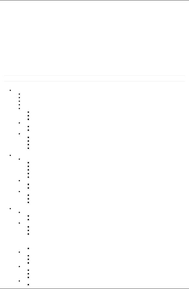
Hesselbach's (inguinal) triangle
Boundaries:
Medially: lateral border of rectus abdominis
Laterally: inferior epigastric vessels
Inferiorly: inguinal ligament
Significance
Onlyperitoneum, endoabdominal fascia and transversalis fascia form the wall
The superficial inguinal ring lies directlyexternal to it
It is thus the site where direct inguinal hernias protrude from the abdominal cavity
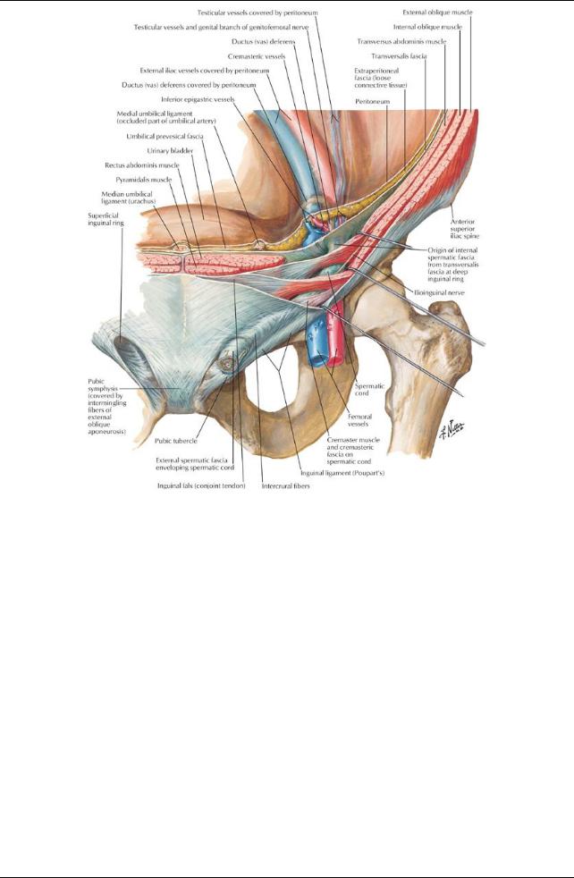
Inguinal canal: A feature of the anterior abdominal wall
[Plate 253, Inguinal Region: Dissections]
199 / 425
[Plate 254, Inguinal Canal and Spermatic Cord]
200 / 425
[Plate 255, Femoral Sheath and Inguinal Canal]
Oblique canal, approximately4 cm long at the inferior margin of the anterior abdominal wall Parallel and superior to the medial half of the inguinal ligament
Deep inguinal ring: internal entrance to canal
Entrance to the canal through the transversalis fascia.
Located 1.25 cm superior to the midpoint of the inguinal ligament
 Occurs lateral to the epigastric vessels Superficial ring: external exit of the canal
Exit through the external oblique aponeurosis
 Located superolateral to the pubic tubercle Boundaries:
Anterior wall: external oblique aponeurosis (and internal oblique laterally)
Posterior wall
Transversalis fascia laterally
Internal oblique and conjoint tendon (joint insertion of aponeuroses of internal oblique and transverses abdominus) medially
Roof: arching fibers of internal oblique
 Floor: inguinal ligament, reinforced mediallybythe lacunar ligament Contents:
Spermatic cord in men
Round ligament in women (Section 5: Pelvis and Perineum)
Ilioinguinal nerve
Blood and lymphatic vessels
201 / 425