

GUIDE
Head and Neck: Bones and Ligaments
Bones of head and neck
Skull
Mandible
Cervical vertebrae
Skull
The skull is divided into the neurocranium or calvaria (contains the brain and its meningeal coverings) and the viscerocranium (facial skeleton). The skull is composed of 22 bones (excluding the middle ear ossicles), with 8 forming the cranium and 14 forming the face. The orbits (eye sockets) lie between the calvaria (skull cap) and the facial skeleton and are formed bycontributions from 7 different bones.
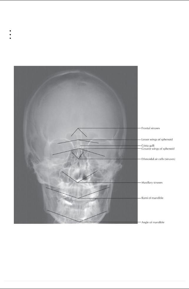
[Plate 5 - Skull: Anteroposterior Radiograph]
Neurocranium |
|
Viscerocranium |
|
Ethmoid |
1 |
Zygomatic |
2 |
Frontal |
1 |
Vomer |
1 |
Occipital |
1 |
Inferior nasal concha |
2 |
Sphenoid |
1 |
Maxilla |
2 |
Parietal |
2 |
Nasal |
2 |
Temporal |
2 |
Palatine |
2 |
|
|
Lacrimal |
2 |
|
|
(Mandible) |
1 |
N=22 |
8 |
+ |
14 |
page 4
page 5
7 / 425

Function of skull
Encloses, supports and protects brain and meninges
Contains foramina for the transmission of nerves and vessels
Forms foundation for the face
Contains specialized cavities and openings for sense organs (e.g., nasal, oral)
Neurocranium
Cranial vault and base of skull
Encloses and protects brain
Composed of 8 bones
Bones united byinterlocking sutures
Can be divided
Calvaria-dome-like roof
Cranial base
Calvaria composed of 4 bones
Frontal bone anteriorly
Occipital bone posteriorly
Two parietal bones laterally
Cranial base formed from
Ethmoid bone
Parts of occipital and temporal bones
Viscerocranium
= facial skeleton
Composed of 14 bones
Encloses orbits, nose, paranasal sinuses, mouth, and pharynx
Maxillae and mandible form upper and lower jaw, respectively, and house the teeth
There are also three auditoryossicles
Malleus, incus, and stapes
Found spanning tympanic cavity
First bones to be completelyossified during development
Major sutures of the skull
Most bones of the skull are bound bysutures, a type of fibrous joint that fuses with age and becomes immobile.
Coronal suture separates frontal and parietal bones
Sagittal suture separates two parietal bones
Lambdoid suture separates parietal and temporal bones from occipital bones
Squamous suture separates squamous part of temporal bone from parietal bone
Sphenosquamous suture separates squamous part of temporal bone from greater wing of the sphenoid
Metopic suture between two frontal bones is largelyobliterated with fusion of frontal bones
8 / 425

[Plate 6, Skull: Lateral View]
9 / 425
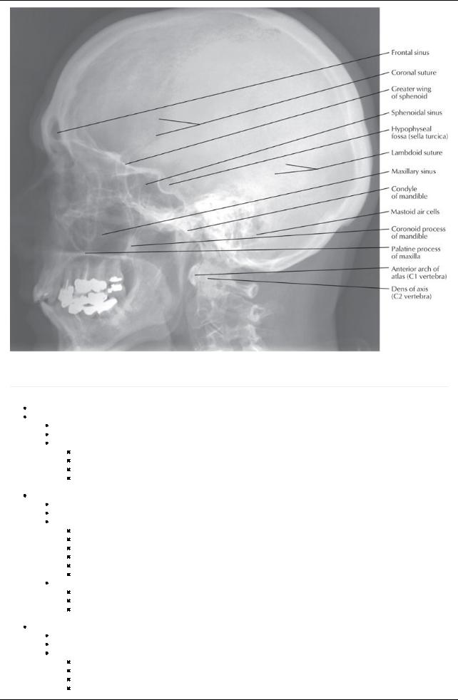
[Plate 7, Skull: Lateral Radiograph]
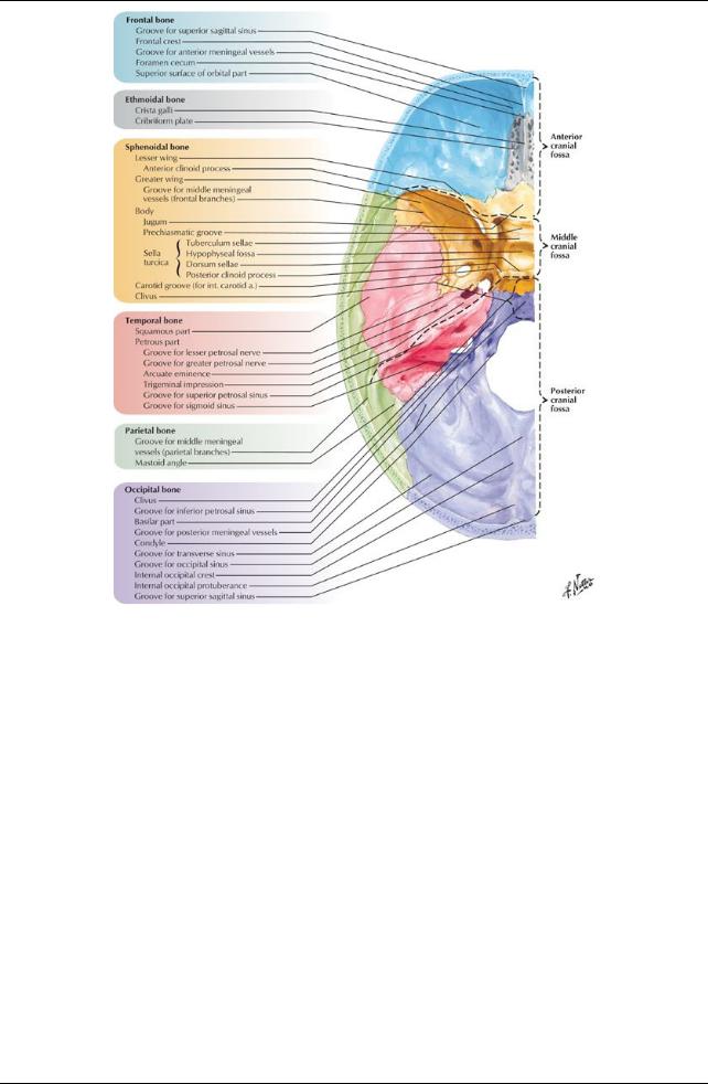
Internal Features of Base of Skull
page 5 page 6
Divided into anterior, middle, and posterior cranial fossae Anterior cranial fossa
Contains frontal lobe of brain
Formed byfrontal bone anteriorly, ethmoid bone medially, and lesser wing of sphenoid posteriorly
Features
Frontal crest-midline bonyextension of frontal bone
Foramen cecum-foramen at base of frontal crest
Crista galli-Midline ridge of bone from ethmoid posterior to foramen cecum
Cribriform plate-Thin, sieve-like plate of bone on either side of crista galli, which transmits olfactorynerves from nasal cavity to olfactorybulbs
Middle cranial fossa
Contains temporal lobe, hypothalamus, and pituitarygland
Formed bygreater wing and bodyof sphenoid, petrous temporal bone, lesser wing sphenoid
Features
Sella turcica-central depression in bodyof sphenoid for pituitarygland
Tuberculum sellae-Swelling anterior to sella turcica
Dorsum sellae-crest on bodyof sphenoid posterior to sella turcica
Anterior clinoid processes-medial projections of lesser wings of sphenoid bones
Posterior clinoid processes-swelling at either end of dorsum sellae
Foramen lacerum (one on each side)-jagged opening closed byplate of cartilage in life, transmits nothing
Contains four foramina in a crescent on either side in the bodyof the sphenoid
Superior orbital fissure
Foramen rotundum
Foramen ovale

 Foramen spinosum Posterior cranial fossa:
Contains cerebellum, pons, and medulla oblongata
Composed largelyof occipital bone, bodyof sphenoid, petrous, and mastoid parts of temporal bone
Features
Foramen magnum-transmits spinal cord
Internal occipital crest-divides posterior fossa into two lateral cerebellar fossae
Grooves for transverse and sigmoid dural venous sinuses
Jugular foramen-transmits sigmoid sinus (internal jugular vein) and several cranial nerves
10 / 425
Internal acoustic meatus-anterior and superior to jugular foramen, transmits facial and vestibulocochlear nerves (CN VII and CN VIII)
Hypoglossal canal-anterolateral and superior to foramen magnum, transmits hypoglossal nerve (CN XII)
Foramina of Skull
Numerous holes appear in the cranial floor and theyare called foramina. Important structures, especiallycranial nerves arising from the brain, pass through the foramen to access the exterior.
[Plate 10, Cranial Base: Inferior View]
11 / 425
[Plate 11, Cranial Base: Superior View]
Foramen/Opening |
Bone |
Structures Transmitted |
Optic canal |
Lesser wing sphenoid |
Optic nerve |
|
|
Ophthalmic artery |
|
|
Sympathetic plexus |
Superior orbital fissure |
Greater and lesser wings sphenoid |
Lacrimal nerve (V1) |
|
|
Frontal nerve (V1) |
|
|
Trochlear nerve (IV) |
|
|
Oculomotor nerve (III) |
|
|
Abducent nerve (VI) |
|
|
Nasociliarynerve (V1) |
|
|
Superior ophthalmic vein |
Inferior orbital fissure |
Between greater wing of sphenoid and zygomatic |
Infraorbital vein |
|
|
Infraorbital artery |
|
|
Infraorbital nerve |
Foramen spinosum |
Greater wing of sphenoid |
Middle meningeal arteryand vein |
Foramen rotundum |
Greater wing of sphenoid |
Maxillarydivision trigeminal nerve (V3) |
Foramen ovale |
Greater wing of sphenoid |
Mandibular division trigeminal nerve |
|
|
Lesser petrosal nerve |
Foramen lacerum |
Between temporal bone (petrous area) and sphenoid bone |
Internal carotid artery |
Foramen magnum |
Occipital bone |
Medulla oblongata |
|
|
Vertebral artery |
|
|
Meninges |
|
|
Spinal roots of accessorynerve |
Hypoglossal canal |
Occipital bone |
Hypoglossal nerve (XII) |
Jugular foramen |
Between temporal bone (petrous area) and occipital bone |
Glossopharyngeal nerve (IX) |
|
|
Vagus nerve (X) |
|
|
Accessorynerve (XI) |
|
|
Inferior petrosal sinus |
|
|
Sigmoid sinus |
12 / 425
Posterior meningeal artery
Mandible
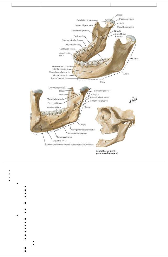
[Plate 17, Mandible]
page 7
page 8
Unpaired bone of lower jaw Largest and strongest bone in face
Articulates with temporal bone at temporomandibular joint Consists of
Body
Can be divided into lower base and upper alveolar part
Has a mental protuberance anteriorlyand inferiorlywhere two sides come together
Mental spine: rough projection on inner surface of bodyin the midline
Mental foramen below second premolar transmits terminal branch of inferior alveolar nerve to supplyskin and mucus membrane of lower lip and chin
Mylohyoid line: a ridge extending upward and backward on internal surface of alveolar part of mandible for attachment mylohyoid muscle
Submandibular fossa: long depression below mylohyoid line, which accommodates submandibular gland
Sublingual fossa: concavities on either side of mental spine for sublingual gland
Rami
Lateral vertical projections from body
Each meets bodyinferiorlyat angle of the jaw
Two processes at superior end: coronoid process and condylar process
Coronoid process-attachment of temporalis muscle
Condylar process-part of temporomandibular joint
Mandibular notch-concavitybetween condylar and coronoid processes
Mandibular foramen
On inner surface of ramus
Entrance to mandibular canal, through which passes the inferior alveolar nerve
Lingula-thin projection of bone overlapping mandibular foramen
Mylohyoid groove-groove leading anteriorlyand inferiorlyfrom mandibular foramen indicating course of mylohyoid nerve and vessels
13 / 425
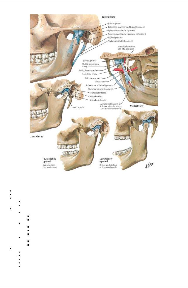
Temporomandibular Joint
[Plate 18, Temporomandibular Joint]
The mandible articulates with the temporal bone and in chewing or speaking, it is onlythe mandible or lower jaw that moves; the upper jaw or maxilla remains stationary. The teeth are contained in the alveolar portion of the mandible.
Articulation between condylar process of mandible, articular tubercle of temporal bone, and mandibular fossa
Modified hinge-type synovial joint
Contains fibrocartilaginous disc, which divides joint cavityinto two compartments
Gliding movements (protrusion and retrusion/retraction) occur in upper compartment
Hinge movements (depression and elevation) occur in lower compartment
Stabilized bythree ligaments:
Lateral temporomandibular ligament
Lateral thickened parts of articular capsule
Prevent posterior dislocation of joint
Sphenomandibular ligament
Primarypassive support
Runs from spine of sphenoid to lingual of mandible
Serves as swinging hinge and check ligament
Stylomandibular ligament
Thickening in capsule of parotid gland
Runs from styloid process to angle of mandible
Movements
Depression-suprahyoid and infrahyoid muscles, gravity
Elevation-temporalis, masseter, and medial pterygoid muscles
Protrusion-lateral pterygoid, masseter, medial pterygoid
Retraction/retraction-temporalis, masseter
Side to side grinding-retractors of same side, protruders of opposite side
Cervical vertebrae
See: Back and Spinal Cord-Bones and Ligaments
14 / 425