


7 Pharynx
STUDYAIMS
At the end of your study, you should be able to:
Know the general anatomyof the pharynx
Describe the anatomyof the nasopharynx
Describe the anatomyof the oropharynx
Describe the anatomyof the laryngopharynx
Know the muscles of the pharynx
Know the vascular supplyand lymphatic drainage of the pharynx
Understand the innervation of the pharynx
Outline the process of swallowing
45 / 425
GUIDE
Head and Neck-Pharynx
Pharynx
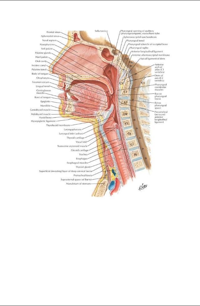
[Plate 63, Pharynx: Median Section]
46 / 425
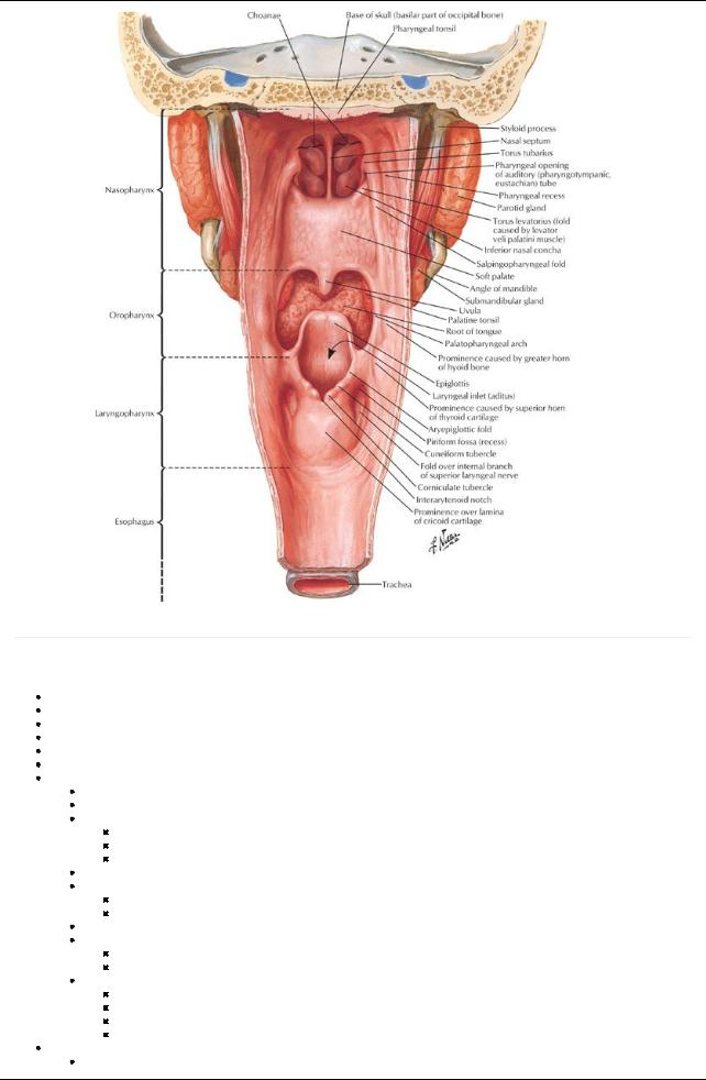
[Plate 66, Pharynx: Opened Posterior View]
page 38
page 39
The pharynxis a muscular tube, which is deficient anteriorlyas a result of the openings nasal and oral cavities and larynx-as revealed when the posterior wall is removed.
Muscular tube
Posterior to nasal and oral cavities
Continuous with both esophagus and larynx
Anterior to superior sixcervical vertebrae and prevertebral muscles and fascia
Retropharyngeal space = potential space between pharynxand prevertebral fascia
Divided into three parts: Nasopharynx, Oropharynx, and Laryngopharynx
Nasopharynx
Posterior to nose and above soft palate
Lined with ciliated epithelia
Boundaries
Anteriorly: continuous with nasal cavities via choanae
Roof and posterior wall: bodyof sphenoid and base of occipital bone
Laterally: superior constrictor muscle
Contains openings of auditory(Eustachian) tubes (from middle ear)
Salpingopharyngeal fold
Extends inferiorlyfrom medial end of auditorytube
Covers salpingopharyngeus muscle-opens tube during swallowing
Ridge over opening = torus tubarius
Pharyngeal recess
Slit-like projection
Posterior to torus
Contains abundant lymphoid tissue
Incomplete ring in superior part of pharynx
Aggregates in certain areas = tonsils
Lymphoid tissue in mucus membrane of roof and posterior wall = adenoids
Lymphoid tissue near opening of auditorytube = tubal tonsil
Oropharynx
From soft palate to superior ends of epiglottis
47 / 425

Boundaries
Anteriorly: oropharyngeal opening posterior one third tongue epiglottis
Laterally: palatoglossal and palatopharyngeal arches (containing palatoglossus and palatopharyngeus muscles)
Superiorly: soft palate
Posteriorly: superior and middle constrictor muscles
Contains palatine tonsils
Found in cleft between palatoglossal and palatopharyngeal arches
Tonsil lies on tonsillar bed = superior constrictor muscle and pharyngobasilar fascia
Epiglottis
United to tongue bymedian and lateral glossoepiglottic folds
 Depression between medial and lateral folds = epiglottic valleculae Laryngopharynx
From superior border of epiglottis to inferior border of cricoid cartilage
Lined with stratified squamous epithelium
Boundaries
Inferiorly: continuous with esophagus
Superiorly: continuous with oropharynx
Anteriorly: larynx
Posteriorly: middle and inferior constrictor muscles deep: Bodies of C4-C6 vertebrae
Laterally: middle and inferior constrictor muscles
Piriform recesses
Small depressions on either side of laryngeal inlet
Separated from inlet byaryepiglottic folds
Bounded mediallybythyroid cartilage and thyrohyoid membrane
Muscles of Pharynx
page 39
page 40
Wall of pharynxis unique
Composed of outer circular and inner longitudinal layers of muscles
External circular layer consists of three constrictor muscles: pharyngeal constrictors
Inner longitudinal layer consists of three paired muscles
Pharyngeal constrictors = three muscles
Superior, middle, and inferior constrictor muscles form a muscular sleeve
Have strong internal facial lining: pharyngobasilar fascia
Contract involuntarilyin sequence = peristalsis
All supplied bypharyngeal plexus of nerves
Inner longitudinal layer = three muscles
Elevate larynx
Shorten pharynx
Act during swallowing and speaking
Stylopharyngeus
Palatopharyngeus
Salpingopharyngeus
Gaps between constrictors
Areas where structures can enter and leave pharynx
Between superior constructor and skull
Levator veli palatini
Auditorytube
Ascending palatine artery
Between superior and middle constructor
Stylopharyngeus muscle
Glossopharyngeal nerve
Stylohyoid ligament
Between middle and inferior constrictor
Internal laryngeal nerve
Superior laryngeal arteryand vein
Below inferior constructor
Recurrent laryngeal nerve
Inferior laryngeal artery
Muscle |
Origin |
Insertion |
Innervation |
Main Actions |
Superior pharyngeal |
Hamulus, pterygomandibular raphe, |
Median raphe |
Vagus via |
Constricts wall of pharynxduring |
constrictor |
mylohyoid line of mandible |
of pharynx |
pharyngeal plexus |
swallowing |
Middle pharyngeal |
Stylohyoid ligament and horns of |
Median raphe |
Vagus via |
Constricts wall of pharynxduring |
constrictor |
hyoid bone |
of pharynx |
pharyngeal plexus |
swallowing |
Inferior pharyngeal |
Oblique line of thyroid cartilage, and |
Median raphe |
Vagus via |
Constricts wall of pharynxduring |
constrictor |
cricoid cartilage |
of pharynx |
pharyngeal plexus |
swallowing |
Salpingopharyngeus |
Auditory(pharyngotympanic) tube |
Side of |
Vagus via |
Elevates pharynxand larynxduring |
|
|
pharynxwall |
pharyngeal plexus |
swallowing and speaking |
Stylopharyngeus |
Medial aspect of styloid process |
Pharyngeal |
Glossopharyngeal |
Elevates pharynxand larynxduring |
|
|
wall |
nerve |
swallowing and speaking |
Arterial supply
Tonsillar artery(from facial) to tonsil
48 / 425
Branches from
Ascending pharyngeal
Lingual
Ascending and descending palatine
Venous drainage
External palatine vein → pharyngeal plexus
Pharyngeal venous plexus → internal jugular vein
Lymphatic drainage
General drainage to deep cervical nodes
From tonsillar tissue to nodes near angle of mandible and tonsillar (jugulodigastric) node
Innervation
page 40
page 41
From pharyngeal plexus (motor and almost all sensory)
Motor
From pharyngeal plexus via vagus nerve from cranial root of accessorynerve (cranial nerve [CN] XI)
To all muscles of pharynxexcept stylopharyngeus (CN V2)
Branches from external and recurrent branches of vagus
To inferior constrictor
Sensory
Mainlyfrom glossopharyngeal nerve (CN IX) via plexus
Also
Maxillarynerve (CN V2) to anterior and superior nasopharynx
Tonsillar nerves from branches of glossopharyngeal and vagus (CN X)
Swallowing (deglutition)
Occurs in three stages 1 = voluntary
Food is in the mouth, breathing occurs through the nasopharynx
Food is chewed (masticated) and mixed with saliva to produce a bolus
Bolus of food is compressed against hard palate
Palatoglossal folds relax
Muscles of tongue and soft palate push bolus into oropharynx
 Cycle lasts 1 to 2 seconds 2 = involuntary
Reflexive, mediated via glossopharyngeal nerve
Nasopharynxis closed off bytension and elevation of the soft palate
Prevents refluxof food/fluids into the nose
Mediated bytensor veli palatine and levator veli palatine muscles
Suprahyoid muscles and longitudinal pharyngeal muscles contract
Elevate larynx
Close epiglottis

 Propel bolus 3 = involuntary
 Food propelled through the pharynxbyperistalsis (sequential contraction of all three constrictors) On reaching the distal end of pharynx, high pressure causes relaxation of terminal part of inferior constrictor
Called cricopharyngeus muscle
Serves as superior esophageal sphincter
Food enters the oesophagus
As bolus passes pressure drops, the sphincter closes Larynxand epiglottis return to normal positions
49 / 425