


FACTS & HINTS
HIGH-YIELD FACTS
Clinical Points
page 218
page 219
Fracture of the Clavicle
Common, especiallyin children
Usuallyresults from a fall on outstretched hand or direct trauma to the shoulder Fractures of middle third are most frequentlyseen
Sternocleidomastoid muscle pulls the proximal fragment superiorlyand the shoulder pulls the distal fragment inferiorly Small lump mayremain after the clavicle has healed
Calcific Supraspinatus Tendonitis
Inflammation and calcification of the subacromial bursa resulting in pain, tenderness and limitation of movement of the shoulder joint Calcium deposits frequentlyalso seen in the supraspinatus tendon
Pain is especiallysevere with the arm abducted between 50 to 130 degrees (the painful arc) as the supraspinatus tendon is in contact with the inferior surface of the acromion here
Shoulder Dislocation
High mobilityand instabilityof the glenohumeral joint leads to frequent dislocation
95% of dislocations are in anteroinferior direction, caused byexcessive extension and lateral rotation of humerus (e.g., in the throwing motion)
Humeral head places stress on joint capsule, which maybe torn anteriorly, with elements of the rotator cuff Axillaryand musculocutaneous nerves mayalso be injured
Posterior dislocation is uncommon, but mayoccur during epileptic seizure or electrocution
Rotator Cuff Injury
Musculotendinous rotator cuff maybe damaged bytrauma or degenerative disease
One or more of tendons maybe torn when the arm is forcefullyabducted, leading to pain in the anterosuperior aspect of the shoulder Supraspinatus tendon is most commonlyinvolved in degenerative tendonitis
Leads patient's arm to drop suddenlyat approximately90-degree abduction, when instructed to lower it slowlyfrom a fullyabducted position
MNEMONICS
Memory Aids
Rotator cuff muscles: |
SITS = Supraspinatus, Infraspinatus, Teres minor, Subscapularis |
343 / 425

45 Arm
STUDYAIMS
At the end of your study, you should be able to:
Identifythe different parts and surface markings of the humerus
Describe the organization of the deep fascia and compartments of the arm
Know the origins, insertions, and actions of the muscles of the arm
Know the cutaneous nerves of the arm
Describe the boundaries and contents of the cubital fossa
344 / 425
GUIDE
Upper Limb: Arm
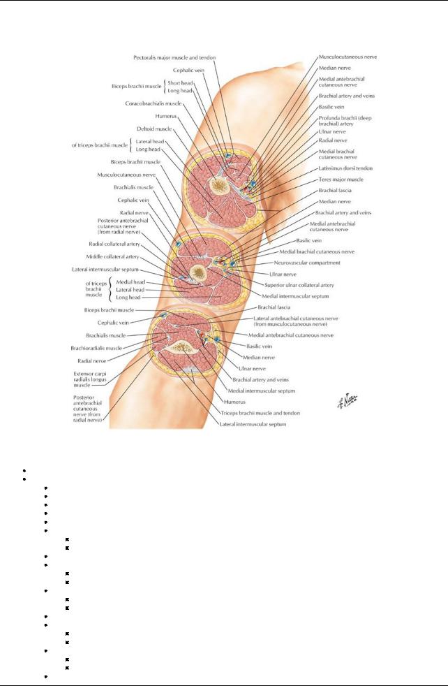
[Plate 423, Arm: Serial Cross Sections]
Humerus
Largest bone of the upper limb
Osteological features
Head
Anatomical neck-circumscribes head above tubercles
Surgical neck: below tubercles-common site of fracture
Greater and lesser tubercles
Intertubercular groove
Bodywith
Deltoid tuberosityfor attachment of deltoid muscle
Radial groove on posterior surface where radial nerve and deep brachial arterytraverse
Medial and lateral supracondylar ridges-widening of humerus distallyas sharp ridges on either side
Medial epicondyle
Prominent medial extension at distal end
Common origin of forearm flexors; ulnar nerve posterior
Lateral epicondyle
Prominent lateral extension at distal end
Common origin of forearm extensors; radial nerve posterior
Condyle-distal end of humerus
Trochlea
Medial articular surface of condyle
For articulation with trochlear notch of ulna
Capitulum
Lateral articular surface of condyle
For articulation with head of radius
Coronoid fossa (see also Section 6-4: Upper Limb: Elbowand Forearm)
345 / 425
Superior to trochlea
Receives coronoid process of ulna
Olecranon fossae
Posterior distal end of humerus
Receives olecranon of ulna during full extension of forearm at elbow
page 220 page 221
Fascia of the Arm
Brachial fascia
Asleeve of deep fascia around the arm
Continuous with the antebrachial fascia of the forearm
Medial and lateral intermuscular septa
Extend from deep surface of brachial fascia to humerus
Divide arm into anterior (flexor) and posterior (extensor) compartments
Medial septum: medial lip of intertubercular sulcus (superiorly) → medial epicondyle
Lateral septum: lateral lip of intertubercular sulcus (superiorly) → lateral epicondyle
Muscles of the Arm
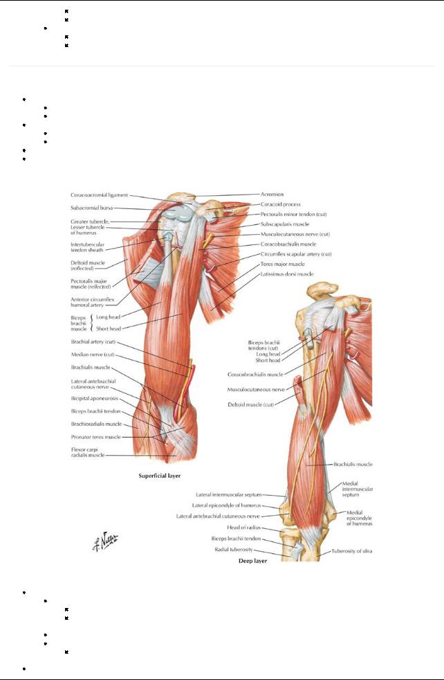
[Plate 419, Muscles of Arm: Anterior Views]
Anterior (flexor) compartment
Biceps brachii
Flexes and supinates
Continues distallyas bicipital aponeurosis: triangular membrane from the biceps tendon across cubital fossa and blends with antebrachial fascia over the flexor muscles of the forearm
Brachialis: main flexor of forearm
Coracobrachialis
Flexes and adducts arm
 Pierced bythe musculocutaneous nerve Posterior (extensor) compartment
346 / 425
Triceps brachii
Main extensor of arm
Stabilizes head of humerus in glenohumeral joint
Anconeus: extends arm, and resists adduction of the ulna during pronation
Muscles |
Origin |
Insertion |
Innervation |
Blood supply |
Action |
Biceps brachii |
Long head-supraglenoid tubercle of |
Radial |
Musculocutaneous |
Muscular |
Supinates |
|
humerus; Short head-tip of coracoid process |
tuberosity, fascia |
nerve (C5,C6) |
branches of |
forearm |
|
of scapula |
of forearm via |
|
brachial |
Flexes |
|
|
bicipital |
|
artery |
forearm when |
|
|
aponeurosis |
|
|
it is supinated |
Coracobrachialis |
Tip of coracoid process of scapula |
Middle third of |
Musculocutaneous |
Muscular |
Flexes and |
|
|
medial surface |
nerve (C5,C6,C7) |
branches of |
adducts arm |
|
|
of humerus |
|
brachial |
|
|
|
|
|
artery |
|
Brachialis |
Distal half of anterior surface of humerus |
Coronoid |
Musculocutaneous |
Radial |
Flexes |
|
|
process and |
nerve (C5,C6) |
recurrent |
forearm |
|
|
tuberosityof ulna |
|
artery, |
|
|
|
|
|
muscular |
|
|
|
|
|
branches of |
|
|
|
|
|
brachial |
|
|
|
|
|
artery |
|
Triceps brachii |
Long head-infraglenoid tubercle of scapula; |
Posterior |
Radial nerve |
Branch of |
Extends |
|
Lateral head -upper half of posterior |
surface of |
(C6,C7,C8) |
profunda |
forearm, long |
|
humerus; Medial head -distal two thirds of |
olecranon |
|
brachii artery |
head |
|
medial and posterior humerus |
process of ulna |
|
|
stabilizes |
|
|
|
|
|
head of |
|
|
|
|
|
abducted |
|
|
|
|
|
humerus |
page 221 page 222
Cutaneous Nerves of Arm
Supraclavicular nerves (C4,C5) supplyskin over shoulder
Superior lateral cutaneous nerve (C5,C6)
Branch of axillary
Supplies skin over upper lateral arm
Inferior lateral cutaneous nerve (C5,C6)
Cutaneous branch of radial nerve
Supplies skin over lower lateral arm
Intercostobrachial nerve (T2)
Lateral cutaneous branch of second intercostal nerve
Supplies upper medial arm anteriorlyand posteriorly
Medial brachial cutaneous (C8-T1)
Branch of brachial plexus
Supplies lower anterior medial arm
Posterior brachial cutaneous nerve (C5-C8)
Branch of radial nerve
Supplies lower posterior medial arm
Cubital Fossa
Boundaries
Superiorly: (imaginary) line connecting the medial and lateral epicondyles
Medially: pronator teres muscle
Laterally: brachioradialis
Floor: brachialis and supinator muscles of arm and forearm
Roof: deep fascia, bicipital aponeurosis, subcutaneous tissue, and skin
Contents
Brachial artery(terminal part)
Biceps brachii tendon
Nerves: median
In subcutaneous connective tissue
Medial and lateral antebrachial cutaneous nerves
Basilic and cephalic veins
Median cubital vein
In floor of fossa
Deep and superficial branches of radial nerve
347 / 425