


39 Testis, Epididymis & Ductus Deferens
STUDYAIMS
At the end of your study, you should be able to:
Describe the gross structure of the testes
Understand the organization of the ducts of the testes: seminiferous tubes, rete testis, efferent ductules Describe the basic structure of the epididymis
Describe the course of the ductus deferens from the testes to the urethra
Understand the anatomyof the seminal vesicles and their ducts in relation to the formation of the ejaculatoryduct Know the location of the ejaculatoryducts and where theyenter the urethra
Know the anatomyand organization of the prostate gland
Outline the contents of semen and the factors that control ejaculation
305 / 425

GUIDE
Pelvis and Perineum: Testes, Epididymis, and Ductus Deferens
[Plate 364, Prostate and Seminal Vesicles]
306 / 425
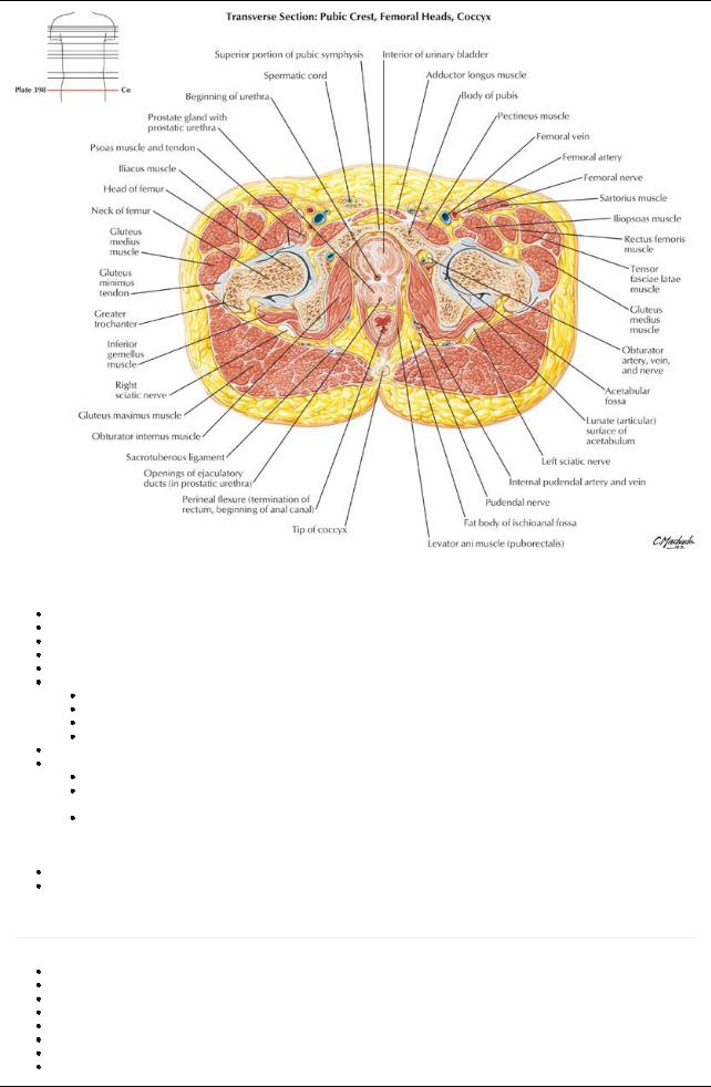
[Plate 398, Male Pelvis: Bladder-Prostate Junction]
Testes
Ovoid structure, approximately5 cm long
Site of sperm production
Contains 200 to 300 lobules separated byconnective tissue septa
Two to 3 highlycoiled seminiferous tubules in each lobule, 1 meter in length
Tubules converge toward posterior testis and discharge contents into duct network of rete testis
Mediastinum testis
Posterior region of testis where vessels and nerves enter and leave
Not covered bytunica vaginalis
Contains rete testis
Contains efferent ductules connecting rete testis to head of epididymis
Suspended byspermatic cord and located in scrotum
Is covered bythree distinguishable layers:
Tunica albuginea-tough fibrous layer
Visceral layer of tunica vaginalis-serous layer
Applied to testis, epididymis, and distal spermatic cord
Parietal tunica vaginalis
Parietal and visceral tunica vaginalis derived from peritoneal outpocketing in embryonic life
Separated bysmall amount of serous fluid that allows testis to move in scrotal sac
Deficient posteriorlyto transmit epididymis and blood vessels
Blood supply: Testicular artery
Venous drainage: Pampiniform plexus
Epididymis
page 195
page 196
Formed from convolutions of narrow duct of the epididymis
Located on posterior aspect testes within scrotum
Consists of a head, a body, and a tail
Head formed from ends of approximately12 efferent ductules from the testis
Tail is continuous with ductus deferens
Where sperm are stored, mature, and become motile
Blood supply: Testicular artery
Venous drainage: Pampiniform plexus
307 / 425
Ductus deferens (Vas deferens)
Twenty-five cm long
Connects tail of epididymis to ejaculatoryduct Course:
Commences at tail of epididymis
Travels in spermatic cord (where it can be palpated) through superficial inguinal ring and through inguinal canal
Emerges at deep inguinal ring and crosses over external iliac vessels to run along the lateral pelvic wall
Crosses above and medial to the ureter where it becomes dilated and forms the ampulla
Ampullae converge with each other on the posterior aspect of the bladder and narrow before uniting with duct of the seminal vesicle to form ejaculatoryduct
Ejaculatoryduct opens into the prostatic urethra
Blood supply: Branch of inferior vesical artery= deferential artery
Venous drainage: Deferential vein(s) to prostatic venous plexus to internal iliac veins.
Seminal vesicles
Elongated, coiled structures between fundus of bladder and rectum
Separated from rectum byrectovesical pouch
Do not store sperm
Secrete alkaline fluid that mixes with sperms in ejaculatoryducts and urethra
Duct of each seminal vesicle joins ductus deferens to form ejaculatoryduct
Supplied bybranches and tributaries of inferior and middle rectal vessels
Ejaculatory ducts
Formed from union of ductus deferens with duct of seminal vesicle
Short, approximately2.5 cm
Converge and open on seminal colliculus of prostatic urethra as two slits
Supplied bydeferential arteries
Drained byprostatic and vesical plexuses
Prostate
page 196
page 197
Encapsulated gland surrounding urethra between neck of bladder and pelvic floor Directlybehind inferior edge of pubic symphysis
Composed of
Two lateral lobes
Anterior lobe or isthmus connecting lateral lobes anteriorly
Posterior lobe below ejaculatoryducts and posterior to urethra
 Middle lobe between urethra and ejaculatoryducts Contains glands that produce 20% of volume of semen
Ducts of glands combine to form prostatic ducts opening into prostatic sinuses on either side of seminal colliculus in prostatic urethra Dense fascia sheath outside capsule contains prostatic venous plexus that drains to internal iliac veins
Supplied byprostatic arteries from several sources including internal iliac or inferior vesical arteries
308 / 425