- •1. Topographic Surface Anatomy
- •Guide
- •Facts & Hints
- •Guide
- •Facts & Hints
- •3. Superficial Face
- •Guide
- •Facts & Hints
- •4. Neck
- •Guide
- •Facts & Hints
- •5. Nasal Region
- •Guide
- •Facts & Hints
- •6. Oral Region
- •Guide
- •Facts & Hints
- •7. Pharynx
- •Guide
- •Facts & Hints
- •Guide
- •Facts & Hints
- •Guide
- •Facts & Hints
- •Guide
- •Facts & Hints
- •Guide
- •Facts & Hints
- •Guide
- •Facts & Hints
- •13. Cerebral Vasculature
- •Guide
- •Facts & Hints
- •14. Topographic Anatomy
- •Guide
- •Facts & Hints
- •Guide
- •Facts & Hints
- •16. Spinal Cord
- •Guide
- •Facts & Hints
- •Guide
- •Facts & Hints
- •Thorax
- •18. Topographic Anatomy
- •Guides
- •Facts & Hints
- •19. Mammary Gland
- •Guides
- •Facts & Hints
- •20. Body Wall
- •Guides
- •Facts & Hints
- •21. Lungs
- •Guides
- •Facts & Hints
- •22. Heart
- •Guides
- •Facts & Hints
- •23. Mediastinum
- •Guides
- •Facts & Hints
- •Abdomen
- •24. Topographic Anatomy
- •Guide
- •Facts & Hints
- •25. Body Wall
- •Guide
- •Facts & Hints
- •26. Peritoneal Cavity
- •Guide
- •Facts & Hints
- •27. Viscera (Gut)
- •Guide
- •Facts & Hints
- •28. Viscera (Accessory Organs)
- •Guide
- •Facts & Hints
- •29. Visceral Vasculature
- •Guide
- •Facts & Hints
- •30. Innervation
- •Guide
- •Facts & Hints
- •Guide
- •Facts & Hints
- •32. Topographic Anatomy
- •Guide
- •Facts & Hints
- •Guide
- •Facts & Hints
- •Guide
- •Facts & Hints
- •35. Urinary Bladder
- •Guide
- •Facts & Hints
- •Guide
- •Facts & Hints
- •Guide
- •Facts & Hints
- •Guide
- •Facts & Hints
- •39. Testis, Epididymis & Ductus Deferens
- •Guide
- •Facts & Hints
- •40. Rectum
- •Guide
- •Facts & Hints
- •41. Vasculature
- •Guide
- •Facts & Hints
- •42. Innervation
- •Guide
- •Facts & Hints
- •Upper Limb
- •43. Topographic Anatomy
- •Guide
- •Facts & Hints
- •Guide
- •Facts & Hints
- •Guide
- •Facts & Hints
- •Guide
- •Facts & Hints
- •Guide
- •Facts & Hints
- •48. Neurovasculature
- •Guide
- •Facts & Hints
- •Lower Limb
- •49. Topographic Anatomy
- •Guide
- •Facts & Hints
- •Guide
- •Facts & Hints
- •51. Knee
- •Guide
- •Facts & Hints
- •Guide
- •Facts & Hints
- •Guide
- •Facts & Hints
- •54. Neurovasculature
- •Guide
- •Facts & Hints
Back and Spinal Cord
page 81
page 82
14 Topographic Anatomy
STUDYAIMS
At the end of your study, you should be able to:
Identifyposteromedian furrow
Identifyexternal occipital protuberance, vertebra prominens, iliac crests, posterior superior iliac spines
Identifydeltoid, latissimus dorsi, trapezius, erector spinae, teres major, infraspinatus, gluteus maximus, and medius Identifymargins of scapula
101 / 425
GUIDE
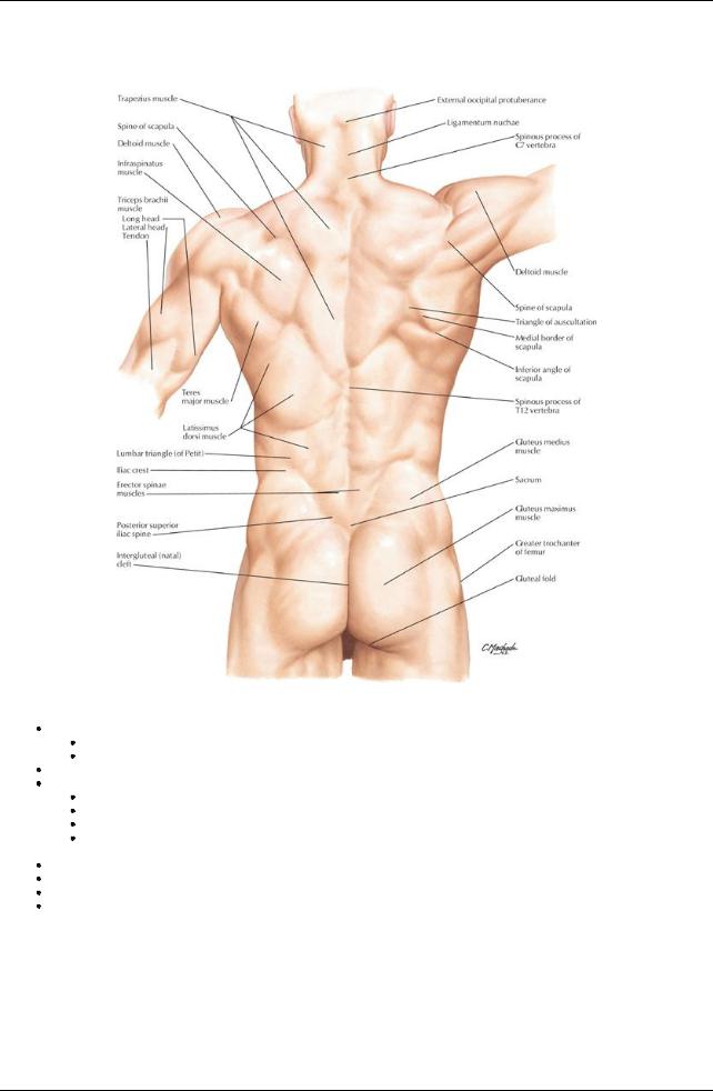
Back and Spinal Cord: Topographic Anatomy
[Plate 149, Topographic Anatomy]
Median line of back: posteromedian furrow overlies tips of spinous processes
Deepest in lower thoracic/upper lumbar region
Bordered byerector spinae
Vertebra prominens = C7 spinous process (T1 may be more prominent) Scapula:
Superior angle at the level of T2
Medial end of scapular spine opposite spinous process of T3
Inferior angle at level of T7
Medial border of scapula parallels the sixth rib and approximates oblique fissure of lung when the arm is abducted byplacing the hand on the head
Iliac crests at level of L4 = supracristal line
S2 spinous process lies level with a line joining the posterior superior iliac spines Tip of coccyxapproximately2.5 cm posterosuperior to the anus
Anatomy of muscles of the back are covered in Section 2-4: Back and Spinal Cord-Muscles and Nerves. (Muscles that are readily visible are trapezius, latissimus dorsi, and teres major).
102 / 425
[Plate 160, Sympathetic Nervous System: Schema]
103 / 425
[Plate 161, Parasympathetic Nervous Sytem: Schema]
Vertebrae and corresponding structures
Level |
Corresponding Structure |
C2-C3 |
Mandible |
C3 |
Hyoid bone |
C4-C5 |
Thyroid cartilage |
C6 |
Cricoid cartilage |
C7 |
Vertebra prominens |
T3 |
Spine of scapula |
T4/5 |
Sternal angle (of Louis) |
T7 |
Inferior angle of scapula |
T8 |
Inferior vena cava pierces diaphragm |
T10 |
Xiphisternal junction |
T10 |
Esophagus enters stomach |
T12 |
Aorta passes behind diaphragm |
L1/2 |
Spinal cord ends (cauda equina starts) |
L3 |
Subcostal plane |
L3-L4 |
Umbilicus |
L4 |
Bifurcation of aorta |
L4 |
Iliac crests-supracristal line |
S2 |
End of dural sac |
S2 |
Level of posterior superior iliac spines |
104 / 425