


19 Mammary Gland
STUDYAIMS
At the end of your study, you should be able to:
Identifythe mammarygland
Identifythe location of the gland
Locate the blood supplyof the breast
Understand the lymphatic drainage of the breast
Know how to palpate the breast
138 / 425
GUIDES
Thorax: Mammary Gland
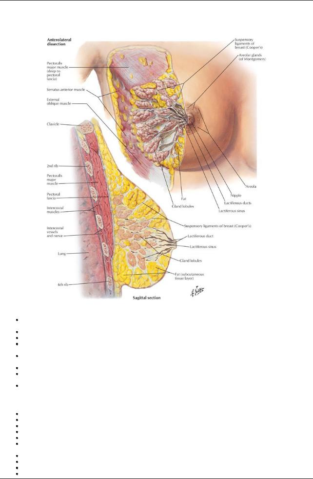
[Plate 176, Mammary Gland]
Consists of glandular tissue in which the majorityis embedded within the tela subcutanea (superficial fascia) of the anterior chest wall overlying the pectoral muscles.
The glands are rudimentaryin males and immature females.
Size and shape of the adult female breast varies; the size is determined bythe amount of fat surrounding the glandular tissue.
The base of the breast is fairlyconsistent extending from the lateral border of the sternum to the midaxillaryline and from the 2nd to the 6th ribs.
The majorityof the breast overlies the deep pectoral fascia of the pectoralis major muscle, with the remainder overlying the fascia of the serratus anterior.
The breast is separated from the pectoralis major muscle bythe retromammaryspace, a potential space filled with loose connective tissue. The breast is firmlyattached to the overlying skin bycondensation of connective tissue called the suspensoryligaments (of Cooper), which help to support the lobules of the breast.
Asmall part of the mammarygland mayextend toward the axilla, called the axillarytail (of Spence).
Structure of the Breast
For descriptive purposes, the breast is divided into four quadrants: upper and lower lateral, and upper and lower medial. The most prominent feature of the breast is the nipple.
The nipple is surrounded bythe areola, a circular pigmented area of skin. The areola is pink in Caucasians and brown inAfrican andAsian people. The pigmentation of the areola increases during pregnancy.
The areola contains sebaceous glands, following a pregnancythese secrete an oilysubstance to protect the mother's nipple from irritation during nursing.
The breast is composed of 15 to 20 lobules of glandular tissue, formed bythe septa of the suspensoryligaments.
The mammaryglands are modified sweat glands that are formed from the development of milk-secreting alveoli, arranged in clusters. Each lobule is drained bya lactiferous duct
Each lactiferous duct opens on the nipple.
139 / 425
Vasculature of the Breast
page 99
page 100
Blood supplyof the breast arises from the perforating branches and anterior intercostal branches of the internal thoracic artery. The breast is also supplied bythe branches of the thoracoacromial and lateral thoracic arteries (from the axillaryartery). Venous drainage parallels the arterial supplyand is mainlyto the axillaryarteryand internal thoracic vein.
Lymphatic Drainage of the Breast
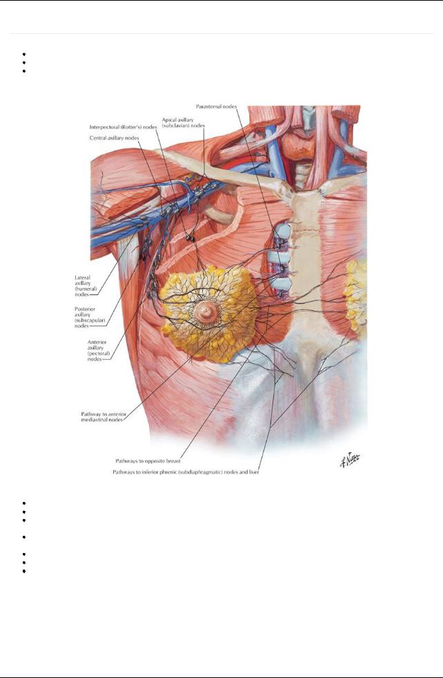
[Plate 178, Lymph Vessels and Nodes of Mammary Gland]
Lymph from the nipple, areola, and lobules of the mammaryglands drains to a subareolar lymphatic plexus. From there, a system of interconnecting lymphatic channels drains lymph to various lymph nodes.
The majorityof the lymph, especiallyfrom the lateral quadrants of the breast, drains to the pectoral nodes, and from there to the axillary nodes.
The remaining amount of lymph, especiallyfrom the medial quadrants of the breast, drains into the parasternal lymph nodes along the internal thoracic vessels.
Some lymph from the lower quadrants of the breast passes to the inferior phrenic nodes.
It is important to note that lymph from the medial quadrants can cross to the opposite breast. Thus secondarymetastases of breast carcinoma can spread to the opposite breast in this way.
140 / 425
FACTS & HINTS
High-Yield Facts
Clinical Points
Examination of the Breast
Clinicallythe breast is divided into quadrants:
UI: upper inner
UO: upper outer (includes axillarytail)
LI: lower inner
LO: lower outer
The breast is palpated in a circular fashion, beginning with the nipple and moving outward. The palpation should extend into the axilla to palpate the axillarytails.
After palpation of one breast, the other should be palpated in the same way.
Examine the skin of the breast for a change in texture or dimpling (peau d'orange sign) and the nipple for retraction, since these signs may indicate an underlying pathology.
Pathologyof the Breast
Fibroadenoma:benign tumor, usuallya solid and solitarymass that moves easilyunder the skin. Often painless although sometimes tender on palpation. More common in young women but can occur at anyage.
Intraductal carcinoma or breast cancer:the commonest type of malignancyin women but can also occur in men.Approximately50% of cancers develop in the upper quadrant of the breast; metastases from these cancers often spread to the axillarylymph nodes. This malignancypresents as a palpable mass that is hard, immobile and sometimes painful.Additional signs can include bloodyor watery nipple discharge if the larger ducts are involved.
Gynecomastia:enlargement of the breasts in males because of aging, drug treatment, and changes in the metabolism of sexhormones by the liver.
141 / 425