


13 Cerebral Vasculature
STUDYAIMS
At the end of your study, you should be able to:
State the main arteries that supplythe brain
Describe the course of the vertebral artery
Identifythe arteries contributing to the circle of Willis
Identifythe regions that each of the cerebral arteries supplies
Describe the venous drainage of the brain
Identifythe branches of the external carotid arteryand structures supplied
Describe the division of the subclavian arterybyscalene anterior and the branches given off byeach part
Understand the organization and major vessels of the venous drainage of the head and neck
Understand the principles and organization of the lymphatic drainage of the face and head and neck
Understand the principles and organization of the lymphatic drainage of the neck
93 / 425
GUIDE
Head and Neck: Cerebral Vasculature
Vascular Supply to the Brain
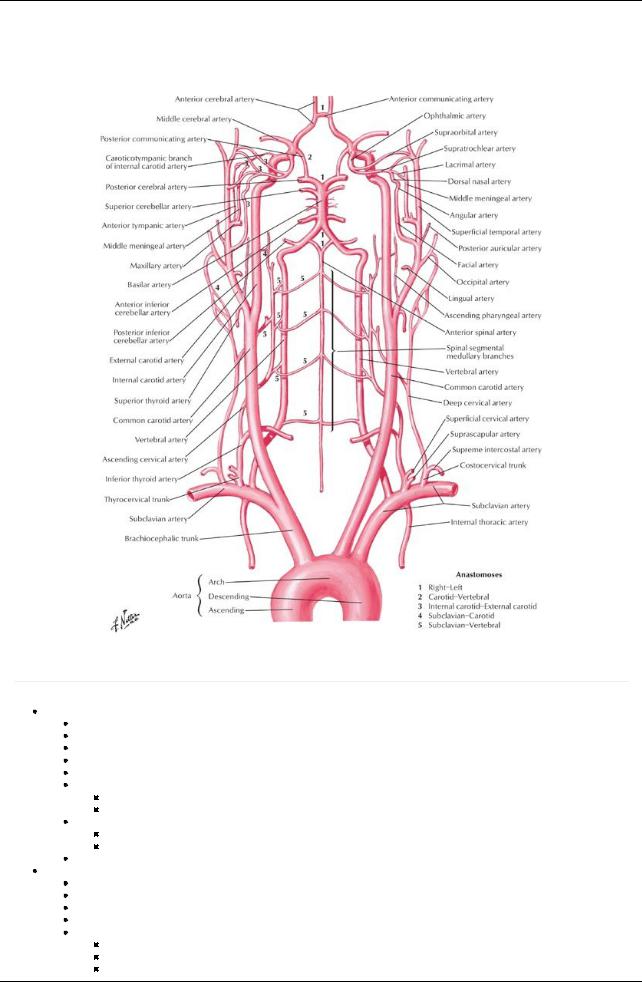
[Plate 136, Arteries to Brain: Schema]
Arterial Supply
page 77
page 78
Internal carotid arteries
Arise from common carotid arteries in neck
Begin at upper border of thyroid cartilage
Have no branches to face or neck
Enter carotid canals in temporal bone, then pass anteriorlyand medially
Run through carotid sinuses in grooves on side of bodyof sphenoid
Terminal branches
Anterior cerebral artery
Middle cerebral artery
Contribute to circle of Willis
United to posterior cerebral arterybyposterior communicating branches
Complete arterial circle around interpeduncular fossa
Provide anterior circulation of brain
Vertebral arteries
First branches of subclavian arteries
Ascend in foramina transversaria of first sixcervical vertebrae
Provide vascular supplyto cervical spinal cord and neck
Pierce dura and enter cranium via foramen magnum
Unite as at caudal end of pons to form basilar artery
Ascends on clivus
Terminates bydividing into two posterior cerebral arteries
Contribute to circle of Willis
94 / 425

Posterior cerebral arteries unite with anterior cerebral arteries via posterior communicating arteries
 Provide posterior circular of brain Cerebral arteries
Each supplies a region of the brain
Anterior cerebral artery
Medial and upper lateral surfaces of cerebral hemisphere
Frontal pole
Middle cerebral artery
Lower and lateral cerebral hemisphere
Temporal pole
Posterior cerebral artery
Inferior surface of cerebral hemisphere

 Occipital pole Cerebral arterial circle (circle of Willis)
Lies in subarachnoid space
Important anastomosis at base of brain
Formed by
Anterior communicating arteries
Anterior cerebral arteries
Internal carotid arteries
Posterior communicating arteries
Posterior cerebral arteries
Components supplybrain via manysmall branches
Artery |
Course and Structures Supplied |
Vertebral |
From subclavian artery, supplies cerebellum |
Posterior inferior cerebellar |
From vertebral artery, goes to posteroinferior cerebellum |
Basilar |
From both vertebrals, goes to brainstem, cerebellum, cerebrum |
Anterior inferior cerebellar |
From basilar, supplies inferior cerebellum |
Superior cerebellar |
From basilar, supplies superior cerebellum |
Posterior cerebral |
From basilar, supplies inferior cerebrum, occipital lobe |
Posterior communicating |
Cerebral arterial circle (of Willis) |
Internal carotid (IC) |
From common carotid, supplies cerebral lobes and eye |
Middle cerebral |
From IC, goes to lateral aspect of cerebral hemispheres |
Anterior communicating |
Cerebral arterial circle (of Willis) |
Anterior cerebral |
From IC, goes to cerebral hemispheres (except occipital lobe) |
95 / 425
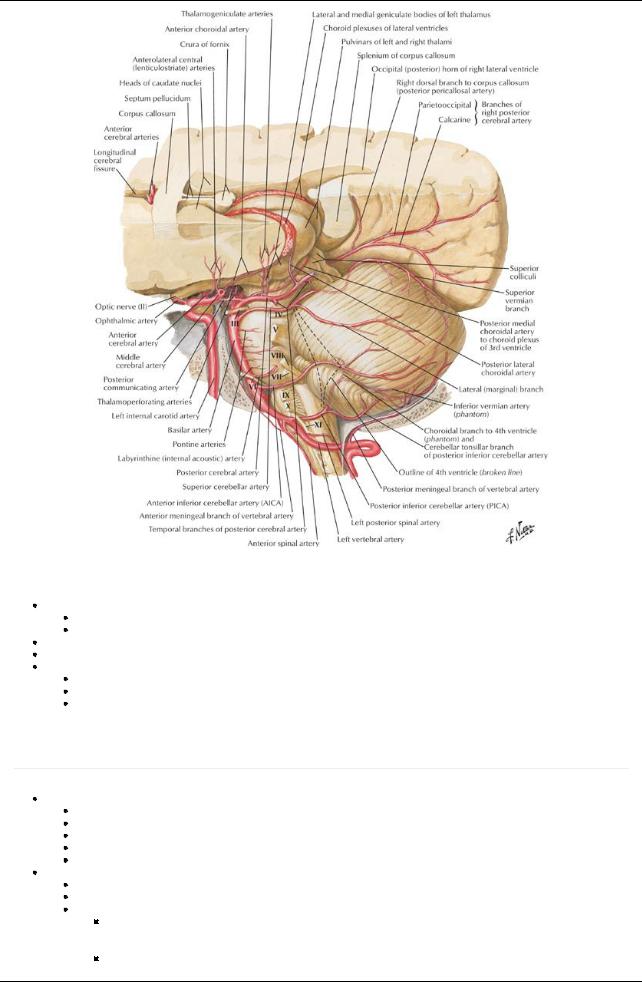
[Plate 141, Arteries of Posterior Cranial Fossa]
Venous Drainage
Dural venous sinuses
Drain venous blood from superficial and deep veins of the brain
Sinuses drain to internal jugular vein via jugular foramen
Veins on superior and lateral surfaces of brain drain to superior sagittal sinus
Basal veins run laterallyand dorsallyaround cerebral peduncle to end in great vein of Galen, which drains to straight sinus Veins on posterior and inferior surfaces of brain, superior cerebellar veins, and transverse sinuses drain to several sinuses
Straight
Transverse Superior petrosal
Vascular Supply to Scalp, Face, and Neck
Arterial Supply
page 78
page 79
Common carotid artery
Branch of aortic arch on left
Branch of brachiocephalic arteryon right
Ascends neck in carotid sheath, beneath anterior border sternocleidomastoid
Bifurcates into internal and external carotid arteries at level thyroid cartilage
Internal carotid arteryhas no branches in the neck
External carotid artery
Begins in upper border thyroid cartilage
Mainlysupplies the face and structures external to the skull, with some branches to the neck
Branches
Ascending pharyngeal
a.Ascends on pharynx
b.Send branches to pharynx, prevertebral muscles, middle ear, and cranial meninges
Superior thyroid
a. Supplies thyroid gland, infrahyoid muscles, and sternocleidomastoid muscle
96 / 425

b. Gives rise to superior laryngeal arterysupplying larynx Lingual
a.Passes deep to hypoglossal nerve, stylohyoid muscle, and posterior bellyof digastric
b.Disappears beneath hyoglossus muscle and becomes deep lingual and sublingual arteries
Facial
a.Branches to tonsil, palate, and submandibular gland
b.Hooks around middle of mandible and enters face
Occipital
a.Passes deep to posterior bellyof the digastric
b.Grooves base of skull
c.Supplies posterior scalp
Posterior auricular
a.Passes posteriorlybetween external acoustic meatus and mastoid process
b.Supplies muscles of region, parotid gland, facial nerve, auricle, and scalp
Maxillary
a.Larger of two terminal branches
b.Branches supplyexternal acoustic meatus, tympanic membrane, dura mater and calvaria, mandible, gingivae and teeth, temporal pterygoid, masseter, and buccinator muscles
Superficial temporal
a.Smaller terminal branch
b.Supplies temporal region of scalp
Carotid Branch |
Course and Structures Supplied |
Superior thyroid |
Supplies thyroid gland, larynx, and infrahyoid muscles |
Ascending pharyngeal |
Supplies pharyngeal region, middle ear, meninges, and prevertebral muscles |
Lingual |
Passes deep to hyoglossus muscle to supplythe tongue |
Facial |
Courses over the mandible and supplies the face |
Occipital |
Supplies SCMand anastomoses with costocervical trunk |
Posterior auricular |
Supplies region posterior to ear |
Maxillary |
Passes into infratemporal fossa (described later) |
Superficial temporal |
Supplies face, temporalis muscle, and lateral scalp |
page 79 page 80
Subclavian artery
Branch of aortic arch on the left
From brachiocephalic trunk on the right
Enters neck between anterior and posterior scalene muscles
Supplies upper limbs, neck and brain
Divided for descriptive purposes into 3 parts, in relation to the anterior scalene muscle
First part
a.Medial to the anterior scalene
b.Has three branches
Second part
a.Posterior to the anterior scalene
b.Has one branch
Third part
a.Lateral to anterior scalene
b.Has one branch
Subclavian Branch |
Course |
Part 1 |
|
Vertebral |
Ascends through C6-C1 transverse foramina and enters foramen magnum |
Internal thoracic |
Descends parasternallyto anastomose with superior epigastric artery |
Thyrocervical trunk |
Gives rise to inferior thyroid, transverse cervical, and suprascapular arteries |
Part 2 |
|
Costocervical trunk |
Gives rise to deep cervical and superior intercostal arteries |
Part 3 |
|
Dorsal scapular |
Is inconstant; mayalso arise from transverse cervical artery |
Venous drainage
Superficial veins
External jugular vein (EJV)
Drains most of scalp and side of face
Formed at angle of mandible byunion of retromandibular vein with posterior auricular vein
Enters posterior triangle and pierces fascia of its roof
Descends to terminate in subclavian vein
Receives
a.Transverse cervical vein
b.Suprascapular vein
c.Anterior jugular vein
Anterior jugular vein
97 / 425

Descends deep to investing fascia
Posterior to sternocleidomastoid (SCM), drains to EJVor subclavian vein
Commonlyunites with anterior jugular on opposite side via a jugular venous arch
Deep veins
Internal jugular vein (IJV)
Most veins in anterior neck are tributaries of IJV
Drains blood from brain, anterior face, cervical viscera, and deep muscles of neck
Begins as dilation of superior bulb just below jugular foramen
Runs inferiorlyin carotid sheath
Inferior end deep to gap between two heads of SCMmuscle
Joins subclavian vein to form brachiocephalic vein
Subclavian vein
Major vein draining upper limb
Passes anterior to anterior scalene muscle
Unites at medial border of muscle with IJVto form brachiocephalic vein
Tributaries of subclavian and IJVtravel with arteries of same name
Lymphatic Drainage
page 80 page 81
Drainage of face and head
Superficial lymphatic vessels accompanyveins
Deep lymphatic vessels accompanyarteries
Lymphatic drainage of face
Drainage from lateral face to parotid nodes
Drainage from upper lip and lateral lower lateral lip to submandibular nodes
Drainage from chin and central lower lip to submental nodes
All drain to parotid, mastoid, or superficial cervical nodes
These drain to deep cervical nodes
Drainage of the neck
Superficial drainage to superficial cervical nodes
Located along course of EJV
Also receive drainage from nodes of face and head
Superficial cervical nodes drain to deep cervical nodes
Deep cervical nodes
Lie along course of IJV, transverse cervical artery, and accessorynerve
Include
Prelaryngeal nodes
Pretracheal nodes
Paratracheal nodes
Retropharyngeal nodes
Drain to jugular lymphatic trunk
Jugular lymphatic trunks
On left
Joins thoracic duct on left
Thoracic duct enters junction of IJVand subclavian vein
On right
Empties directlyinto IJVor brachiocephalic vein
Or forms short right lymphatic duct which enters either of these vessels
98 / 425
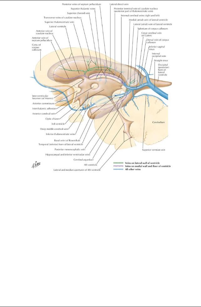
[Plate 144, Subependymal Veins of Brain]
99 / 425