


20 Body Wall
STUDYAIMS
At the end of your study, you should be able to:
Identifymuscles of the anterior chest wall and know their attachments, actions, and innervation
Identifythe intercostal muscles
Identifythe ribs and their parts
Count ribs
Understand the organization of a typical intercostal space and clinical significance of its contents
142 / 425
GUIDES
Thorax-Body Wall Organization
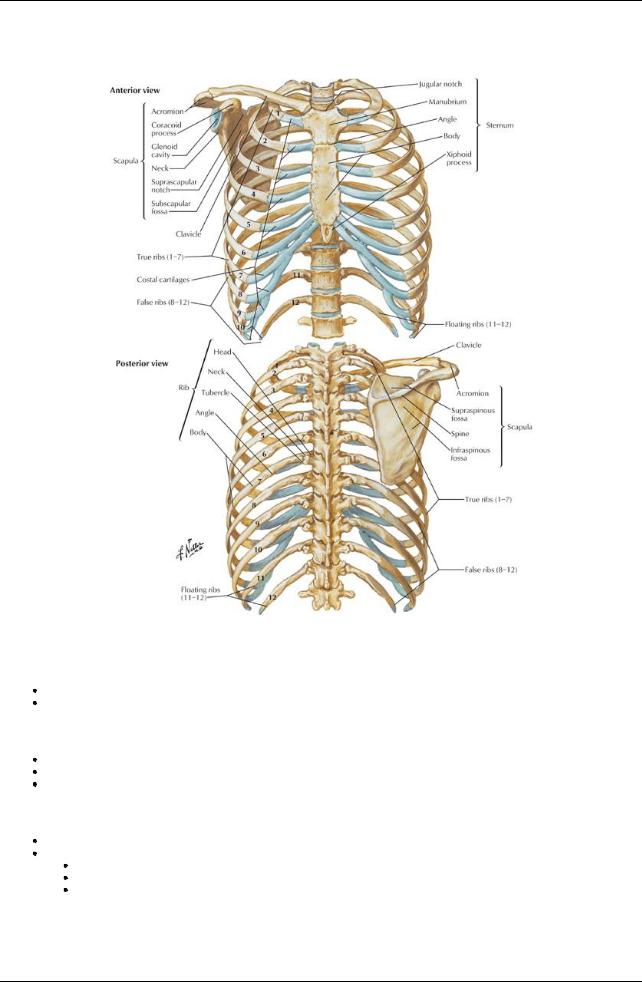
[Plate 179, Bony Framework of Thorax]
Layers
First and second layers
Skin
Tela subcutanea (superficial fascia), including the breasts
Third layer-muscles moving the upper limb
pectoralis major pectoralis minor serratus anterior
Fourth layer-includes muscles of the chest wall
Ribs
Intercostal muscles
External intercostal muscles
Internal intercostal muscles
Innermost intercostal muscles
Intercostal muscles
143 / 425
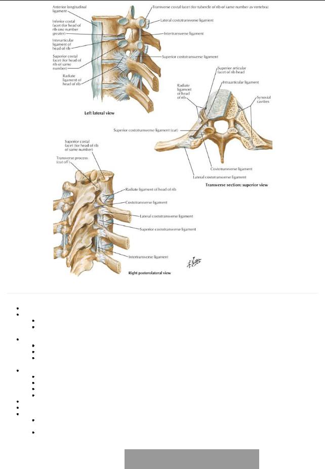
[Plate 181, Costovertebral Joints]
page 101 page 102
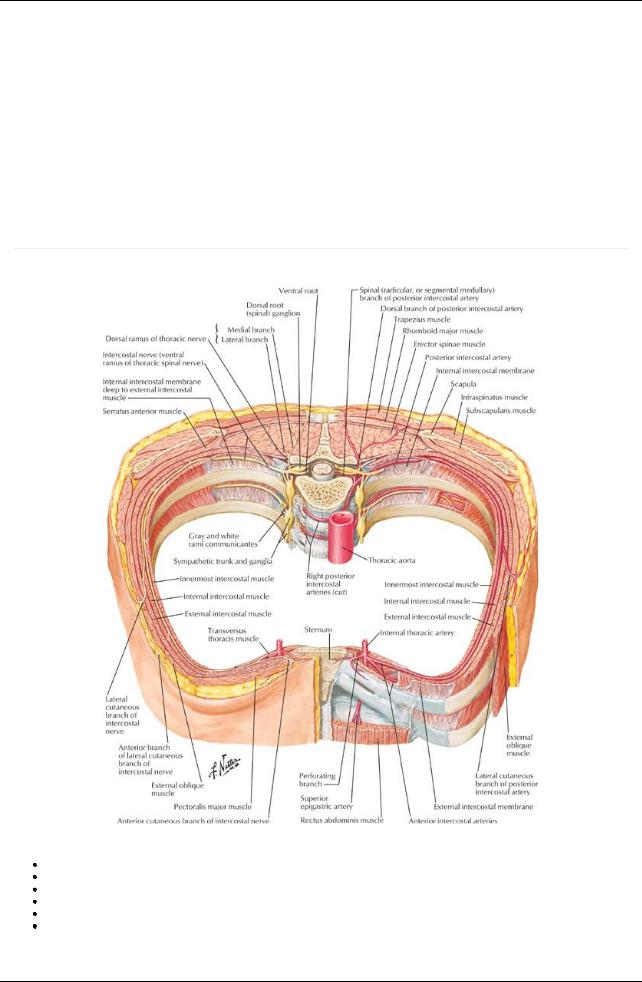
These muscles are arranged in three layers. External intercostal muscles:
Have fibers that slope down and medially.
Extend from the posterior tubercle of the rib to the junction of the rib and its costal cartilage anteriorly.
 Anteriorly, are replaced byexternal intercostal membranes that extend from the costochondral junctions to the sternum. Internal intercostal muscles:
Lie internal to the external intercostal muscles
Their fibers lie at right angles to those of the external intercostal muscles and run inferiorlyand laterally.
Anteriorlyextend to the lateral border of the sternum.
 Posteriorlyextend onlyto the angles of the ribs; medial to the angles, are replaced bythe internal intercostal membranes. Innermost intercostal muscles
Lie deep to the internal intercostal muscles
Are separated from the internal intercostals bythe intercostal vessels and nerves
Occupythe middle parts of the intercostal spaces
Connect inner surfaces of adjacent ribs
All intercostal muscles are supplied byintercostal nerves corresponding in number to their intercostal space. Main action of the intercostals is to maintain the space between the ribs during inspiration and expiration. Other muscles of the rib cage
Subcostal muscles-internal to the internal intercostals, cross from the angle of one rib to internal surface of the rib 1 to 2 spaces below.
Transversus thoracis-4 to 5 slips of muscle that attach to the xiphoid process and inferior sternum and pass superiorlyand laterally to attach to the 2nd through 6th costal cartilages.
|
Muscle |
Superior |
Inferior |
InnervationBlood Supply |
Main Actions |
|
|
|
|
Attachment |
Attachment |
|
|
|
|
|
|
(Origin) |
(Insertion) |
|
|
|
|
|
External |
Inferior border |
Superior border |
Intercostal |
Aorta, posterior intercostals and their collaterals, |
Elevate ribs |
|
|
intercostal |
of rib |
of rib below |
nerve |
costocervical trunk, anterior intercostals of internal |
|
|
|
|
|
|
|
thoracic and musculophrenic arteries |
|
|
|
Internal |
Inferior border |
Superior border |
Intercostal |
Anterior intercostals, posterior intercostals, |
Elevate ribs (upper |
|
144 / 425
intercostal |
of rib |
of rib below |
nerve |
musculophrenic arteryand costocervical trunk |
four and five); |
|
|
|
|
|
others depress |
|
|
|
|
|
ribs |
Innermost |
Inferior border |
Superior border |
Intercostal |
Posterior intercostals and collaterals |
Act similar to |
intercostal |
of rib |
of rib below |
nerve |
|
internal |
|
|
|
|
|
intercostals |
Transversus |
Internal surface |
Posterior |
Intercostal |
Anterior intercostals, internal thoracic artery |
Depress ribs and |
thoracis |
of costal |
surface of lower |
nerve |
|
costal cartilages |
|
cartilages 2-6 |
sternum |
|
|
|
Subcostal |
Internal surface |
Superior |
Intercostal |
Posterior artery, musculophrenic artery |
Depress ribs |
|
of lower rib near |
borders of |
nerve |
|
|
|
their angles |
second or third |
|
|
|
|
|
ribs below |
|
|
|
Levator |
Transverse |
Subjacent ribs |
Dorsal |
Posterior intercostals |
Elevate ribs |
costarum |
processes of |
between |
primary |
|
|
|
C7 and T1-T11 |
tubercle and |
rami of |
|
|
|
|
angle |
C8-T11 |
|
|
page 102
page 103
Intercostal nerves
[Plate 185, Intercostal Nerves and Arteries]
Intercostal nerves arise from the ventral rami of the upper eleven thoracic spinal nerves Each intercostal nerve divides to give a lateral cutaneous branch near the midaxillaryline
Anterior cutaneous branches innervate the skin on the anterior abdomen and thoraxand divide into medial and lateral branches. Muscular branches supplythe intercostal, levatores costarum, transversus thoracis, and serratus posterior muscles.
The lower five intercostal nerves supplythe skin and muscles of the abdominal wall
Contain general somatic afferent and efferent fibers, as well as general visceral efferent fibers from the sympathetic trunk via white and grey rami communicantes and general visceral afferent fibers.
Ribs
145 / 425
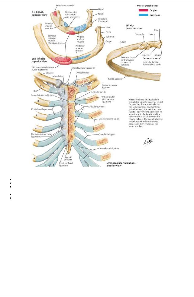
[Plate 180, Ribs and Sternocostal Joints]
All ribs contain bone marrow.
Ribs 1 through 7 are vertebrocostal because theyattach to the sternum via a costal cartilage.
Ribs 8 through 10 are vertebrochondral because their cartilages are joined to the cartilage of the rib above and via that connection to the sternum.
Ribs 11 and 12 are free or floating ribs, which do not connect even indirectlywith the sternum but which have a costal cartilage on their tips. First rib is broad and sharplycurved and has a tubercle of the attachment of scalene muscles.
146 / 425
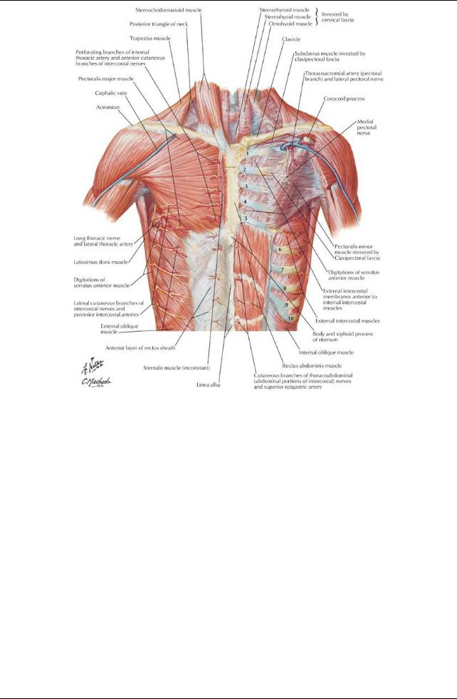
[Plate 182, Anterior Thoracic Wall]
147 / 425
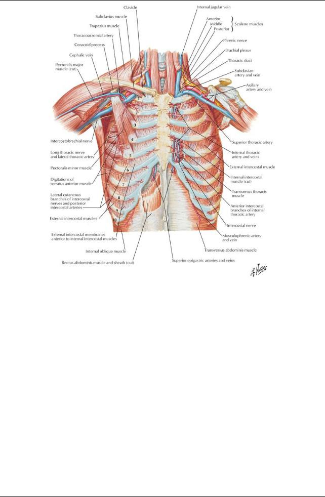
[Plate 183, Anterior Thoracic Wall (Continued)]
148 / 425