

FACTS & HINTS
High-Yield Facts
Clinical Points
Meningitis
Inflammation of the arachnoid and pia mater
Can be caused bydrugs or malignancy, but usuallybypathogenic bacteria
Infection can be due to meningococcal or pneumococcal bacteria
Patient maypresent with fever, nonblanching purpuric rash, neck stiffness, and photophobia
Mortalityfrom bacterial causes can be up to 30%
Diagnosis is byexamination and lumbar puncture
Hydrocephalus
Caused byexcess cerebrospinal fluid (CSF) production or, more likely, byabnormal absorption
Classified as obstructive, communicating or normal pressure
Blockage usuallyin cerebral aqueduct bynarrowing
Can be a result of tumor, hemorrhage, and infection
Communicating blocks movement of CSF from ventricles
Can be caused byabsence of arachnoid granulations or subarachnoid hemorrhage
Dilates ventricles, thins cerebral cortex, separates bones of calvaria in infants
Brain tumors
Twenty-five percent of all brain tumors arise from a different site (metastasis) Common sites of original tumor include: breast, bronchus, prostate, thyroid, and kidney
Primarybrain tumors can be benign, such as meningiomas and neurofibromas, or malignant, such as astrocytomas and oligodendrogliomas
Can present as epilepsy, focal neurology, or signs of raised intracranial pressure
Diagnosis is byhistory, examination, and computed tomography(CT) or magnetic resonance imaging (MRI) of the brain
86 / 425

12 Cranial and Cervical Nerves
STUDYAIMS
At the end of your study, you should be able to:
Know the names and functions of the cranial nerves
State the foramen through which the cranial nerves emerge from the skull
Outline formation of the cervical plexus
Know the sensorynerves arising from the cervical plexus and their distribution
Know the muscles innervated bymotor branches of the cervical plexus
Understand the formation of the ansa cervicalis and know the muscles innervated byits branches
Describe the formation of, and fibers composing, the phrenic nerve
Know the structures innervated bythe various components of the phrenic nerve
87 / 425

GUIDE
Head and Neck: Cranial and Cervical Nerves
Cranial Nerves
12 pairs of cranial nerves arise from the brain, and theyare identified both bytheir names and byRoman numerals I through XII. The cranial nerves are somewhat unique and can contain multiple functional components:
General: same general functions as spinal nerves
Special: functions found onlyin cranial nerves
Afferent and efferent: sensoryor motor functions, respectively
Somatic and visceral: related to skin and skeletal muscle (somatic), or to smooth muscle and glands (visceral)
Hence, each cranial nerve maypossess multiple functional components, such as GSA(general somatic afferents), meaning it contains nerve fibers that are sensoryfrom the skin, not unlike those of the spinal nerve; GVE (general visceral efferents), meaning it contains motor fibers to visceral structures (smooth muscle and/or glands) like a parasympathetic fiber from the sacral spinal cord (S2-S4 gives rise to parasympathetics); or SSA(special somatic afferents), meaning it contains special sensoryfibers, such as those for vision or hearing.
[Plate 126, Accessory Nerve (XI): Schema]
88 / 425
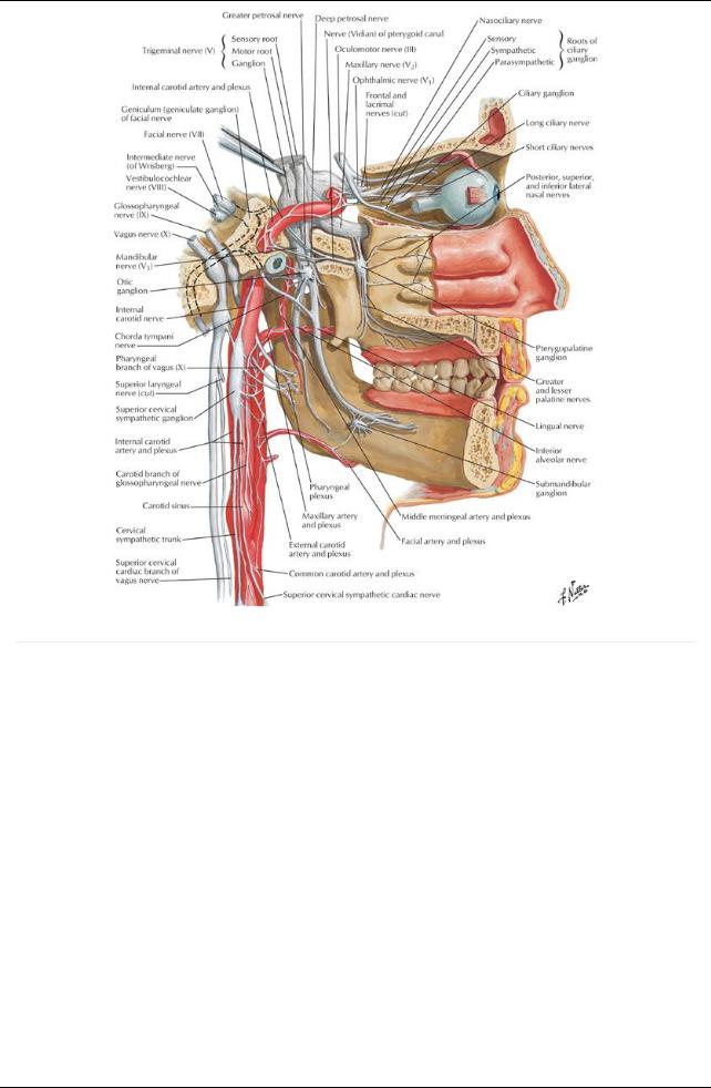
[Plate 130, Autonomic Nerves in Head]
page 72 page 73
Cranial Nerve |
Functional Component |
Cranial Opening |
I Olfactorynerve |
SVA(special sense of smell) |
Foramina in cribriform plate |
II Optic nerve |
SSA(special sense of sight) |
Optic canal |
III Oculomotor |
GSE (motor to extraocular muscles) |
Superior orbital fissure |
nerve |
GVE (parasympathetic to smooth muscle in eye) |
|
IVTrochlear nerve |
GSE (motor to 1 extraocular muscle) |
Superior orbital fissure |
VTrigeminal nerve |
GSA(sensoryto face, orbit, nose, anterior |
three branches: ophthalmic, maxillary, and mandibular travel |
|
tongue) |
through multiple openings |
|
SVE (motor to skeletal muscles) |
|
VIAbducens nerve |
GSE (motor to 1 extraocular muscle) |
Superior orbital fissure |
VII Facial nerve |
GSA(sensoryto skin of ear) |
Internal acoustic meatus Facial canal Stylomastoid foramen |
|
SVA(special sense of taste to anterior tongue) |
|
|
GVE (motor to glands-salivary, nasal, lacrimal) |
|
|
SVE (motor to facial muscles) |
|
VIII |
SSA(special sense of hearing and balance) |
Internal acoustic meatus |
Vestibulocochlear |
|
|
nerve |
|
|
IX |
GSA(sensoryto posterior tongue) |
Jugular foramen |
Glossopharyngeal |
SVA(special sense of taste-posterior tongue) |
|
nerve |
GVA(sensoryfrom middle ear, pharynx, carotid |
|
|
body, and sinus) |
|
|
GVE (motor to parotid gland) |
|
|
SVE (motor to 1 muscle of pharynx) |
|
XVagus nerve |
GSA(sensoryexternal ear) |
Jugular foramen |
|
SVA(special sense of taste-epiglottis) |
|
|
GVA(sensoryfrom pharynx, larynx, and thoracic |
|
|
and abdominal organs) |
|
|
GVE (motor to thoracic and abdominal organs) |
|
89 / 425
|
SVE (motor to muscles of pharynx/larynx) |
|
XI Spinal |
SVE (motor to 2 muscles) |
Jugular foramen |
accessorynerve |
|
|
XII Hypoglossal |
GSE (motor to tongue muscles) |
Hypoglassal canal |
nerve |
|
|
page 73
page 74
In general, CN I and II arise from the forebrain and are reallytracts of the brain for the special senses of smell and sight. CN III, IV, and VI move the extraocular skeletal muscles of the eyeball. CN Vhas three divisions: V1 and V2 are sensory, and V3 is both motor to skeletal muscle and sensory. CN VII, IXand Xare both motor and sensory. CN VIII is the special sense of hearing and balance. CN XI and XII are motor to skeletal muscle. CN III, VII, IXand Xalso contain parasympathetic fibers of origin (visceral), although manyof theANS fibers will "jump" onto the branches of CN Vto reach their targets. The following table summarizes the types of fibers in each cranial nerve and where each passes through the cranium:
Cranial nerves emerge through foramina or fissures in the cranium
Twelve pairs
Numbered in order of origin from the brain and brain stem, rostral to caudal
Contain one or more of sixdifferent types of fibers
Motor fibers to voluntarymuscles
Somatic motor fibers to striated muscles (1)
a.Orbit
b.Tongue
c.Neck (sternocleidomastoid and trapezius)
Branchial motor (or special visceral efferent fibers) to striated muscles derived from pharyngeal arches (example: muscles of mastication) (2)
Motor fibers to involuntarymuscles = general visceral efferent (parasympathetic fibers) (3) Sensoryfibers
General visceral afferent fibers (4)
a.Carrysensation from viscera
b.Originate in carotid body, sinus, heart, lungs, and gastrointestinal tract
General somatic afferent fibers carrying pain, pressure, temperature, touch information (5)
Special sensoryafferent fibers conveying taste, smell, vision, hearing, and balance (6)
Can be sensory, motor, or mixed
Sensory Innervation of the Dura
Dura of the cranial fossae innervated bymeningeal branches of cranial and cervical nerves Anterior cranial fossa
Anterior meningeal branches of the ethmoidal nerves from ophthalmic nerves (CNV1)
Meningeal branches of the maxillarynerves (cranial nerve [CN] V2)
 Meningeal branches of the mandibular nerves (CN V3) Middle cranial fossa
Meningeal branches of the maxillarynerves (CN V2)
 Meningeal branches of the mandibular nerves (CN V3) Posterior cranial fossa
Tentorial nerve from ophthalmic nerve (CN V1)
Meningeal branches directlyfrom C2 and C3 spinal nerves or carried byCN X(vagus) or CN XII (hypoglossal)
Cervical Plexus
page 74
page 75
Formed from anterior rami of C1-C4 spinal nerves Consists of a series of loops and branches from the loops
Lies deep to sternocleidomastoid (SCM) and anteromedial to levator scapulae and middle scalene muscles Cutaneous branches of the cervical plexus
Emerge from posterior border of SCM
Nerves from loop formed between anterior rami of C2 and C3
Lesser occipital (C2) to skin of neck and scalp posterior to auricle
Great auricular (C2 and C3) to skin over parotid gland, mastoid process, auricle, and between angle of mandible and mastoid process
Transverse cervical nerve (C2 and C3) to skin over anterior cervical region
Supraclavicular nerves
Arise from C3-C4 loop
Emerge from under SCM
 Supplyskin over clavicle, superior thoracic wall, and shoulder Motor branches
Are considered deep branches
Innervate prevertebral muscles
Sternocleidomastoid (C2 and C3)
Trapezius (C3 and C4)
Levator scapulae (C3 and C4)
Motor fibers from C1 travel with hypoglossal nerve
Some C1 fibers leave hypoglossal nerve and innervate
a.Thyrohyoid muscle
b.Geniohyoid muscle
Rest leave the hypoglossal as its descending branch
90 / 425
Motor fibers from C1 and C2 directlyinnervate thyrohyoid (an infrahyoid strap muscle) Motor fibers from C2, 3 form the descending cervical nerve
Ansa cervicalis
Loop formed bydescending branch from hypoglossal nerve (superior root) (C1) and descending cervical nerve (inferior root) (C2,C3)
Branches from ansa innervate remaining infrahyoid strap muscles
a.Omohyoid
b.Sternohyoid
c.Sternothyroid
 Motor fibers from C3, C4, and C5 contribute to roots of phrenic nerve Phrenic nerve
Formed bybranches of anterior rami of C3, C4, and C5 spinal nerves
Contains a mixof fibers
Sole motor supplyto the diaphragm
Sensoryfibers from the central part of the diaphragm (sensoryfibers from peripheryprovided byintercostal nerves) Sympathetic nerve fibers from the cervical sympathetic ganglia to smooth muscle of blood vessel walls
91 / 425