


FACTS & HINTS
High-Yield Facts
Clinical Points
Lumbar Puncture:Performed for retrieval of cerebrospinal fluid (CSF) from the lumbar spinal cistern. The patient is placed in the left decubitus position, flexed in the fetal posture with the supracristal line vertical. Puncture should be made at the L3/4 (immediatelysuperior) or L4/5 (immediatelyinferior) interspace in the midline of the back, to avoid the spinal cord.
Mnemonics
Memory Aids
Lumbar puncture: To keep the cord alive, keep the needle between L3 and L5!
105 / 425

15 Bones and Ligaments
STUDYAIMS
At the end of your study, you should be able to:
Identifythe significant parts of a typical vertebra and understand regional variations
Identifythe specialized vertebrae
Know the attachments and function of the vertebral ligaments
Describe the spine, its curvatures, and gross vertebral column movements
Describe the type, location, and movements of the joints of the vertebral column
106 / 425
GUIDE
Back and Spinal Cord: Bones and Ligaments
Vertebral Column
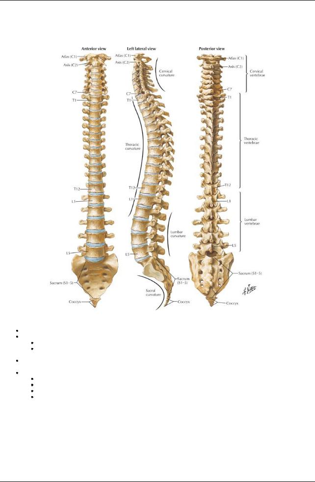
[Plate 150, Vertebral Column]
approximately72 to 75 cm long (25% of length because of intervertebral discs) 33 vertebrae (can vary 32-34)
7 cervical, 12 thoracic, 5 lumbar, 5 sacral, 4 (3-5) coccygeal
Typicallyhave: body; vertebral arch (2 laminae, 2 pedicles) and foramen; spinous (1) and transverse (2) processes; articular processes (4)
Fibrocartilaginous intervertebral discs
 Allow movement between vertebral bodies (in cervical, thoracic, and lumbar regions) Curvature (may be primary or secondary-see below)
Cervical anterior convexity(2°)
Thoracic anterior concavity(1°)
Lumbar anterior convexity(2°) Sacral anterior concavity(1°)
Vertebrae
107 / 425
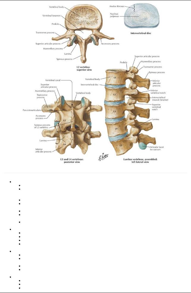
[Plate 152, Lumbar Vertebrae]
page 84 page 85
Cervical vertebrae
Permit forward/lateral flexion, extension, rotation
C1 (Atlas): No bodyor spinous process; articulates with occipital condyles via paired lateral masses and with the axis via the superior articular facets and dens of the axis; groove on superior aspect of the posterior arch for vertebral arteries and dorsal ramus of C1
C2 (Axis): Dens (odontoid process), large superior articular facets for C1
C3-C5: Short bifid spinous processes (anterior tubercle of C6 is the carotid tubercle, which the carotid artery can be compressed against to control bleeding)
C6-7: long, nonbifid spinous processes
C7 (Vertebra prominens): long (nonbifid) spinous process; small transverse foramina that transmit accessoryvertebral veins NOT vertebral artery
Have transverse processes with anterior and posterior tubercles and foramina (foramina transversaria), which transmit vertebral arteries and veins and sympathetic nerves plexuses
Thoracic vertebrae
Are relativelyrigid, mainlyallow rotation of the trunk
T1-T4:Atypical-have some features of cervical vertebrae
T5-T8: Typical
T9-T12:Atypical-have tubercles similar to mamillaryand accessoryprocesses of lumbar vertebrae
 Have long transverse processes that extend posterolaterally Lumbar vertebrae
Are relativelymobile, permit forward/lateral flexion and extension but little rotation
Have accessoryprocess found on posterior surface of the base of each transverse process for attachment of medial intertransverse lumborum muscle
Have mamillaryprocess for attachment of multifidus and medial intertransverse muscles
L5: massive bodyand transverse processes and is thicker anteriorly-contributes to the lumbosacral angle (usually130°-160°) and carries the weight of the upper body
Sacrum (sacral vertebrae)
Composed of five vertebrae that fuse at about 20 years of age, inferior portion is nonweightbearing
Articulates with "hip" bones at sacroiliac joints Has concave pelvic surface
108 / 425

Is wider in females than males
Has a sacral canal (continuation of vertebral canal) that contains cauda equine
Has the following features:
Sacral hiatus (termination of sacral canal) that contains filum terminale
Median crest: fused spinous processes
Paired medial crests: fused articular processes
Paired lateral crests: fused tips of the transverse processes
Sacral corneae that project inferiorlyon either side of sacral hiatus
Coccyx(coccygeal vertebrae)
Consist of three to five coccygeal vertebrae, inferior three fuse as coccyxin midlife
Has coccygeal corneae that articulate with sacral corneae
Provides site of attachment for gluteus maximus, coccygeus muscles, and anococcygeal ligament
Is joined to the sacrum bythe sacrococcygeal symphysis
Summary of vertebral characteristics
Vertebrae Distinctive Features
Cervical |
Small bodies, large vertebral foramina, foramina in transverse processes, anterior and posterior tubercles, bifid |
|
spinous processes |
Thoracic |
Heart-shaped bodies, long spinous processes angled posteroinferiorly; costal facets for rib articulation on bodies and |
|
transverse processes |
Lumbar |
Large kidney-bean-shaped bodies, sturdylaminae, thick and short spinous processes, mammillaryprocesses on the |
|
posterior surface of the superior articular facets (processes) |
Sacral |
Fused as sacrum, four pairs of dorsal and ventral foramina for nerve exit and triangular sacral canal |
Coccygeal |
Fused as small triangular bone-the coccyx |
Joints
[Plate 21, Cervical Vertebrae: Uncovertebral Joints]
page 85
page 86
Intervertebral (IV) discs
Connect articulating surfaces of adjacent vertebral bodies
109 / 425

Are integral part of secondarycartilaginous joints between vertebral bodies (except C1/2)
Are composed of a tough annulus fibrosus surrounding an avascular, gelatinous nucleus pulposus
 Act as "shock absorbers" and semifluid ball bearings to provide small movements between individual vertebrae Zygapophysial (or facet) joints
Are synovial joints between the superior and inferior articular processes
Are surrounded bythin, loose articular capsule
 Permit gliding movements between vertebrae Atlanto-occipital joints
Are synovial joints between the lateral masses of atlas and occipital condyles
 Permit flexion/extension and some lateral bending and rotation Atlantoaxial joints
Are three synovial joints between the inferior lateral masses of C1 and the superior facets of C2 and between the anterior arch of C1 and the dens of C2
 Permit rotation of C1 (and the head), which is limited bythe alar ligaments Costovertebral joints Synovial, between the vertebrae and ribs (see: Thorax) Sacroiliac (SI) joints. Synovial joints (see: Pelvis and Perineum)
Ligament |
Features |
Anterior longitudinal |
Limits extension |
|
Maintains stabilityof IVdiscs |
Posterior longitudinal |
Limits flexion |
|
Prevents IVdisc herniation |
Intertransverse |
Limits lateral bending |
Interspinous |
Limits flexion |
Supraspinous |
Limits flexion |
Ligamenta flava |
Limits flexion |
|
Preserves curvature of column |
|
Prevents injuryto the IVdiscs |
Ligamentum nuchae |
Prevents cervical hyperflexion |
|
Attachment site for trapezius and rhomboid minor |
[Plate 22, External Craniocervical Ligaments]
110 / 425
[Plate 23, Internal Craniocervical Ligaments]
111 / 425

[Plate 153, Lumbar Vertebrae: Radiographs]
112 / 425
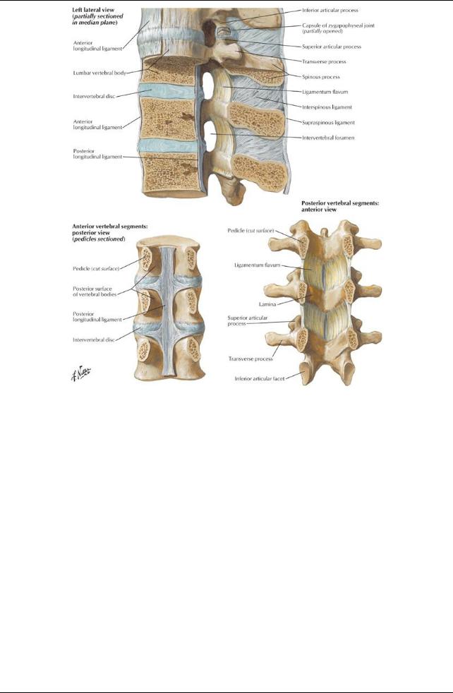
[Plate 156, Vertebral Ligaments: Lumbar Region]
113 / 425