


FACTS & HINTS
HIGH-YIELD FACTS
Clinical Points
page 222
page 223
Fracture of the Humerus
Fractures of the humerus most common at the surgical neck
Especiallycommon in elderlyindividuals with osteoporotic bone, falling on an outstretched arm
The axillarynerve vulnerable to damage here
Direct blow to the arm may
Fracture humerus through its midshaft, with risk of injuryto the radial nerve
Fracture humerus at distal end, risking damage to the median nerve
Biceps Tendonitis
Is inflammation of the tendon of the long head of the biceps
Tendon is susceptible to wear and tear as it moves back and forth within the intertubercular groove
Degenerative wear a common cause of shoulder pain
Inflammation also caused byrepetitive microtrauma, seen in certain sports such as tennis
Rupture of the Biceps Brachii
This produces "Popeye deformity" with muscle forming a ball in distal part of the anterior arm
Tendon of long head has the highest rate of spontaneous rupture of anytendon in the body
Rupture of the tendon on background of chronic tendonitis usuallyaffects those older than 40
Traumatic rupture mayoccur in younger individuals, but is rare
MNEMONICS
Memory Aids
Cubital fossa (from medial to lateral): |
Madeline |
Median Nerve |
|
Brown's |
Brachial Artery |
|
Big |
Biceps tendon |
|
Red |
Radial Nerve |
|
Purse |
Posterior interosseous nerve |
348 / 425

46 Elbow and Forearm
STUDYAIMS
At the end of your study, you should be able to:
Identifythe different parts and surface markings of the radius and ulna
Describe the articulations of the elbow joint, its movements, and supporting ligaments
Describe the distal radioulnar joint, its movements, and supporting ligaments
Describe the organization of the deep fascia and compartments of the arm
Know the origins, insertions, and actions of the major muscles of the forearm
349 / 425
GUIDE
Upper Limb: Elbow and Forearm
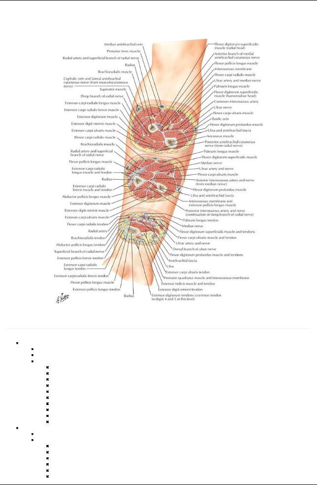
[Plate 437, Forearm: Serial Cross Sections]
Bones
page 224
page 225
Ulna
Stabilizing bone of the forearm
Medial to and longer than radius
Osteological features
Olecranon-projection of proximal posterior end
Coronoid process-projection from proximal anterior end
Trochlear notch-anterior surface of olecranon, articulates with trochlea of humerus
Radial notch-rounded concavityon lateral side of coronoid process for radial head
Ulnar tuberosity-inferior to coronoid process for attachment of brachialis muscle
Supinator crest-crest inferior to radial notch for attachment of supinator muscle
Supinator fossa-concavitybetween supinator crest and coronoid process, for attachment of supinator muscle
Body-thicker proximally, tapering distally
Head-at distal end
Styloid process-conical process from the head
Radius
Lateral and shorter bone of the forearm
Osteologic features
Head-at proximal end, concave for articulation with capitulum of humerus
Neck-constricted region between head and tuberosity
Radial tuberosity-oval protuberance below head and neck for attachment of biceps brachii
Body-convexlaterallyand enlarging distally
Styloid process-projection from lateral aspect of distal end of radius
Dorsal tubercle-dorsal projection at distal end between groves for tendon of extensor carpi radialis longus and brevis and tendon of extensor pollicis longus
350 / 425
Ulnar notch-concavityon medial side of distal end for head of ulna
Radius and ulna connected by
Interosseous membrane
Separates forearm into anterior and posterior compartments
Provides attachment for deep muscles of forearm
Fibers slope downward from radius to ulna
Is most tense when hand is in mid-prone position
Has a gap distal to neck of radius for posterior interosseus vessels
Oblique cord
Crosses gap in interosseus membrane
Attaches neck of radius to ulnar tuberosity
Radioulnar joints-proximal and distal (see below)
Elbow Joint
[Plate 425, Elbow: Radiographs]
351 / 425
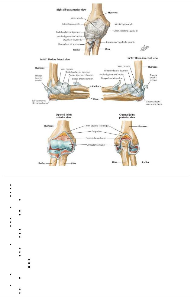
[Plate 426, Ligaments of Elbow]
page 225 page 226
Compound synovial joint in which radius and ulna articulate with humerus Joint cavitycontinuous with radioulnar joint
Fibrous capsule encloses all three articulations Humeroulnar joint
Uniaxial, synovial hinge joint
 Between trochlea of humerus and trochlear notch of ulna Humeroradial joint
Uniaxial, synovial hinge joint
Between capitulum of humerus and head of radius
Blood supplyto humeroulnar and humeroradial joints: anastomoses around elbow from brachial, radial, and ulnar arteries Nerve supplyto these joints: musculocutaneous, radial, and ulnar nerves
Proximal radioulnar joint
Uniaxial, synovial pivot joint
Between head of the radius and radial notch of the ulna
Blood supply: anterior and posterior interosseous arteries
 Nerve supply: musculocutaneous, median and ulnar nerves Ligaments reinforcing elbow joint
Ulnar collateral ligament: medial epicondyle to coronoid process and olecranon
Radial collateral ligament: lateral epicondyle to annular ligament
Annular ligament
Strong fibrous circle
From margins of radial notch of ulna
Encircles head of radius
 Maintains position of the radial head during pronation and supination Movements at the elbow joint
Flexion ( 180 degrees) and extension at humeroulnar and humeroradial joints
 Pronation and supination with rotation of the head of the radius within annular ligament at proximal radioulnar joint Distal radioulnar joint
Synovial pivot joint
Articulation between ulnar notch and articular facet of radius at its distal end
352 / 425
Afibrocartilaginous articular disc binds the ends of the radius and ulna and is strengthened byanterior and posterior ligaments
Disc separates cavityof joint from cavityof wrist joint
Pronation and supination: distal end of radius moves anteriorlyand mediallyacross the ulna
Blood supply: anterior and posterior interosseous arteries
 Nerve supply: posterior interosseous nerve Carrying angle
The angle between the long axis of the humerus and the long axis of the ulna with forearm fullyextended and supinated in the anatomical position
Ten to 15 degrees in males, more than 15 degrees in females (figures are for deviations from 180 degrees)
Bursae (important ones)
Subcutaneous olecranon bursa: Overlies olecranon in subcutaneous tissue
Subtendinous olecranon bursa: Between the olecranon and triceps tendon Bicipitoradial bursa: Between biceps tendon and anterior part of radial tuberosity
Fascial Compartments of Forearm
Forearm divided into anterior and posterior compartments byinterosseous membrane Antebrachial fascia is thickened at distal end of radius
Posteriorlyforms extensor retinaculum
Anteriorlyforms palmar carpal ligament
Also forms flexor retinaculum (transverse carpal ligament) distal and deeper to palmar carpal ligament
Carpal tunnel formed between flexor retinaculum, running between tubercles of scaphion and trapezium radiallyto pisiform and hook of hamate on ulnar side, and anterior concavityof the carpus
Muscles of the Forearm (listed from lateral to medial)
[Plate 429, Individual Muscles of Forearm: Extensors of Wrist and Digits]
353 / 425
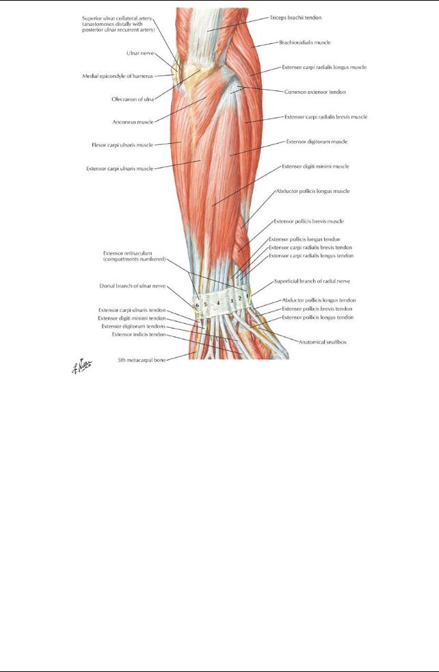
[Plate 432, Muscles of Forearm (Superficial Layer): Posterior View]
354 / 425
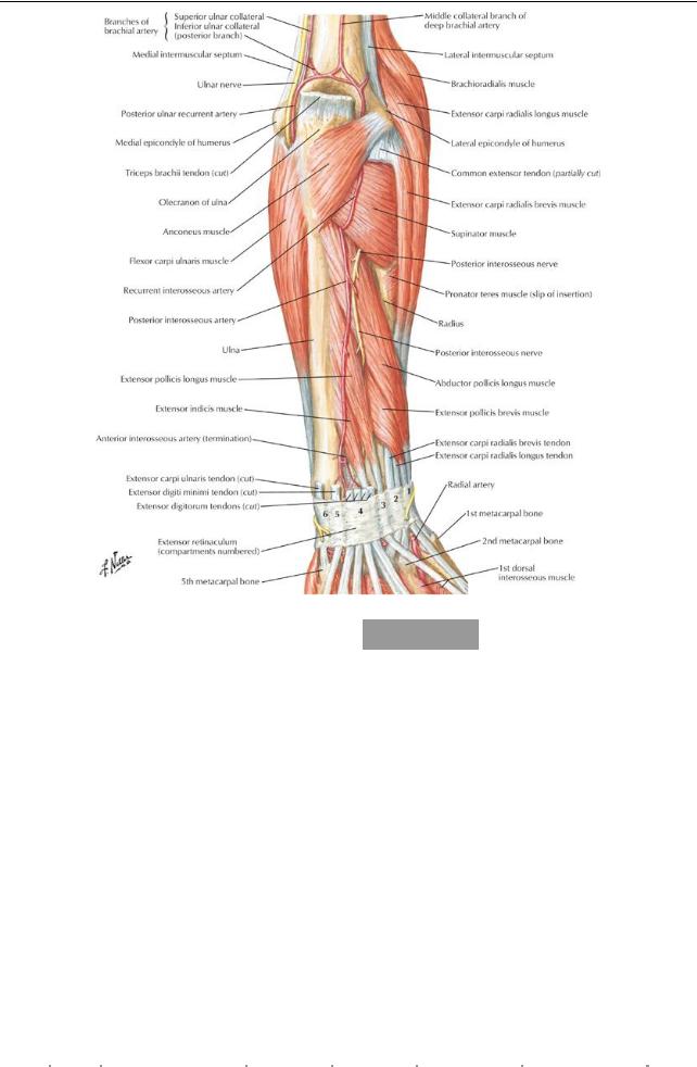
[Plate 433, Muscles of Forarm (Deep Layer): Posterior View]
|
|
Muscle |
|
Origin |
|
Insertion |
|
InnervationBlood |
Action |
|
|
|
|
|
|
|
|
|
|
|
|
|
|
supply |
|
|
|
|
|
|
|
Pronator |
|
Two heads: medial epicondyle |
Midwayalong lateral |
Median |
Anterior |
Pronates forearm, assists in flexion |
|
|
|
|||
|
|
Teres |
|
of humerus and coronoid |
|
surface of radius |
|
nerve |
ulnar |
|
|
|
|
|
|
|
|
|
process of ulna |
|
|
|
(C6,C7) |
recurrent |
|
|
|
|
|
|
|
|
|
|
|
|
|
|
artery |
|
|
|
|
|
|
|
Flexor carpi |
Medial epicondyle of humerus |
|
Base of second |
|
Median |
Radial |
Flexes and abducts hand at wrist |
|
|
|
||
|
|
radialis |
|
|
|
metacarpal |
|
nerve |
artery |
|
|
|
|
|
|
|
|
|
|
|
|
|
(C6,C7) |
|
|
|
|
|
|
|
|
Palmaris |
|
Medial epicondyle of humerus |
|
Distal half of flexor |
Median |
Posterior |
Flexes hand and tenses palmar |
|
|
|
||
|
|
longus |
|
|
|
retinaculum and |
|
nerve |
ulnar |
aponeurosis |
|
|
|
|
|
|
|
|
|
|
palmar aponeurosis |
(C7,C8) |
recurrent |
|
|
|
|
|
|
|
|
|
|
|
|
|
|
|
artery |
|
|
|
|
|
|
|
Flexor carpi |
Humeral head: medial |
|
Pisiform bone, hook of |
Ulnar |
Posterior |
Flexes and adducts hand at wrist |
|
|
|
|||
|
|
ulnaris |
|
epicondyle of humerus; |
|
hamate, base of fifth |
nerve |
ulnar |
|
|
|
|
|
|
|
|
|
|
Ulnar head: olecranon and |
|
metacarpal |
|
(C7,C8 |
recurrent |
|
|
|
|
|
|
|
|
|
posterior border of ulna |
|
|
|
and T1) |
artery |
|
|
|
|
|
|
|
Flexor |
|
Humeroulnar head: medial |
|
Bodies of middle |
|
Median |
Ulnar and |
Flexes middle and proximal |
|
|
|
|
|
|
digitorum |
|
epicondyle and coronoid |
|
phalanges of medial |
nerve (C8- |
radial |
phalanges of medial four digits; |
|
|
|
||
|
|
superficialis |
process of ulna |
|
four digits |
|
T1) |
arteries |
also flexes hand at wrist |
|
|
|
||
|
|
|
|
Radial head: anterior border |
|
|
|
|
|
|
|
|
|
|
|
|
|
|
of radius |
|
|
|
|
|
|
|
|
|
|
|
|
|
|
|
|
|
|
|
|
|
|
page 226 |
||
|
|
|
|
|
|
|
|
|
|
|
|
page 227 |
|
|
|
|
Muscle |
Origin |
Insertion |
Innervation |
Blood supply |
|
Action |
|
|
|
|||
|
|
Deep |
|
|
|
|
|
|
|
|
|
|
|
|
|
|
Flexor |
Medial and anterior surface of |
Anterior base of |
Medial part: ulnar |
Anterior interosseous |
Flexes distal phalanges of |
|
|
|||||
|
|
digitorum |
proximal three-quarters of |
distal phalanges |
nerve (C8-T1); |
arteryand muscular |
medial four digits; assists |
|
|
|||||
|
|
profundus |
ulna, and interosseous |
of medial four |
Lateral part: |
branches of ulnar |
in flexion of hand at wrist |
|
|
|||||
|
|
|
membrane |
digits |
medial nerve |
artery |
|
|
|
|
|
|||
|
|
|
|
|
|
|
(C8-T1) |
|
|
|
|
|
|
|
|
|
|
|
|
|
|
|
|
|
|
355 / 425 |
|
||

|
Flexor |
Anterior surface of radius and |
Palmar base of |
Anterior |
|
Anterior interosseous |
Flexes phalanges of |
|
|
||||||
|
pollicis |
interosseous membrane |
distal phalanxof |
interosseous |
artery |
|
|
thumb |
|
|
|
||||
|
longus |
|
|
thumb |
|
branch of median |
|
|
|
|
|
|
|
|
|
|
|
|
|
|
|
nerve (C7-8) |
|
|
|
|
|
|
|
|
|
|
Pronator |
Distal fourth of anterior ulna |
Distal fourth of |
Anterior |
|
Anterior interosseous |
Pronates forearm |
|
|
||||||
|
quadratus |
|
|
anterior radius |
interosseous |
artery |
|
|
|
|
|
|
|
||
|
|
|
|
|
|
branch of median |
|
|
|
|
|
|
|
|
|
|
|
|
|
|
|
nerve (C7-8) |
|
|
|
|
|
|
|
|
|
|
Muscle |
|
Origin |
|
Insertion |
|
Innervation |
Blood supply |
|
Action |
|
|
|
||
|
Brachioradialis |
Proximal two thirds of lateral |
Lateral side of distal |
Radial nerve |
Radial |
|
Flexes forearm |
|
|
||||||
|
|
|
supracondylar ridge of humerus |
end of radius |
(C5-C6) |
recurrent |
|
|
|
|
|
||||
|
|
|
|
|
|
|
|
|
artery |
|
|
|
|
|
|
|
Extensor carpi |
Distal one-third of lateral |
|
Dorsal base of |
Radial nerve |
Radial artery |
|
Extends and abducts |
|
|
|||||
|
radialis longus |
supracondylar ridge of humerus |
second metacarpal |
(C6-C7) |
and radial |
|
hand at wrist |
|
|
||||||
|
|
|
|
|
|
|
|
|
recurrent |
|
|
|
|
|
|
|
|
|
|
|
|
|
|
|
artery |
|
|
|
|
|
|
|
Extensor carpi |
Lateral epicondyle of humerus |
Dorsal base of third |
Deep branch |
Radial artery |
|
Extends and abducts |
|
|
||||||
|
radialis brevis |
(common extensor tendon) |
metacarpal |
of radial nerve |
and radial |
|
hand at wrist |
|
|
||||||
|
|
|
|
|
|
|
(C7-C8) |
recurrent |
|
|
|
|
|
||
|
|
|
|
|
|
|
|
|
artery |
|
|
|
|
|
|
|
Extensor |
|
Lateral epicondyle of humerus |
Extensor expansions |
Posterior |
Posterior |
|
Extends medial four |
|
|
|||||
|
digitorum |
|
(common extensor tendon) |
of medial four digits |
interosseous |
interosseous |
|
digits; assists in wrist |
|
|
|||||
|
|
|
|
|
|
|
nerve (C7-C8) |
artery |
|
extension |
|
|
|||
|
Extensor digiti |
Lateral epicondyle of humerus |
Extensor expansion |
Posterior |
Posterior |
|
Extends fifth digit |
|
|
||||||
|
minimi |
|
(common extensor tendon) |
of fifth digit |
|
interosseous |
interosseous |
|
|
|
|
|
|||
|
|
|
|
|
|
|
nerve (C7-C8) |
artery |
|
|
|
|
|
||
|
Extensor carpi |
Lateral epicondyle of humerus |
Dorsal base of fifth |
Posterior |
Posterior |
|
Extends and abducts |
|
|
||||||
|
ulnaris |
|
and posterior border of ulna |
metacarpal |
interosseous |
interosseous |
|
hand at wrist |
|
|
|||||
|
|
|
(common extensor tendon) |
|
|
nerve (C7-C8) |
artery |
|
|
|
|
|
|||
|
Anconeus |
|
Posterior surface of lateral |
|
Lateral surface of |
Radial nerve |
Deep brachial |
|
Assists triceps in |
|
|
||||
|
|
|
epicondyle of humerus |
|
olecranon and |
(C5-7) |
|
artery |
|
extending elbow; abducts |
|
|
|||
|
|
|
|
|
posterior proximal |
|
|
|
|
|
ulna during pronation |
|
|
||
|
|
|
|
|
ulna |
|
|
|
|
|
|
|
|
|
|
|
Muscle |
Origin |
|
Insertion |
|
|
Innervation |
|
Blood supply |
|
|
Action |
|
|
|
|
Supinator |
Lateral epicondyle of humerus, |
Lateral, posterior and |
Deep branch of |
|
Radial recurrent and |
Supinates |
|
|
||||||
|
|
supinator crest |
anterior surfaces of proximal |
radial nerve (C6- |
posterior interosseous |
forearm |
|
|
|||||||
|
|
|
|
third of radius |
|
C7) |
|
|
arteries |
|
|
|
|
|
|
|
Abductor |
Posterior surface of ulna, |
Base of first metacarpal |
Posterior |
|
Posterior interosseous |
Abducts and |
|
|
||||||
|
pollicis |
radius, and interosseous |
|
|
|
interosseous |
|
artery |
|
|
extends thumb |
|
|
||
|
longus |
membrane |
|
|
|
nerve (C7-C8) |
|
|
|
|
|
|
|
||
|
Extensor |
Posterior surface of radius and |
Base of proximal phalanxof |
Posterior |
|
Posterior interosseous |
Extends |
|
|
||||||
|
pollicis |
interosseous membrane |
thumb |
|
|
interosseous |
|
artery |
|
|
proximal |
|
|
||
|
brevis |
|
|
|
|
|
nerve (C7-C8) |
|
|
|
|
phalanxof |
|
|
|
|
|
|
|
|
|
|
|
|
|
|
|
|
thumb |
|
|
|
Extensor |
Posterior surface of middle |
Base of distal phalanxof |
Posterior |
|
Posterior interosseous |
Extends distal |
|
|
||||||
|
pollicis |
third of ulna and interosseous |
thumb |
|
|
interosseous |
|
artery |
|
|
phalanxof |
|
|
||
|
longus |
membrane |
|
|
|
nerve (C7-C8) |
|
|
|
|
thumb |
|
|
||
|
Extensor |
Posterior surface of ulna and |
Extensor expansion of |
Posterior |
|
Posterior interosseous |
Extends |
|
|
||||||
|
indicis |
interosseous membrane |
second digit |
|
interosseous |
|
artery |
|
|
second digit |
|
|
|||
|
|
|
|
|
|
|
nerve (C7-C8) |
|
|
|
|
|
|
|
|
|
Anterior (flexor) compartment |
|
|
|
|
|
|
|
|
|
|
|
|
||
|
Superficial group (origin at medial epicondyle) |
|
|
|
|
|
|
|
|
|
|
||||
|
|
Pronator teres (pronates forearm) |
|
|
|
|
|
|
|
|
|
|
|
||
|
|
Flexor carpi radialis (flexes and abducts wrist) |
|
|
|
|
|
|
|
|
|
|
|||
|
|
Palmaris longus (absent in some individuals; tenses palmar aponeurosis) |
|
|
|
|
|
||||||||
|
|
Flexor carpi ulnaris (flexes and adducts wrist) |
|
|
|
|
|
|
|
|
|
|
|||
|
|
Flexor digitorum superficialis (flexes PIP joints via four tendons) |
|
|
|
|
|
|
|
|
|||||
|
Deep group |
|
|
|
|
|
|
|
|
|
|
|
|
||
|
|
Flexor digitorum profundus (flexes at DIP joint) |
|
|
|
|
|
|
|
|
|
|
|||
|
|
Flexor pollicis longus (flexes thumb) |
|
|
|
|
|
|
|
|
|
|
|
||
|
|
Pronator quadratus (pronates forearm) |
|
|
|
|
|
|
|
|
|
|
|||
|
Posterior (extensor) compartment |
|
|
|
|
|
|
|
|
|
|
|
|
||
|
Superficial group (origin at lateral epicondyle) |
|
|
|
|
|
|
|
|
|
|
||||
|
|
Brachioradialis (flexes elbow) |
|
|
|
|
|
|
|
|
|
|
|
||
|
|
Extensor carpi radialis longus and brevis (extend and abduct wrist) |
|
|
|
|
|
|
|
|
|||||
|
|
Extensor digitorum (extends fingers) |
|
|
|
|
|
|
|
|
|
|
|
||
|
|
Extensor digiti minimi (extends fifth digit) |
|
|
|
|
|
|
|
|
|
|
|||
|
|
Extensor carpi ulnaris (extends and adducts wrist) |
|
|
|
|
|
|
|
|
|
||||
|
|
Anconeus (extends elbow) |
|
|
|
|
|
|
|
|
|
|
|
|
|
|
Deep group (originate from shaft of radius, ulna, and interosseous membrane) |
|
|
|
|
|
|
|
|||||||
|
|
Supinator (supinates forearm) |
|
|
|
|
|
|
|
|
|
|
|
||
|
|
Abductor pollicis longus (abduct thumb) |
|
|
|
|
|
|
|
|
|
|
|||
|
|
|
|
|
|
|
|
|
|
|
|
|
356 / 425 |
|
|

Extensor pollicis brevis and longus (extend thumb)
Extensor indicis (extend indexfinger)
357 / 425