

FACTS & HINTS
High-Yield Facts
Clinical Points
Disc Herniation
Protrusion of the nucleus pulposus through the annulus fibrosus:
Ninety-five percent at the L4/5 or L5/S1 level
Usuallyposterolateral herniation where annulus is thinnest
Herniation into the vertebral canal maycompress the nerve root below the disc and cause pain in the related dermatome Age-related dehydration of nucleus pulposus contributes to loss of height and narrowing of IVforamina
Lumbar Spinal Stenosis
Narrowing of the vertebral canal
Compression of spinal cord caused byage-related degenerative changes such as bulging of the IVdiscs or arthritis Surgical laminectomyor removal of the entire vertebral arch maybe necessaryto alleviate symptoms
Clinical Points
Spinal cord development:In the fetus, the spinal cord extends down to the sacral vertebrae.As a fetus matures, the cord shortens relative to the rest of the body, so at birth the conus medullaris reaches the L2/3 level, and byadulthood onlyaround the level of the L1/2 IVdisc, where the cauda equina begins
Epidural block:Anaesthetic injected into epidural space of the sacral canal either via the sacral hiatus (caudal epidural) using the sacral corneae as landmarks, or via the posterior sacral foramina (transsacral epidural). The anesthetic solution spreads superiorlyto act on spinal nerves S2-Co. The height to which the anesthetic ascends is affected bythe amount of solution injected and the position of the patient.
Spinal block:Introduction of an anesthetic directlyinto the CSF (in the subarachnoid space) utilizing a lumbar puncture (see above). Onset of anesthesia is rapid <1 minute (unlike epidural anesthesia that may take up to 20 minutes). Subsequent leakage of CSF maycause a headache in some individuals.
Mnemonics
Memory Aids
Dermatomes: |
T-ten over your belly but-ten |
|
L3 over the knee |
|
Sit on Sacral dermatomes |
123 / 425

17 Muscles and Nerves
STUDYAIMS
At the end of your study, you should be able to:
Describe the origins, insertions, major functions, and innervation of the superficial, intermediate, and deep muscles of the back Identifystructures of the back as seen in transverse section
Understand the anatomyof the suboccipital triangle Describe the typical organization of the thoracic spinal nerves
124 / 425
GUIDE
Back and Spinal Cord: Muscles and Nerves
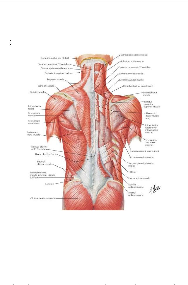
The muscles of the back are divided into the extrinsic muscles that connect the upper limb to the trunk and the intrinsic (deep or true) muscles that specificallyact on the vertebral column to produce movements and maintain posture.
Extrinsic muscles of the back
Superficial: Trapezius, latissimus dorsi, levator scapulae, rhomboid minor and major
Intermediate: Serratus posterior superior and posterior inferior (muscles of respiration)
[Plate 168, Muscles of Back: Superficial Layers]
|
Muscle |
Proximal Attachment (Origin) |
Distal Attachment |
Innervation |
Main Actions |
|
|
|
|
|
(Insertion) |
|
|
|
|
|
Trapezius |
Superior nuchal line, external occipital |
Lateral third of |
Accessorynerve |
Elevates, retracts, and rotates |
|
|
|
|
protuberance, nuchal ligament, and |
clavicle, acromion, |
(cranial nerve XI) and |
scapula; lower fibers depress |
|
|
|
|
spinous processes of C7-T12 |
and spine of |
C3-C4 (proprioception) |
scapula |
|
|
|
|
|
scapula |
|
|
|
|
|
Latissimus |
Spinous processes of T7-T12, |
Humerus |
Thoracodorsal nerve |
Extends, adducts, and |
|
|
|
dorsi |
thoracolumbar fascia, iliac crest, and |
(intertubercular |
(C6-C8) |
mediallyrotates humerus |
|
|
|
|
last 3-4 ribs |
sulcus) |
|
|
|
|
|
Levator |
Transverse processes of C1-C4 |
Medial border of |
C3-C4 and dorsal |
Elevates scapula and tilts |
|
|
|
scapulae |
|
scapula |
scapular (C5) nerve |
glenoid cavityinferiorly |
|
|
|
Rhomboid |
Minor: nuchal ligament and spinous |
Medial border of |
Dorsal scapular nerve |
Retract scapula, rotate it to |
|
|
|
minor and |
processes of C7-T1 |
scapula |
(C4-C5) |
depress glenoid cavity, and fix |
|
|
|
major |
Major: spinous processes of T2-T5 |
|
|
scapula to thoracic wall |
|
|
|
Serratus |
Ligamentum nuchae, spinous |
Superior aspect of |
T1-T4 |
Elevate ribs |
|
|
|
posterior |
processes of C7-T3 |
ribs 2-4 |
|
|
|
|
|
superior |
|
|
|
|
|
|
|
|
|
|
|
|
|
|
|
|
|
|
|
125 / 425 |
||
Serratus |
Spinous processes of T11-L2 |
Inferior aspect of |
T9-T12 |
Depress ribs |
posterior |
|
ribs 9-12 |
|
|
inferior |
|
|
|
|
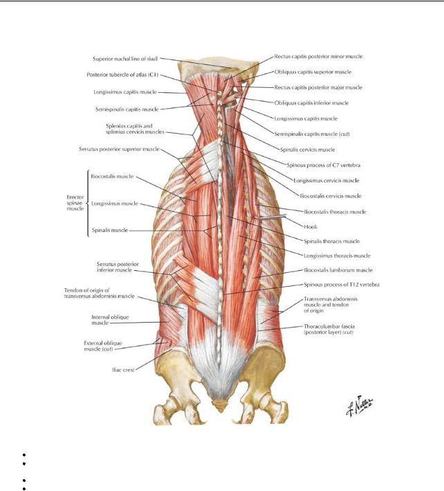
Intrinsic muscles of the back
[Plate 169, Muscles of Back: Intermediate Layers]
Superficial: Splenius (capitus/cervicus)
Intermediate: Erector spinae (sacrospinalis) group-iliocostalis (lumborum/thoracis/cervicis) longissimus (thoracis/cervicis/capitis), spinalis (thoracis/cervicis/capitis)
Deep: Transversospinal group-semispinalis (thoracis/cervicis/capitis), multifidus, rotatores Minor deep: interspinales, intertransversarii, levatores costarum (brevis and longus)
|
Muscle |
Proximal Attachment (Origin) |
Distal Attachment (Insertion) |
Innervation |
Main actions |
|
|
Superficial Layer |
|
|
|
|
|
|
Splenius |
Nuchal ligament, spinous process C7-T3 |
Mastoid process of temporal bone and |
Middle |
Bilaterally: extend |
|
|
capitis |
|
lateral third of superior nuchal line |
cervical |
head |
|
|
|
|
|
nerves |
Unilaterally: |
|
|
|
|
|
|
laterallybend (flex) |
|
|
|
|
|
|
and rotate face to |
|
|
|
|
|
|
same side |
|
|
Splenius |
Spinous process T3-T6 |
Transverse process (C1-C3) |
Lower |
Bilaterally: extend |
|
|
cervicis |
|
|
cervical |
neck |
|
|
|
|
|
nerves |
Unilaterally: |
|
|
|
|
|
|
laterallybend (flex) |
|
|
|
|
|
|
and rotate neck |
|
|
|
|
|
|
toward same side |
|
|
Intermediate Layer |
|
|
|
|
|
|
Erector |
Posterior sacrum, iliac crest, |
Iliocostalis: angles of lower ribs and |
Respective |
Extend and laterally |
|
|
spinae |
sacrospinous ligament, supraspinous |
cervical transverse processes |
spinal |
bend vertebral |
|
|
|
ligament, and spinous processes of lower |
Longissimus: between tubercles and |
nerves of |
column and head |
|
|
|
lumbar and sacral vertebrae |
angles of ribs, transverse processes of |
each |
|
|
|
|
|
|
|
|
|
126 / 425
|
|
thoracic and cervical vertebrae, and |
region |
|
|
|
mastoid process |
|
|
|
|
Spinalis: spinous processes of upper |
|
|
|
|
thoracic and midcervical vertebrae |
|
|
Semispinalis |
Transverse processes C4-T12 |
Spinous processes of cervical and |
Respective |
Extend head, neck, |
|
|
thoracic regions |
spinal |
and thoraxand |
|
|
|
nerves of |
rotate them to |
|
|
|
each |
opposite side |
|
|
|
region |
|
Multifidi |
Sacrum, ilium, and transverse processes |
Spinous processes of vertebrae above, |
Respective |
Stabilizes spine |
|
of T1-T12, and articular processes of C4- |
spanning two to four segments |
spinal |
|
|
C7 |
|
nerves of |
|
|
|
|
each |
|
|
|
|
region |
|
Rotatores |
Transverse processes |
Lamina and transverse process or |
Respective |
Stabilize, extend, |
|
|
spine above, spanning one or two |
spinal |
and rotate spine |
|
|
segments |
nerves of |
|
|
|
|
each |
|
|
|
|
region |
|
Fascia
Encloses deep muscles of the back
Attached mediallyto the nuchal ligament, tips of the spinous processes, supraspinous ligament, and median line of sacrum Attached laterallyto the cervical and lumbar transverse processes
Thickened as the thoracolumbar fascia toward lumbar region and extends between the twelfth rib and the iliac crest
Vascular supply to muscles and skin of the back
Arteries
Cervical: branches from occipital, ascending cervical, vertebral, and deep cervical
Thoracoabdominal: branches of posterior intercostals, subcostal, and lumbar
Pelvic: iliolumbar and lateral sacral branches of the internal iliac
Veins drain via the valveless vertebral venous plexus
Lymph
Neck: Drains to the anterior, lateral, and deep cervical nodes
Trunk: drains to axillarynodes above umbilicus and superior inguinal nodes below it
Suboccipital region
Inferior to the occiput, deep to the trapezius and semispinalis capitis, overlying C1 and C2 Muscles
Rectus capitis posterior minor and major
Obliquus capitis superior and inferior
All laterally flex, extend, and rotate the head
All supplied by the suboccipital nerve (dorsal ramus of C1)
Contains the dorsal rami of C1-C4 Suboccipital triangle
Contains the vertebral artery, suboccipital nerve and suboccipital venous plexus.
Bounded byrectus capitis posterior major, obliquus capitis superior and obliquus capitis inferior, floor-atlantooccipital membrane, roof-semispinalis capitis
Deep Layer |
|
|
|
|
Muscle |
Proximal Attachment |
Distal Attachment |
Innervation |
Main Actions |
|
(Origin) |
(Insertion) |
|
|
Rectus capitis posterior |
Spine of axis |
Lateral inferior nuchal |
Suboccipital nerve |
Extends head and rotates to |
major |
|
line |
(C1) |
same side |
Rectus capitis posterior |
Tubercle of posterior arch |
Median inferior nuchal |
Suboccipital nerve |
Extends head |
minor |
of atlas |
line |
(C1) |
|
Obliquus capitis |
Transverse process of |
Occipital bone |
Suboccipital nerve |
Extend head and bend it laterally |
superior |
atlas |
|
(C1) |
|
Obliquus capitis inferior |
Spine of axis |
Atlas transverse |
Suboccipital nerve |
Rotates atlas to turn face to |
|
|
process |
(C1) |
same side |
Branches of spinal nerves
Ventral rami innervate the muscles and overlying skin of the anterior thoracic, abdominal and pelvic wall and contribute to
Cervical plexus [C1-C4] (see: Head and Neck)
Brachial plexus [C5-T1] (see: Upper Limb)
Thoracic intercostal nerves (see also: Thorax)
Lumbar plexus [T12-L4] (see: Pelvis and Perineum and Lower Limb)
 Sacral plexus [L4-S5] (see: Lower Limb) Dorsal rami
C1: Suboccipital nerve-pierces the atlantooccipital membrane and is motor to the suboccipital muscles C2: Greater occipital nerve-passes inferior to OCI and is sensoryto skin over neck and occipital bone
127 / 425

C3-Co: Segmentallyinnervate the intrinsic muscles of the back and overlying skin
128 / 425