


74 Adverse Drug Effects
Cutaneous Reactions
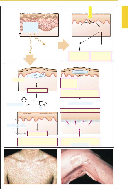
Upon systemic distribution, many drugs evoke skin reactions that are caused on an immunological basis. Moreover, cutaneous injurycan also arise from nonimmunological mechanisms. Cutaneous side effects vary in severity from harmless to lethal. Cutaneous reactions are a common form of drug adverse reaction. Nearly half of themare attributed to antibiotics or sulfonamides, and onethird to nonsteroidal anti-inflammatory agents, with many other pharmaceuticals joining the list.
The following clinical pictures are noted:
Toxic erythema with a maculopapular rash similar to that of measles and scarlet fever (B, left). Urticaria with itchy swellings as part of a Type 1 reaction including anaphylactic shock.
Fixed eruptions (drug exanthemas) with mostly few demarcated, painful lesions, usually located in intertriginous skin regions (genital area, mucous membranes). With repeated exposure, these typically recur at the same sites.
Steven–Johnson syndrome (SJS, erythema multiforme) and toxic epidermal necrolysis (TEN or Lyell syndrome) with apoptosis of keratinocytes and bullous detachment of the epidermis from the dermis. When more than 30% of the body surface is affected, TEN is present. Its course is dramatic and the outcome not
rarely fatal.
The aforementioned reactions are thought to involve the following pathogenetic mechanisms.
With penicillins, opening of the β-lactam bond is possible. The resulting penicilloyl group binds as a hapten to a protein. This may lead to an Ig-E mediated anaphylactic reaction, manifested on the skin as urticaria.
Biotransformation via cytochrome oxidase may yield reactive products. Presumably keratinocytes are capable of such metabolic reactions. In this way, the
para-amino group of sulfonamides can be converted into a hydroxyl amine group, which then acts as a hapten to induce a Type 4 reaction in the skin. Fixed maculopapular lesions are thought to arise on this basis.
Pemphiguslike manifestations with formation of blisters. The development of cutaneous manifestations is not as ominous as in SJS or TEN, the blisters being located intraepidermally. This condition involves the formation of autoantibodies directed against adhesion proteins (desmogelin) of desmosomes, which link keratinocytes to each other. D-Penicillamine and rifampin are inducers of the rare drug-associated pemphigus (p.308).
Photosensitivity reactions result from exposure to sunlight, in particular the UVA component. In phototoxic reactions, drug molecules absorb photic energy and turn into reactive compounds that damage skin cells at their site of production. In photoallergic reactions, photoreaction products bind covalently to proteins as haptens and trigger Type 4 allergic responses. The type and localization are difficult to predict.
Luellmann, Color Atlas of Pharmacology © 2005 Thieme
All rights reserved. Usage subject to terms and conditions of license.
|
|
|
|
|
|
Cutaneous Reactions |
75 |
||
A. Adverse drug effect: cutaneous reaction |
|
|
|
|
|||||
|
|
|
|
|
|
|
Sunlight (UVA) |
|
|
Epidermal |
|
|
|
|
|
|
|
|
|
keratinocytes |
Drug |
|
|
|
|
|
|
|
|
|
|
|
|
|
|
|
|
|
|
|
or |
|
|
|
|
|
Drug |
Metabolite |
|
|
metabolite |
|
|
|
|
||||
Dermis |
|
|
|
|
|
|
|||
|
|
|
|
|
|
|
|
|
|
|
|
|
|
|
Photo- |
|
|
|
|
|
|
|
|
|
sensi- |
Radical formation |
Immune reaction |
|
|
|
|
|
|
|
tization |
|
|||
|
|
|
|
|
|
|
|
|
|
Immune reaction |
|
|
Phototoxicity, |
Photoallergy, |
|
||||
|
|
Sunburn |
Type 4 |
|
|||||
|
|
|
|
|
|
reaction |
reaction |
|
|
Urticaria Edema of upper dermis |
Pemphigus- |
Intraepidermal |
|
||||||
|
|
|
|
|
|
like |
blisters |
|
|
|
|
|
|
|
|
reaction |
|
|
|
|
Type 1 reaction |
Autoantibody against |
|
|
|||||
|
|
|
|
|
|
|
|
||
Penicilloyl |
|
O |
|
|
|
desmosomal adhesion proteins |
|
||
group |
|
|
|
|
|
|
|
|
|
CH2 |
C |
NH |
S |
CH3 |
|
|
|
|
|
|
|
|
|
|
|||||
|
|
|
|
e.g., penicillamine |
|
|
|||
e.g., penicillin |
|
|
O |
N |
CH3 |
|
|
||
|
|
|
|
|
|
||||
|
|
|
H |
|
|
|
|
|
|
|
|
|
Protein COOH |
Stevens–Johnson syndrome, TEN |
|
||||
|
|
|
|
|
|
|
|||
|
Production of metabolites |
Blisters at the epidermis/dermis |
|
||||||
|
|
boundary |
|
|
|||||
|
in keratinocytes |
|
|
|
|
|
|||
e.g., sulfonamide |
|
|
|
|
Apoptosis of keratinocytes |
|
|||
|
|
? |
|
|
|
|
|||
|
|
|
|
|
|
|
|
|
|
? |
Type 4 reaction |
|
|
|
|
||||
|
|
|
|
|
|||||
Maculopapular drug exanthema, |
Cell-mediated immune reaction |
|
|||||||
fixed eruption |
|
|
|
e.g., sulfonamide |
|
|
|||
|
|
|
|
|
|
|
|
||
Drug exanthema |
|
|
|
|
|
Toxic epidermal necrolysis (TEN) |
|
||
Luellmann, Color Atlas of Pharmacology © 2005 Thieme
All rights reserved. Usage subject to terms and conditions of license.

76 Adverse Drug Effects
Drug Toxicity in Pregnancy and Lactation
Drugs taken by the mother can be passed on transplacentally or via breast milk and can adversely affect the unborn or the neonate.
Pregnancy (A). Limb malformations induced by the hypnotic thalidomide (Contergan) first focused attention on the potential of drugs to cause malformations (teratogenicity). Drug effects on the unborn fall into two basic categories:
1.Predictable effects that derive from the known pharmacological drug properties. Examples include masculinization of the female fetus by androgenic hormones; brain hemorrhage due to oral anticoagulants; bradycardia due to β-blockers.
2.Effects that specifically affect the developing organism and that cannot be predicted on the basis of the known pharma-
cological activity profile.
In assessing the risks attending drug use during pregnancy, the following points have to be considered:
aTime of drug use. The possible sequelae of exposure to a drug depend on the stage of fetal development, as shown in (A). Thus, the hazard posed by a drug with a specific action is limited in time, as illustrated by the tetracyclines, which produce effects on teeth and bones only after the third
month of gestation, when mineralization begins.
b Transplacental passage. Most drugs can pass in the placenta from the maternal into the fetal circulation. The syncytiotrophoblast formed by the fusion of cytotrophoblast cells represents the major diffusion barrier. It possesses a higher permeability to drugs than suggested by the term “placental barrier.” Accordingly, all centrally-acting drugs administered to a pregnant woman can easily reach the fetal organism. Relevant examples include antiepileptics, anxiolytics, hypnotics, antidepressants, and neuroleptics.
c Teratogenicity. Statistical risk estimates are available for familiar, frequently used drugs. For many drugs, teratogenic potency cannot be demonstrated; however, in the case of novel drugs it is usually not yet possible to define their teratogenic hazard.
Drugs with established human teratogenicity include derivatives of vitamin A (etretinate, isotretinoic acid [used internally in skin diseases]). A peculiar type of damage results from the synthetic estrogenic agent diethylstilbestrol following its use during pregnancy: daughters of treated mothers have an increased incidence of cervical and vaginal carcinoma at the age of about 20 years. Use of this substance in pregnancy was banned in the United States in 1971.
In assessing the risk–benefit ratio, it is also necessary to consider the benefit for the child resulting from adequate therapeutic treatment of its mother. For instance, therapy with antiepileptic drugs is indispensable, because untreated epilepsy endangers the unborn child at least as much as does administration of anticonvulsants.
Drug withdrawal reactions are liable to occur in neonates whose mothers are ingesting drugs of abuse or antidepressants of the SSRI type (p.228).
Lactation (B). Drugs present in the maternal organism can be secreted in breast milk and thus be ingested by the infant. Evaluation of risksshould be based on factorslisted in B. In case of doubt, potential danger to the infant can be averted only by weaning.
Luellmann, Color Atlas of Pharmacology © 2005 Thieme
All rights reserved. Usage subject to terms and conditions of license.

Drug Toxicity in Pregnancy and Lactation |
77 |
A. Pregnancy: fetal damage due to drugs |
|
|
|
|||
Ovum |
1 day |
|
|
|
|
|
Sperm |
~3 days |
|
|
|
|
|
cells |
|
|
|
|
|
|
Endometrium |
|
|
|
|
|
|
|
Blastocyst |
|
|
|
|
|
Age of fetus |
1 |
21 2 |
|
12 |
38 |
|
(weeks) |
|
|
||||
|
|
|
|
|
|
|
Development |
Nidation |
Embryo: |
organ |
Fetus: |
growth |
|
stage |
|
|
|
develop- |
|
and |
|
|
|
|
ment |
|
maturation |
|
|
Fetal death |
Malformation |
Functional disturbances |
||
|
|
|
Artery |
|
Uterus wall |
Vein |
Sequelae |
|
|
|
|
|
Mother |
of |
|
|
|
|
|
|
damage |
|
|
|
|
|
|
by drug |
|
|
Transfer of |
|
|
Syncytio- |
|
|
|
|
|
||
|
|
|
metabolites |
|
|
trophoblast |
|
|
|
Capillary |
|
|
“Placental |
|
|
|
|
|
barrier” |
|
|
|
|
|
|
|
|
Placental transfer of metabolites |
|
|
|
Intervillous space |
||
|
Fetus |
To umbilical cord |
||||
|
|
|
|
|||
B. Lactation: maternal intake of drugs |
|
|
|
|
||
Drug |
|
|
Extent of |
Distribution |
||
|
|
|
transfer of |
of drug |
||
|
|
|
drug into |
in infant |
||
|
|
|
milk |
|
|
|
Therapeutic |
|
|
Infant dose |
|
|
|
effect in |
|
|
|
|
Rate of |
|
mother |
|
|
|
|
elimination |
|
|
|
|
|
|
of drug |
|
? |
|
|
|
|
from infant |
|
|
|
|
|
|
|
|
Drug concentration in infant’s blood
Unwanted |
|
|
effect |
|
|
in child |
Sensitivity of site of action |
Effect |
Luellmann, Color Atlas of Pharmacology © 2005 Thieme
All rights reserved. Usage subject to terms and conditions of license.