

charcterristic of phiologyical type of occlussion of different type of mallolusion wiki
Occlusion: An Orthodontic Perspective By Paul M. Kasrovi, DDS, MS; Michael Meyer, DDS; Gerald D. Nelson, DDS Copyright 2000 Journal of the California Dental Association.
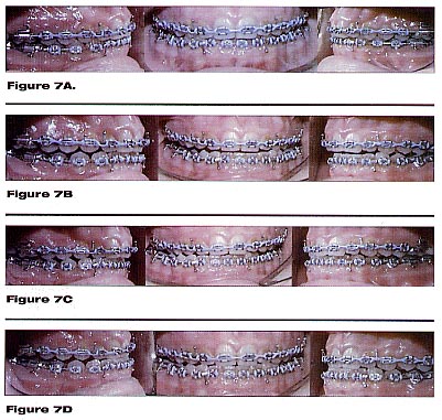
Edward H. Angle, the father of modern orthodontics, is credited with the definition of normal occlusion as well as classification of malocclusions.1 According to Dr. Angle, normal occlusion exists when the mesiobuccal cusp of the upper first molar occludes with the buccal groove of the lower first molar. This definition of occlusion, based on a static position of the teeth in closure, launched orthodontics as the specialty dedicated to treating deviations from Angle’s definition of normal occlusion.2 B.B. McCullum, the father of modern occlusion, and the founder of the Gnathological Society, introduced a more dynamic concept of occlusion.3 His concept of occlusion, along with that of Stallard, Stuart, Huffman, and Regenos, focused on cusp contact during functional movements.4-6 The dynamic concept was readily adopted by prosthodontists and restorative dentists whose challenge was to restore teeth to ideal anatomic form. The dynamic concept of occlusion provided practical guidelines for building cusp heights and placing grooves to facilitate functional jaw movements without functional interferences. There is more acceptance than ever for a dynamic concept of occlusion; however, the concept remains controversial. There is no scientific evidence to show whether such a concept is valid. In such circumstances, one must be guided by clinical judgment. It seems prudent when planning and executing orthodontic treatment to include a consideration of mandibular movements. Producing fine orthodontic results is a major technical challenge, and producing an occlusion that coordinates with temporomandibular joint function increases the complexity. The introduction of pre-adjusted orthodontic brackets, new wires with remarkable range of motion and resiliency, and the diagnostic techniques to relate occlusion to skeletal landmarks and mandibular movements have increased the sophistication of treatment goals and the ability to achieve them. Cusp-Fossa vs. Cusp-Embrasure Occlusion Charles Stuart5 stated that there was a mesial and distal limit to a well-functioning occlusion. The mesial limit was referred to as a "tooth-to-tooth" and the distal limit was referred to as the "tooth-to-two-teeth" occlusion. Huffman and Regenos6 later summarized the two types of occlusion described by Stuart, referring to them as "cusp-fossa" and "cusp-embrasure" occlusion. In a cusp-fossa occlusion, as also advocated by Thomas,7 each mandibular buccal cusp occludes into the fossa of the maxillary counterpart and each maxillary lingual cusp occludes into the fossa of the mandibular counterpart (Figure 1). This occlusal scheme, which places maxillary teeth slightly mesial of Angle Class I relationship, very effectively directs forces along the long axis of teeth, an arrangement that encourages positional stability. Furthermore, food impaction between teeth is minimized since no cusp tips strike opposing embrasures, and there is less tendency for wear of cusp tips and occlusal attrition. In a cusp-embrasure occlusal scheme, all teeth, with the exception of mandibular central incisors and maxillary third molars, have a tooth-to-two teeth relationship (Figure 2). Each maxillary tooth is distal and facial to the mandibular counterpart (Figure 3). Mandibular buccal cusps contact maxillary embrasures. Maxillary lingual cusps are in a fossa relationship with the mandibular counterpart (except for the distolingual cusps of the maxillary first and second molars). It is useful that there is a mesiodistal range for a nicely functioning occlusion. Anatomic considerations of teeth (e.g., tooth size discrepancies, atypical teeth, and over-retained primary teeth in place of congenitally missing teeth), skeletal disharmonies, and atypical extraction patterns may favor one occlusal scheme (Figure 4). The terms distal-normal, mesial-normal, and median-normal (an occlusal relationship midway between the other two) have been used to describe a satisfactory finished orthodontic occlusion.8 Current orthodontic goals of incisor overlap and overjet and of interincisal angle lead the orthodontist to prefer cusp-embrasure as the goal. This scheme provides for freedom of movement in lateral and protrusive excursions with protective cuspid and incisor function. Six Keys to Ideal Static Occlusion As discussed above, from a static occlusal standpoint, Stuart’s distal limit (tooth-to-two teeth), also referred to as the Angle Class I occlusion, is the typical goal in orthodontic treatment. In a 1972 landmark article, Andrews9 described six significant characteristics in his study of 120 cases of non-orthodontic normal occlusions that have since become additional goals for a well-finished orthodontic case. It is important to note that the subjects in Andrews’ study all had naturally straight teeth with pleasing smiles; their casts occluded in a generally ideal position, and they had never received any orthodontic treatment. The six characteristics, which are referred to as the six keys to normal occlusion, are as follows: * The first key: molar relationship. The distobuccal cusp of the upper first molar contacts the mesiobuccal cusp of the lower second molar. The mesiobuccal cusp of the upper first molar falls in the groove between the mesiobuccal and middle-buccal cusps of the lower first molar. The proper relationship allows the cuspids and bicuspids to enjoy a cusp-embrasure relationship buccally and a cusp-fossa relationship lingually (Figure 5A). * The second key: crown angulation (tip). The gingival portion of the crowns of all the teeth is more distal than the incisal or occlusal portion of the crowns. The long axis of all teeth, except molars, is the mid-developmental ridge of the facial surfaces of the teeth. The long axis of the molar is considered to be the buccal groove and its extension to the gingival (Figure 5B). * The third key: crown inclination (torque). This refers to labiolingual inclination of the anterior teeth or buccolingual inclination of the posterior teeth. The upper incisors have a labial crown inclination (positive torque). The lower incisors have a slight lingual inclination (negative torque). The upper cuspids and posterior teeth have lingual crown inclination (negative torque). The inclination is similar for upper cuspids and bicuspids and slightly more pronounced in the molars. Lingual crown inclination (negative torque) also exists in the lower cuspids and posterior teeth and progressively increases from the cuspids through the second molars (Figure 5C). * The fourth key: no rotations. Rotated teeth occupy more space within the dental arch (Figure 5D). * The fifth key: no spaces. All contact points are tight (Figure 5E). * The sixth key: the occlusal plane. The occlusal plane may vary from generally flat to a slight curve of Spee (Figure 5F). These important findings gave rise to pre-adjusted orthodontic brackets, incorporating specific built-in adjustments that produce ideal tip, torque, in/out, and rotational tooth positions, significantly reducing the need for archwire adjustments throughout the course of orthodontic treatment. Goals of Ideal Dynamic Occlusion Dynamic occlusion requires that the teeth function during jaw movements free from premature contacts and interferences. To achieve this, one must pay particular attention to static tooth positions, to condylar position upon closure into occlusion, and to mandibular border movements as determined by the temporomandibular joints. Roth10 has demonstrated that if the orthodontist is able to finish cases to Andrew’s six keys and establish maximum intercuspation in harmony with centric relation, then Stuart’s goals of ideal functional occlusion can be achieved. Although still not universally accepted and practiced by all orthodontists, many of these functional occlusion principles have received significant recognition in the specialty. Essential to the stability of an ideal occlusion is the relationship between centric relation (CR) and maximum intercuspation (MI). Centric relation will permit seating of the condyles into the glenoid fossa against the articular discs at the most superior position against the eminentia and also centered in the transverse plane of space (Figure 6). In an ideal orthodontic finish, this condylar position should occur when upper and lower teeth are closed into MI. It has been shown that the displacement of the condyles from the above position caused by intercusping of teeth (i.e., CR-MI discrepancy) should ideally be less than 1 mm vertically and horizontally and less than 0.5 mm transversely.11,12 To achieve optimal functional occlusion, the teeth should be positioned so that they create what has been termed a mutually protected occlusal scheme. In this scheme, posterior teeth protect anterior teeth from lateral stress at full closure; and the anterior teeth protect posterior teeth from lateral stress during mandibular movements, as long as the condyles are permitted a gliding movement along the eminentia. In MI, there are simultaneous occlusal contacts around centric cusps of posterior teeth directing the closure stress along their long axes. In addition, there is no actual contact of the anterior teeth in MI, but a slight clearance detectable by a 0.0005-inch thickness shimstock. Upon movement of the mandible in any direction from full closure (left and right lateral excursions and straight protrusion), the anterior teeth and canines provide a gentle guide ramp that allows the condyles to traverse the eminentia, and disclude the posterior teeth on the working and balancing sides (Figures 7A through D). The condylar guidance expressed by the morphology of the joints is the major determinant of the overbite-overjet relationship of anterior teeth and canines, and the major determinant of posterior tooth positioning and archform.11 Generally speaking, when teeth are in MI with the condyles seated in CR, there must be 4 mm of vertical overbite and 2 to 3 mm of overjet from the incisal edges of the maxillary incisors to the facial surfaces of the mandibular incisors and 1 mm of overjet from the tip of the maxillary canine to the facial surface of the mandibular canine. Mounted Models in Diagnosis and Treatment Planning For diagnosis and treatment planning, orthodontists have traditionally relied on hand-held study casts trimmed with an MI interocclusal wax registration (Figure 8). Many orthodontists analyze the functional status of the patient’s occlusion as part of their diagnosis and treatment plan. To that end, study casts are mounted on a semi-adjustable articulator with a CR interocclusal wax registration. To produce such a wax registration, the operator must guide the mandible into centric relation by seating the condyles in a superior-anterior direction within the glenoid fossa (Figure 6). This is accomplished by supporting the angles of the mandible in a superior direction while pushing gently down on the chin and asking the patient to relax and close slowly (Figure 9A). Although different CR interocclusal wax registration techniques have been proposed, the two-piece "power centric" technique has shown merit in seating the condyles as close to CR as possible (Figure 9B).12-14 The term power centric refers to the use of the patient’s closure muscles (masseter, medial pterygoid, and the superior head of the lateral pterygoid) to seat the condyles in the desired direction.11 Using centric relation mounted study casts (Figure 9C), one can evaluate the following aspects of the dental-orthopedic complex: * Relate the position of the maxilla to the cranial base; * Relate the mandible to the maxilla with the condyles seated in centric relation; * Plan treatment to a reproducible condylar position, allowing examination of excursive mandibular movements; and * Detect the point of initial tooth contact, unmasking occlusal interferences and evaluating the cant of the occlusal plane.15 The degree of discrepancy between CR and MI can not only be qualitatively assessed, but also quantitatively measured in three dimensions of space using additional instrumentation. The condylar position indicator and mandibular position indicator are instruments used with the Panodent and SAM articulators, respectively. They are designed to record the position of the condylar axis and measure CR/MI discrepancy in three planes of space and have been demonstrated to be both accurate and reliable (Figures 10A and B).16-19 One can bring this information to the lateral cephalogram, which is typically taken in MI, and correct the mandibular position to CR on the tracing of the cephalogram prior to cephalometric analysis (Figure 10C). This, in addition to the CR mounted models, will allow the orthodontist to develop a diagnosis and treatment plan while accounting for centric relation. Mounted study casts are only as accurate as the interocclusal and facebow record that was used to construct the mounting. The accuracy of the interocclusal record (wax bite registration) is only as good as the ability of the practitioner in registering the patient’s bite. The possibility of correctly registering that bite is directly dependent on whether the patient’s muscles of mastication are relaxed (i.e., without spasm or splinting), and the condyle is balanced or stable, without strain or stress within the articular fossa.20 It is critical to identify the patient’s proper CR before any orthodontic treatment is initiated, rather than see it surface halfway through the treatment. As the position of teeth is altered with orthodontic appliances, the patient’s MI adapts continuously, and the only reliable reference point is centric relation. There are, however, times when capturing a reliable CR record on a patient is unlikely, due to muscular interventions. In such cases, a period of splint (orthotic) therapy, prior to active orthodontic treatment, can deprogram the patient’s muscular and proprioceptive influences, allowing uninhibited condylar seating (Figure 11). Developing Malocclusions: Six Warning Signs in a 7-Year-Old Most referrals for orthodontic treatment are occlusion-related.21 Many warning signs of developing malocclusions appear in the early mixed dentition. Ideally, children should be screened for orthodontic treatment no later than age 7, when the posterior occlusion has been established by the eruption of the six-year molars. This allows the orthodontist to evaluate anteroposterior and transverse relationships of the occlusion, as well as any functional mandibular shifts. The presence of permanent maxillary and mandibular central and lateral incisors reveals arch length discrepancies, habit patterns, and vertical dimension problems such as deep bite, open bite, or gummy smile. Most facial asymmetries appear by age 7. To raise the awareness of the public and establish an easy-to-follow set of guidelines for general practitioners, the California Association of Orthodontists has introduced a brochure called "Bite Down Early" that outlines the six warning signs in 7-year-olds (this brochure may be ordered by calling the California Association of Orthodontists at [415] 441-2416). 22 Six Warning Signs * Is there excessive horizontal overlap (overjet)? Protrusion of the upper front teeth (Figure 12A). * Is there excessive vertical overlap (deep bite)? Lower incisors near the palatal tissue (Figure 12B). * Is there a crossbite of the anterior or posterior teeth? The upper teeth fit inside the lower teeth (Figures 12C and D). * Is there an open bite? The child can stick his/her tongue between the upper and lower front teeth when back teeth are together. This may indicate an underlying habit such as tongue thrust, finger sucking or mouth breathing due to respiratory dysfunctions or blocked airways (enlarged tonsils and adenoids) (Figure 12E). * Is there spacing/crowding between the teeth? Teeth are overlapped and rotated or there are noticeably large gaps between them (Figures 12F and G). * Do the upper and lower midlines coincide? The maxillary and mandibular midlines should be coincident and should both also align with the facial midline (mid-sagittal plane). If the midlines do not coincide, there may be many causes, including impacted teeth, functional mandibular shift, uneven mandibular growth, and midface asymmetries (Figures 12H and I). Close attention to these early warning signs and timely referral to the orthodontist can prevent more complicated later treatments for the patient. Many unfavorable growth patterns may be altered if early treatment is rendered with growth-modification appliances. Arch development with expansion devices can be provided in the early mixed dentition to correct crossbites and relieve crowded arches and in some cases prevent extraction of permanent teeth. Elimination of habits is also significantly more successful and stable if addressed in the early mixed dentition. Referral for an orthodontic screening at age 7 does not always result in immediate treatment but allows the orthodontist to determine how and when a child’s particular problem should be treated for maximum improvement with the least amount of time and expense. Many orthodontic problems will best await treatment until the late mixed dentition or permanent dentition. Orthodontic Treatment and Temporomandibular Disorders Does orthodontic treatment cause temporomandibular dysfunction (TMD)? Is occlusion an etiologic factor in signs and symptoms of TMD? Do occlusal changes created by orthodontic treatment predispose an individual to increased risk for TMD? Several studies have been conducted to answer these questions, but there is still a great deal of controversy. Many studies conducted have suffered from flaws in research technique. This combined with the complexity and the multifactorial nature of TMD has contributed to some uncertainty. One particular shortcoming is the lack of quantitative information at the condylar level. Most measurement criteria have been based on changes at the occlusal level, either intraorally or with hand-held study casts, or on tomographic assessment of the condyles. Researchers have looked at the tooth end of the mandible and have examined anatomical factors such as molar classification, overjet, overbite, crowding and subjective clinical inspection to determine a correlation with TMD signs and symptoms. Such occlusal characteristics are poor indicators of condylar position.23-25Tomography has been shown to be unreliable in evaluating condylar position.26,27 In most studies that have shown a lack of correlation between occlusion and TMD, no instrumentation has been used to evaluate condylar position as dictated by the occlusion. Conclusions have been based on chin point manipulation, intraoral inspection and hand-held study casts. In a recent study,28Crawford showed a reduction in TMD signs and symptoms using a mutually protected occlusal scheme with CR and MI relatively coincidental as measured by condylar position indicator readings. A review of the literature reveals many opinions on these topics. Most reports suggest the lack of any correlation between occlusion, orthodontic treatment, and TMD. Signs and symptoms of TMD after orthodontic treatment were found to occur in some cases, but no connection was observed between TMJ-related functional disturbances and a well-planned and well-executed orthodontic therapy.29 Several long-term research studies have indicated that orthodontics is not a cause of TMD.30-32 Conversely, no data exist to support the notion that orthodontic treatment of children or adults prevents or lowers the risk of subsequently developing TMD.33 Further, studies have found that orthodontic treatment does not appear to pose an increased risk for development of TMJ sounds or symptoms, regardless of whether extraction or non-extraction treatment has been rendered.34 Orthodontic patients are not more likely to develop TMD signs and symptoms while undergoing treatment.35 Post-orthodontic patients have been shown to have no more signs and symptoms of TMD than those with untreated malocclusion or those with normal occlusion.33Studies have failed to connect a period of orthodontic treatment to either the onset of TMD or a change in TMD signs and symptoms.36 If TMD arises during orthodontic treatment, observation and palliative care should precede irreversible treatment approaches. The onset of pain during treatment will lead the orthodontist to modify active therapy, reduce forces, stop headgear wear, eliminate distalizing forces, minimize or eliminate interarch elastics use, or eliminate gross occlusal interferences by occlusal coverage splint therapy.33 Patients who present with TMD signs and symptoms, a significant CR/MI shift, or with severe postural habits, may benefit from a period of behavior modification, physical therapy, and even CR-positioning splint therapy before any active orthodontic treatment begins. The Importance of Occlusion in Orthodontic Treatment Can the orthodontist achieve good functional occlusion routinely with orthodontic treatment? Yes, in most cases. Is it easy to achieve? No. Striving for excellence in treating each case to functional ideals is challenging. The goals are excellent functional occlusion, stable tooth position, periodontal health, and a beautiful smile with balanced facial features, but they are not always possible to achieve. Do ideal occlusion goals apply to the adult patient? Yes, but treatment planning for the adult patient is more complicated and requires input from the patient’s dentist and perhaps other specialists. Orthodontists view freeway space in the growing patient as an important ally, allowing changes in the dentition not possible in the adult. The lack of jaw growth in adults means that goals requiring a change in jaw shape or size must involve orthognathic surgery. Adolescents are quite resistant to periodontal damage in the face of patient neglect. Adults are not. However, with careful interdisciplinary planning, remarkable improvements are possible for the adult patient, and ideal occlusion goals need not be set aside. Although there are conflicting views on the role of malocclusion as a potential risk factor for TMD,32,37,38 there are other reasons for orthodontists to be concerned with functional occlusion. Collision of cusps, if allowed to occur, will cause trauma with potential sequela of pulpitis, tooth mobility, attrition, and periodontal breakdown.39 Muscles can fatigue if the neuromuscular protection mechanism must restrict mandibular movement to avoid cusp collision. Such working and/or balancing interferences during mandibular border movements are also major culprits for significant deviation of MI from CR. Orthodontists want to detect such interferences and eliminate them during orthodontic therapy. Is an ideal functional orthodontic result stable? If only that were so. Patients have to be told that teeth shift throughout their lives, whether or not there has been orthodontic treatment, and that the changes are typically inconsequential. Orthodontic treatment cannot prevent these changes. Orthodontists try to evaluate the stability of the cases after treatment and make decisions about which patients should indefinitely continue with some sort of retention devices. Conclusion As technologic advances in orthodontic therapy have evolved, orthodontists have widened their diagnostic criteria and treatment goals to include an accurate determination of mandibular position and function. The treatment goal of an ideal static occlusion remains important but is now considered only part of a broader view of an ideal final result. An ideal final result will also include the functional criteria of mutually protected occlusion with centric relation closely coincident with maximum intercuspation. Ideal final results are not always possible. Orthodontic screening by age 7 allows good planning for ideal correction of a malocclusion. Correction of many orthodontic problems in the developing or adolescent dentition is preferable to waiting until the adult dentition. Treatment during this younger period improves the chance of achieving excellent results and better post-treatment stability. In adults, lack of growth and periodontal risk present the orthodontist some diagnostic challenges, but ideal functional occlusion remains a workable goal. The relationship between occlusion and TMD is still unclear. Research has not established a connection, but reliable analysis of condylar position has not been addressed in such research. Excellent static occlusal goals and functional goals are now understood to be critical elements in long-term stability of orthodontic treatment. Authors Paul M. Kasrovi, DDS, MS, is an assistant clinical professor in the Division of Orthodontics at the University of California at San Francisco. Michael Meyer, DDS, is an associate clinical professor in the Division of Orthodontics at UCSF School of Dentistry. Gerald D. Nelson, DDS, is an associate clinical professor in the Division of Orthodontics at UCSF and a diplomate of the American Board of Orthodontics. References 1. Angle EH, Classification of malocclusion. D Cosmos 41:248, 1899. 2. Aronowitz HI, Orthodontics and occlusion: a historical perspective. J Cal Dent Assoc 24(10):16-8, 1996. 3. McCollum BB and Stuart CE, A research report. California Scientific Press, South Pasadena, 1955. 4. Stallard H and Stuart CE, Concepts of occlusion. Dent Clin N Am 1963:591-606. 5. Stuart CE, Good occlusion for natural teeth. J Prosth Dent 14:316-24,1964. 6. Huffman RW, Regenos JW, Principles of Occlusion, Laboratory and Clinical Teaching Manual. Department of Operative Dentistry, Ohio State University, 1973. 7. Thomas PK, Syllabus on full mouth waxing technique for rehabilitation: tooth-to-tooth cusp fossa concept of organic occlusion, 2nd ed. Instant printing services, 1967, pp. 1-10. 8. Moore T, Gnathology and orthodontics. Unpublished thesis, Angle Society Of Northern California. 9. Andrews LF, The six keys to normal occlusion. Am J Orthod 62:296-309, 1972. 10. Roth RH, Functional occlusion for the orthodontist. J Clin Orthod part 1, 15:32-51; part 2, 15:100-23, part 3, 15:174-98; part 4, 15:246-65, 1981. 11. Roth RH, The Roth functional occlusion approach to orthodontics, Roth/Williams Center for Functional Occlusion. Course syllabus, 1999. 12. Williamson EH, Steinke RM, et al, Centric relation: A comparison of muscle-determined position and operator guidance. Am J Orthod 77(2):133-45, 1980. 13. Wood DP, Floreani KJ, et al, The effect of incisal bite force on condylar seating. Angle Orthod 64(1):53-62, 1994. 14. Lundeen HC, Centric relation records: The effect of muscle action. J Prosthet Dent 31(3):244-53, 1974. 15. Kasrovi PM, Mounted models, tomorrow’s technology? Pacific Coast Society of Orthodontists Bulletin, pp 43-44, Fall 1999. 16. Slavicek, R, Part 4 instrumental analysis of mandibular casts using the mandibular position indicator. J Clin Orthod 22:566-575, 1988. 17. Utt TW, Meyers CE, Wierzba TF, A three-dimensional comparison of condylar position changes between centric relation and centric occlusion using the mandibular position indicator. Am J Orthod Dentofacial Orthop 107:298-308, 1995. 18. Wood DP, Korne PH, Estimated and true hinge axis: a comparison of condylar displacements. Angle Orthod 62(3):167-75, 1992. 19. Lavine DS, Kulbersh R, Bonner PT, Reliability of the condylar position indicator. University of Detroit, In press. 20. Lundeen HC. Centric relation records: The effect of muscle action. J Prosthet Dent 31(3):244-253, 1974. 21. Nurminen L, Pietilea T, Motivation for and satisfaction with orthodontic-surgical treatment: a retrospective study of 28 patients. Eur J Orthodont 21(1):79-87, 1999 22. Bite down early: A parent’s guide to detecting bite problems. Brochure by the California State Society Of Orthodontists, 1993. 23. Lundeen HC, Gibbs CH, Advances in Occlusion. John Wright PSG, Boston, 1982, pp 2-32. 24. Shildkraut M, Wood DP, Hunter WS, The CR-CO discrepancy and its effects on cephalometric measurements. Angle Orthod 64(5):333-42, 1994. 25. Wood DP, Elliot RW, Reproducibility of the centric relation bite registration technique. Angle Orthod 64(3):211-20, 1994. 26. Hatcher DC, Blom RJ, Baker CG, Temporomandibular joint spatial relationships: Osseous and soft tissues. J Prosthet Dent 56(3):344-53, 1986. 27. Roth RH, Occlusion and condylar position. Am J Orthod Dentofac Orthop 107(3):315-8, 1995. 28. Crawford SD, Condylar axis position, as determined by the occlusion and measured by the CPI instrument, and signs and symptoms of temporomandibular dysfunction. Angle Orthod69(2):103-15, 1999. 29. Keb K, Bakopulos K, Witt E, TMJ function with or without orthodontic treatment. Eur J Orthod 13:192-6, 1991. 30. Baker RW, Catania JA, Baker RW, Occlusion as it relates to TMJ: A study of the literature. NY State Dent J 57:36-9, 1991. 31. Sadowsky C, Poulson AM, TMD and functional occlusion after orthodontic treatment: Results of two long-term studies. Am J Orthod 86:386-90, 1984. 32. Sadowsky C, BeGole E, Long-term status of TMJ function and functional occlusion after orthodontic treatment. Am J Orthod 78:201-12, 1980. 33. Greene CS, Orthodontics, orthodontists and TMD. Summarized by Crouch DL. Pacific Coast Society of Orthodontists Bulletin, Winter 1990, 33-5. 34. Sadowsky C, Theisen TA, Sakols EI, Orthodontic treatment and TMJ sounds -- A longitudinal study. Am J Ortho Dentofacial Orthop 99:441-7, 1991. 35. Hirata RH, Heft NW, Hernandez B, Longitudinal study of signs of TMD in orthodontically treated and nontreated groups. Am J Orthop Dentofacial Orthop 101:35-40, 1992. 36. Rendell JK, Norton LA, Gay T, Orthodontic treatment and temporomandibular joint disorders. Am J Orthod Dentofacial Orthop 101:84-7, 1992. 37. Righellis EG, Commentary: Condylar axis position and temporomandibular dysfunction, Angle Orthod 69(2):115-6, 1999. 38. McNamara JA, Okeson SA, Occlusion, orthodontic treatment, temporomandibular joint disorders: a review. J Orofacial Pain 9:73-90, 1995. 39. Burgett FG, Trauma from occlusion, periodontal concerns. Dent Clin N Am 39(2):301-11, 1995. To requested a printed copy of this article, please contact: Paul M. Kasrovi, DDS, MS, 8201 Edgewater Drive, Suite 101, Oakland, CA 94621. Legends
Figure 1. Cusp-fossa (tooth-to-tooth) occlusion (From McNeill C, ed, Science and Practice of Occlusion. Quintessence Publishing, Chicago, 1997, p 408).
Figure 2. Cusp embrasure occlusion (tooth-to-two-teeth).
Figure 3. Cusp-fossa (above) vs. cusp-embrasure occlusion (From McNeill C, ed, Science and Practice of Occlusion. Quintessence Publishing, Chicago, 1997, page 315).
Figure 4. A finished orthodontic case, which required extraction of upper bicuspids and a lower incisor, presents with an atypical, yet functionally acceptable, occlusion.
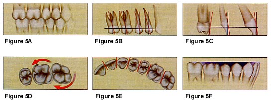
Figure 5A. The first key: molar relationship (courtesy of GAC International). Figure 5B. The second key: crown angulation (tip). Figure 5C. The third key: crown inclination (torque). Figure 5D. The fourth key: no rotations. Figure 5E. The fifth key: no spaces. Figure 5F. The sixth key: the occlusal plane.
Figure 6. Centric relation allows seating of the condyles superiorly and anteriorly within the glenoid fossa (From McNeill C, ed, Science and Practice of Occlusion. Quintessence Publishing, Chicago, 1997, Page 507).
Figure 7A. Maximum intercuspation. Figure 7B. Left working, right balancing. Figure 7C. Right working, left balancing. Figure 7D. Left and right protrusive.
Figure 8. Hand-held study casts.
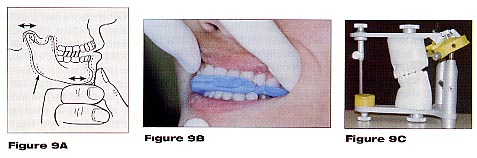
Figure 9A. Proper manipulation of the mandible to seat the condyles in a superior-anterior direction (From McNeill C, ed, Science and Practice of Occlusion. Quintessence Publishing, Chicago, 1997, Page 507). Figure 9B. Taking a "power centric" relation wax registration. Figure 9C. Study casts mounted in centric relation.
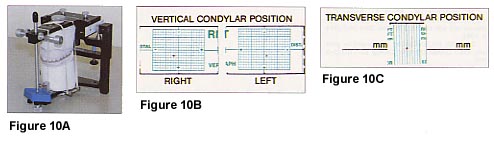
Figure 10A. Determination of CR-MI discrepancy using a condylar position indicator (CPI). Figure 10B. For this patient, the measurements indicate that when in maximum intercuspation, the right condyle is displaced 2 mm downward and 2 mm backward and the left condyle is displaced 2.5 mm downward and 1 mm backward from their respective centric relation positions. There is very minimal transverse shift between CR and MI. Figure 10C. CR converted lateral cephalogram tracing based on the CPI measurements.
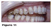
Figure 11. Full arch hard plastic CR repositioning splint (orthotic).
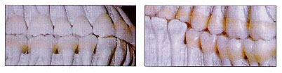
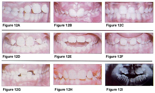
Figure 12A. Excess overjet. Figure 12B. Deep bite. Figure 12C. Anterior crossbite Figure 12D. Posterior crossbite Figure 12E. Anterior open bite Figure 12F. Dental spacing Figure 12G. Crowding Figure 12H. Midline displacement Figure 12I. Midline displacement causes blocked canine
|


![]()
JOURNAL OF THE CALIFORNIA DENTAL ASSOCIATION ©2000 CALIFORNIA DENTAL ASSOCIATION
Occlusal trauma
From Wikipedia, the free encyclopedia
Occlusal trauma is a dental term that refers to the damage incurred when teeth are left in traumatic occlusion without proper treatment.[1]
When the maxillary and mandibular dental arches approach each together, as they do, for example, during chewing or at rest, the relationship between the opposing teeth is referred to as occlusion. If this occlusal relationship is not balanced properly it may result in pain, tenderness and even mobility of the affected teeth.[1]
When the natural course of trauma, disease and dental treatment alters an individual's occlusion by removing or changing the occlusal (biting) surface of any of the teeth, that individual's teeth will come together, or occlude, differently, and their occlusion will change.[2] When that change is detrimental to the manner in which the teeth occlude, the patient is said to possess a traumatic occlusion.[3] Traumatic occlusion may cause a thickening of the cervical margin of the alveolar bone[4] and widening of the periodontal ligament, although the latter is not pathognomonic for this condition.[5]
Contents [hide]
|
[edit]Histologic features associated with occlusal trauma
Microscopically, there will be a number of features that accompany occlusal trauma[6]:
Hemorrhage
Necrosis
Widening of the periodontal ligament, or PDL (also serves as a very common radiographic feature)
Bone resorption
Cementum loss and tears
It was concluded that widening of the periodontal ligament was a "functional adaptation to changes in functional requirements".[7]
[edit]Clinical signs and symptoms associated with occlusal trauma
Clinically, there are a number of physiologic results that serve as evidence of occlusal trauma[8]:
Tooth mobility
Fremitus
Tooth migration
Pain
Wear facets
[edit]Primary vs. secondary occlusal trauma
There are two types of occlusal trauma, primary and secondary.
[edit]Primary occlusal trauma
Primary occlusal trauma occurs when greater than normal occlusal forces are placed on teeth, as in the case of parafunctional habits, such as bruxism or various chewing or biting habits, including but not limited to those involving fingernails and pencils or pens.
The associated excessive forces can be grouped into three categories. Excesses of[9]:
Duration
Frequency and
Magnitude
Primary occlusal trauma will occur when there is a normal periodontal attachment apparatus and, thus, no periodontal disease.[10]
[Edit]Secondary occlusal trauma
![]()
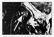
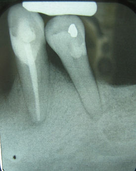
An example of secondary occlusal trauma. This X-ray film displays two lone-standing mandibular teeth, #21 and #22, or the lower left first premolar and canine. As the remnants of a once full complement of 16 lower teeth, these two teeth have been alone in opposing the forces associated with mastication for some time, as can be evidenced by the widened PDL surrounding the premolar Because this trauma is occurring on teeth that have 30-50% bone loss, this would be classified assecondary oclcusal trauma.
Secondary occlusal trauma occurs when normal occlusal forces are placed on teeth with compromised periodontal attachment, thus contributing harm to an already damaged system. As stated, secondary occlusal trauma occurs when there is a compromised periodontal attachment and, thus, a pre-existing periodontal condition.[10]