


30 Distribution in the Body
Binding to Plasma Proteins
Having entered the blood, drugs may bind to the protein molecules that are present in abundance, resulting in the formation of drug–protein complexes.
Protein binding involves primarily albumin and, to a lesser extent, β-globulins and acidic glycoproteins. Other plasma proteins (e.g., transcortin, transferrin, thyroxin-bind- ing globulin) serve specialized functions in connection with specific substances. The degree of binding is governed by the concentration of the reactants and the af nity of a drug for a givenprotein. Albumin concentration in plasma amounts to 4.6 g/100 ml, or 0.6 mM, and thus provides a very high binding capacity (two sites per molecule). As a rule, drugs exhibit much lower af nity (KD ~ 10-5–10-3 M) for plasma proteins than for their specific binding sites (receptors). In the range of therapeutically relevant concentrations, protein binding of most drugs increases linearly with concentration (exceptions: salicylate and certain sulfonamides).
The albumin molecule has different binding sites for anionic and cationic ligands, but van der Waals forces also contribute (p.58). The extent of binding correlates with drug hydrophobicity (repulsion of drug by water).
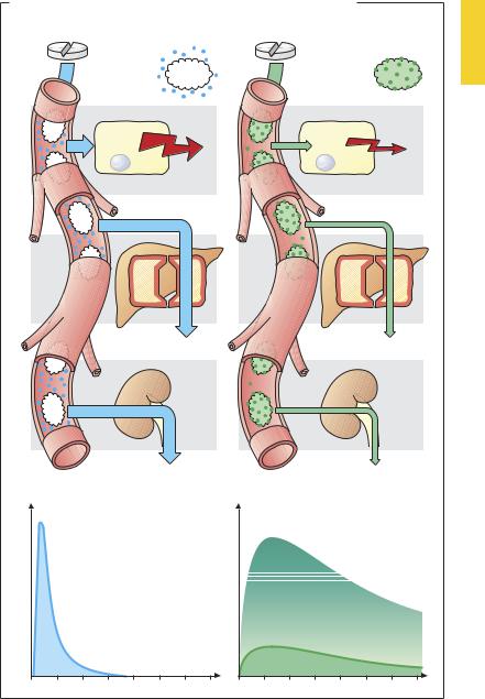
Binding to plasma proteins is instantaneous and reversible, i.e., any change in the concentration of unbound drug is immediately followed by a corresponding change in the concentration of bound drug. Protein binding is of great importance because it is the concentration of free drug that determines the intensity of the effect. At a given total plasma concentration (say, 100 ng/ml) the effective concentration will be 90 ng/ml for a drug 10% bound to protein, but 1 ng/ml for a drug 99% bound to protein. The reduction in concentration of free drug resulting from protein binding affects not only the intensity of the effect but also biotransformation (e.g., in the liver) and elimination from the kidney, because only free drug will enterhepatic sites ofmetabolismor undergo
glomerular filtration. When concentrations of free drug fall, drug is resupplied from binding sites on plasma proteins. Binding to plasma protein is equivalent to a depot in prolonging the duration of the effect by retarding elimination, whereas the intensity of the effect is reduced. If two substances have af nity for the same binding site on the albumin molecule, they may compete for that site. One drug may displace another from its binding site and thereby elevate the free (effective) concentration of the displaced drug (a form of drug interaction). Elevation of the free concentration of the displaced drug means increased effectiveness and accelerated elimination.
A decrease in the concentration of albumin (in liver disease, nephrotic syndrome, poor general condition) leads to altered pharmacokinetics of drugs that are highly bound to albumin.
Plasma protein-bound drugs that are substrates for transport carriers can be cleared from blood at high velocity; e.g., p-amino- hippurate by the renal tubule and sulfobromophthalein by the liver. Clearance rates of thesesubstancescanbeusedtodetermine renal or hepatic blood flow.
Luellmann, Color Atlas of Pharmacology © 2005 Thieme
All rights reserved. Usage subject to terms and conditions of license.
Binding to Plasma Proteins |
31 |
A. Importance of protein binding for intensity and duration of drug effect |
|
Drug is not |
Drug is |
bound |
strongly bound |
to plasma |
to plasma |
proteins |
proteins |
Effect |
Effect |
Effector cell |
Effector cell |
Biotransformation |
Biotransformation |
Renal elimination |
Renal elimination |
Plasma concentration |
Plasma concentration |
Free drug |
Bound drug |
|
Free drug |
Time |
Time |
Luellmann, Color Atlas of Pharmacology © 2005 Thieme
All rights reserved. Usage subject to terms and conditions of license.

32 Drug Elimination
The Liver as an Excretory Organ
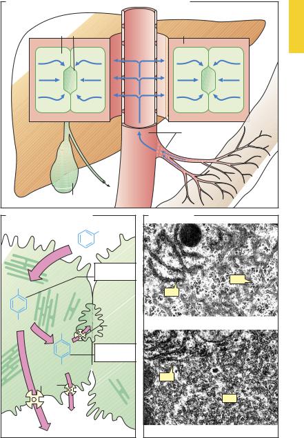
Asthemajororganofdrugbiotransformation, the liver is richly supplied with blood, of which 1100 ml is received each minute from the intestines through the portal vein and 350 ml through the hepatic artery, comprising nearly 1/3 of cardiac output. The blood content of hepatic vessels and sinusoids amounts to 500 ml. Owing to the widening of the portal lumen, intrahepatic blood flow decelerates (A). Moreover, the endothelial lining of hepatic sinusoids (p.24) contains pores large enough to permit rapid exit of plasma proteins. Thus, blood and hepatic parenchyma are able to maintain intimate contact and intensive exchange of substances, which is further facilitated by microvilli covering the hepatocyte surfaces abutting Disse’s spaces.
The hepatocyte secretes biliary fluid into the bile canaliculi (dark green), tubular intercellular clefts that are sealed off the blood spaces by tight junctions. Secretory activity in the hepatocytes results in movement of fluid toward the canalicular space (A).
The hepatocyte is endowed with numerous metabolically important enzymes that are localized in part in mitochondria and in part on membranes of the rough (rER) and smooth (sER) endoplasmic reticulum. Enzymes of the sER play a most important role in drug biotransformation. At this site, direct consumption of molecular oxygen (O2) takes place in oxidative reactions. Because these enzymes can catalyze either hydroxylation or oxidative cleavage of –N–C– or –O–C– bonds, they are referred to as “mixed-func- tion” oxidases or hydroxylases. The integral component of this enzyme system is the iron-containing cytochrome P450 (see p.38). Many P450 isozymes are known and they exhibit different patterns of substrate specificity. Interindividual genetic differences in isozyme make-up (e.g., CYP2D6) underlie subject-to-subject variations in drug biotransformation. The same holds for other enzyme systems; hence, the phenom-
enon is generally referred to as genetic polymorphism of biotransformation.
Compared with hydrophilic drugs not undergoing transport, lipophilic drugs are more rapidly taken up from the blood into hepatocytes and more readily gain access to mixed-function oxidases embedded in sER membranes. For instance, a drug having lipophilicity by virtue of an aromatic substituent (phenyl ring) (B) can be hydroxylated and thus become more hydrophilic (phase I reaction, p.36). Besides oxidases, sER also contains reductases and glucuronyltransferases. The latter conjugate glucuronic acid with hydroxyl, carboxyl, amine, and amide groups and hence also phenolic products of phase I metabolism (phase II conjugation). PhaseIandphaseII metabolitescan betransported back into the blood—probably via a gradient-dependent carrier—or actively secreted into bile via the ABC transporter (ATP-binding cassette transporter). Different transport proteins are available: for instance, MRP2 (the multidrug resistance associated protein 2) transports anionic conjugates into the bile canaliculi, whereas MRP3 can route these via the basolateral membrane of the hepatocyte toward the general circulation.
Prolonged exposure to substrates of one of the membrane-bound enzymes results in a proliferation of sER membranes in the liver (cf. C and D). The molecular mechanism of this sER “hypertrophy” has been elucidated for some drugs: thus, phenobarbital binds to a nuclear receptor (constitutive androstane receptor) that regulates the expression of cytochromes CYP2C9 and CYP2D6. Enzyme induction leads to accelerated biotransformation,notonlyoftheinducingagentbutalsoof other drugs (a form of drug interaction). With continued exposure, it develops in a few days, resulting in an increase in reaction velocity, maximally 2–3-fold, that disappears after removal of the inducing agent.
Luellmann, Color Atlas of Pharmacology © 2005 Thieme
All rights reserved. Usage subject to terms and conditions of license.
Biotransformation of Drugs |
33 |
A. Flow patterns in portal vein, Disse’s space, and hepatocyte
Hepatocyte Biliary capillary |
Disse’s space |
Intestine
Portal vein
Gallbladder
B. Fate of drugs undergoing hepatic hydroxylation
|
R |
|
|
Phase I |
|
|
metabolite |
|
R |
|
|
|
Biliary |
|
|
capillary |
|
OH |
ABC |
|
trans- |
||
R |
||
porter |
Phase II
metabolite
O-Glucuronide
Carrier
C . Hepatocyte
sER
rER
a) Normal
rER
sER
b) After phenobarbital administration
Luellmann, Color Atlas of Pharmacology © 2005 Thieme
All rights reserved. Usage subject to terms and conditions of license.