


58 Drug–Receptor Interaction
Types of Binding Forces
Unless a drug comes into contact with intrinsic structures of the body, it cannot affect body function.
Covalent Bonding
Two atoms enter a covalent bond if each donates an electron to a shared electron pair (cloud). This state is depicted in structural formulas by a dash. The covalent bond is “firm,” that is, not reversible or poorly so. Few drugs are covalently bound to biological structures. The bond, and possibly the effect, persist for a long time after intake of a drug has been discontinued, making therapy difficult to control. Examples include alkylating cytostatics (p.300) or organophosphates (p.311). Conjugation reactions occurring in biotransformation also represent covalent linkages (e.g., to glucuronic acid).
Noncovalent Bonding
In noncovalent bonding there is no formation of a shared electron pair. The bond is reversible and is typical of most drug–recep- tor interactions. Since a drug usually attaches to its site of action by multiple contacts, several of the types of bonds described below may participate.
Electrostatic attraction (A). A positive and a negative charge attract each other.
Ionic interaction: An ion is a particle charged either positively (cation) or negatively (anion), i.e., the atom is deficient in electrons or has surplus electrons, respectively. Attraction between ions of opposite charge is inversely proportional to the square of the distance between them; it is the initial force drawing a charged drug to its binding site. Ionic bonds have a relatively high stability.
Dipole–ion interaction: When bonding electrons are asymmetrically distributed over the atomic nuclei involved, one atom will bear a negative (δ–), and its partner a positive (δ+) partial charge. The molecule
thus presents a positive and a negative pole, i.e., it has polarity or is a dipole. A partial charge can interact electrostatically with an ion of opposite charge.
Dipole-dipole interaction is the electrostatic attraction between opposite partial charges. When a hydrogen atom bearing a partial positive charge bridges two atoms bearing partial negative charges, a hydrogen bond is created.
van der Waals bonds (B) are formed between apolar molecular groups that have come into close proximity. Spontaneous transient distortion of electron clouds (momentary faint dipole, δδ) may induce an opposite dipole in the neighboring molecule. The van der Waals bond, therefore, is also a form of electrostatic attraction, albeit of very low strength (inversely proportional to 7th power of distance).
Hydrophobic interaction (C). The attraction between the water dipoles is strong enough to hinder intercalation of any apolar (uncharged) molecules. By tending toward each other, H2O molecules squeeze apolar particles from their midst. Accordingly, in the organism, apolar particles such as fatty acid chains of cell membranes or apolar regions of a receptor have an increased probability of remaining in nonaqueous, apolar surroundings.
Luellmann, Color Atlas of Pharmacology © 2005 Thieme
All rights reserved. Usage subject to terms and conditions of license.
Types of Binding Forces |
59 |
A. Electrostatic attraction
|
Drug |
|
|
|
+ |
Binding site |
|
|
Complex |
|
||||
H |
|
|
|
|
|
|
O |
|
|
|
H |
|
O |
|
D +N |
|
|
50 nm |
|
–O P O |
D + N |
–O P O |
|||||||
H |
H |
|
|
|
|
|
OH |
|
H |
H |
|
OH |
||
Ion |
|
|
|
|
|
Ion |
|
|
Ionic bond |
|
||||
|
|
δ – δ + |
|
|
|
O |
|
δ – δ + |
|
O |
|
|||
|
|
|
|
–O |
|
|
|
–O |
|
|
||||
|
D |
O |
H |
1,5 nm |
P |
O |
D |
O |
H |
P |
O |
|||
|
|
|
|
|
|
|
OH |
|
|
|
|
OH |
||
|
Dipole (permanent) |
Ion |
|
|
|
|
|
|
||||||
|
|
|
D |
δ – δ + |
|
δ |
– |
D |
δ – δ + |
δ – |
||||
|
|
|
O |
H |
0,5 nm |
O |
O |
H |
|
O |
||||
D = Drug |
|
|
|
|
|
δ +H |
|
|
|
|
δ + H |
|||
|
|
|
|
|
|
|
|
|
|
|
|
|
||
|
|
|
|
Dipole |
Dipole |
Hydrogen bond |
|
|||||||
B. van der Waals’ bonding |
|
|
|
|
|
|
|
|
|
|
|
|||
|
|
|
|
|
CH |
CH |
|
|
δδ |
+CH |
CH |
δδ |
– |
|
|
|
|
|
|
2 |
2 |
|
|
|
|
2 |
2 |
|
|
|
|
|
|
|
CH |
CH |
|
|
δδ |
–CH |
CH |
δδ |
+ |
|
|
|
|
|
D |
2 |
2 |
|
|
D δδ |
|
2 |
2 |
|
|
|
|
|
|
CH |
CH |
|
|
–CH |
CH |
δδ |
+ |
|||
|
|
|
|
|
2 |
2 |
|
|
|
|
2 |
2 |
|
|
|
|
|
|
|
CH |
CH |
|
|
δδ |
+CH |
CH |
δδ |
– |
|
|
|
|
|
|
2 |
2 |
|
|
|
|
2 |
2 |
|
|
|
|
|
|
|
|
|
|
|
|
|
Induced |
|
|
|
|
|
|
|
|
|
|
|
|
|
|
transient |
|
||
|
|
|
|
|
|
|
|
|
|
|
fluctuating dipoles |
|||
C. Hydrophobic interaction |
|
|
||
δ + |
|
|
“Repulsion” of apolar |
|
H |
H |
|
|
|
δ −O |
|
apolar |
particle in polar solvent ( H2O) |
|
polar |
|
|
|
|
membrane |
Apolar |
|
|
|
acyl chain |
|
|
|
|
Phospholipid |
|
|
|
|
|
Insertion in apolar membrane interior |
Adsorption |
||
|
|
|
to apolar surface |
|
|
|
|
|
|
Luellmann, Color Atlas of Pharmacology © 2005 Thieme
All rights reserved. Usage subject to terms and conditions of license.

60 Drug–Receptor Interaction
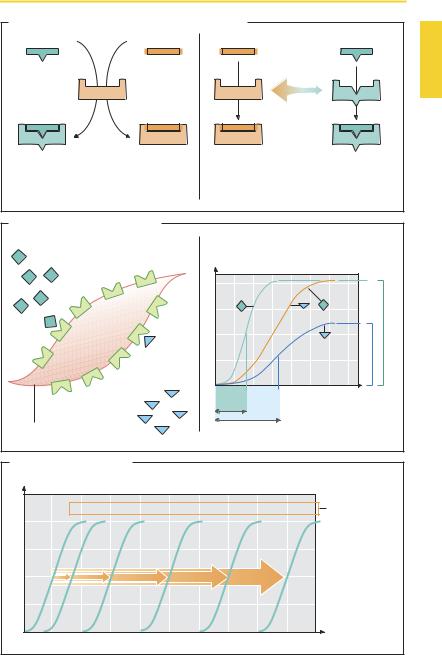
Agonists—Antagonists
An agonist (A) has af nity (tendency to adhere) for a receptor and affects the receptor protein in such a manner as to cause a change in cell function—“intrinsic activity.” The biological effect of the agonist (i.e., the change in cell function) depends on the effectiveness of signal transduction steps (p.66) associated with receptor activation. The maximal effect of an agonist may alreadyoccurwhenonlyafractionoftheavailable receptors is occupied (B, agonist A). Another agonist (agonist B), possessing equal af nity but less ability to activate the receptor and the associated signal transduction steps (i.e., less intrinsic activity), will produce a smaller maximal effect even if all receptors are occupied—smaller ef cacy. Agonist B is a partial agonist. The potency of an agonist is characterized by the concentration (EC50) at which a half-maximal effect is attained.
Antagonists (A) attenuate the effect of agonists: they act “antiagonistically.” Competitive antagonists possess af nity for the receptors, but their binding does not elicit a change in cell function. In other words, they aredevoidofintrinsicactivity. Whenpresent simultaneously, an agonist and a competitive antagonist vie for occupancy of the receptor. The af nities and concentrations of both competitors determine whether binding of agonist or antagonist predominates. By increasing the concentration of the agonist, blockade induced by an antagonist can be surmounted (C): that is, the concentra- tion–effect curve of the agonist is shifted “right”—to higher concentrations—with preservation of the maximal effect.
Models of the Molecular Mechanism of Agonist/Antagonist Action (A)
Agonist induces an active conformation.
The agonist binds to the inactive receptor and thereby causes the resting conformation to change into the active state. The antago-
nist attachesto the inactive receptor without altering its conformation.
Agonist stabilizes spontaneously occurring active conformation. The receptor may spontaneously “flip” into the active conformation. Usually, however, the statistical probability of such an event is so small that a spontaneous excitation of thecells remains undetectable. Selective binding of the agonist can occur only to the active conformation and thus favors the existence of this state. The antagonist shows af nity only for theinactivestate,promotingexistenceofthe latter. If the system has little spontaneous activity, no measurable effect will result from adding an antagonist. However, if the system displays high spontaneous activity, the antagonist is liable to produce an effect opposite to that of an agonist: inverse agonist. A “true” antagonist without intrinsic activity (“neutral antagonist”) displays equal af nity for the active and inactive conformations of the receptor and does not interfere with the basal activity of the cell. According to this model, a partial agonist has less selectivity for the active state; however, to a certain extent it binds also to the inactive state.
Other Forms of Antagonism
Allosteric antagonism. The antagonist is bound outside the agonist’s site of attachment at the receptor and induces a decrease in agonist af nity. The latter is increased in the case of allosteric synergism.
Functional antagonism. Two agonists acting via different receptors affect the same variable (e.g., luminal diameter of bronchi) in opposite directions (epinephrine † dilation; histamine † constriction).
Luellmann, Color Atlas of Pharmacology © 2005 Thieme
All rights reserved. Usage subject to terms and conditions of license.
|
|
Agonists—Antagonists |
61 |
|
A. Molecular mechanisms of drug–receptor interaction |
|
|
||
Agonist |
Antagonist |
Antagonist |
Agonist |
|
|
|
Rare spontaneous |
|
|
|
|
transition |
|
|
|
Receptor |
|
|
|
|
|
inactive |
active |
|
Agonist |
Antagonist |
Antagonist |
Agonist |
|
induces active |
occupies |
selects inactive |
selects active |
|
conformation of |
receptor |
receptor |
receptor |
|
receptor protein |
without effects |
conformation |
conformation |
|
B. Potency and efficacy of agonists
Receptors |
Increase in tension |
|
|
|
||
|
|
|
||||
Agonist A |
|
|
Receptor occupation |
|
|
|
|
|
|
||||
|
|
|
|
|
||
|
|
|
|
|
|
|
Efficacy
EC50
EC50
Concentration (log) of agonist
Smooth-muscle cell
|
|
Agonist B |
Potency |
|
|
|
C. Competitive antagonism |
|
|
|
|
||
Agonist effect |
|
|
|
|
|
|
0 |
1 |
10 |
100 |
1000 |
10 000 |
Concentration |
|
|
|
|
|
|
of |
|
|
|
|
|
|
antagonist |
|
|
Agonist concentration (log) |
|
|
|
|
Luellmann, Color Atlas of Pharmacology © 2005 Thieme
All rights reserved. Usage subject to terms and conditions of license.