
- •VOLUME 1
- •CONTRIBUTOR LIST
- •PREFACE
- •LIST OF ARTICLES
- •ABBREVIATIONS AND ACRONYMS
- •CONVERSION FACTORS AND UNIT SYMBOLS
- •ABLATION.
- •ABSORBABLE BIOMATERIALS.
- •ACRYLIC BONE CEMENT.
- •ACTINOTHERAPY.
- •ADOPTIVE IMMUNOTHERAPY.
- •AFFINITY CHROMATOGRAPHY.
- •ALLOYS, SHAPE MEMORY
- •AMBULATORY MONITORING
- •ANALYTICAL METHODS, AUTOMATED
- •ANALYZER, OXYGEN.
- •ANESTHESIA MACHINES
- •ANESTHESIA MONITORING.
- •ANESTHESIA, COMPUTERS IN
- •ANGER CAMERA
- •ANGIOPLASTY.
- •ANORECTAL MANOMETRY
- •ANTIBODIES, MONOCLONAL.
- •APNEA DETECTION.
- •ARRHYTHMIA, TREATMENT.
- •ARRHYTHMIA ANALYSIS, AUTOMATED
- •ARTERIAL TONOMETRY.
- •ARTIFICIAL BLOOD.
- •ARTIFICIAL HEART.
- •ARTIFICIAL HEART VALVE.
- •ARTIFICIAL HIP JOINTS.
- •ARTIFICIAL LARYNX.
- •ARTIFICIAL PANCREAS.
- •ARTERIES, ELASTIC PROPERTIES OF
- •ASSISTIVE DEVICES FOR THE DISABLED.
- •ATOMIC ABSORPTION SPECTROMETRY.
- •AUDIOMETRY
- •BACTERIAL DETECTION SYSTEMS.
- •BALLOON PUMP.
- •BANKED BLOOD.
- •BAROTRAUMA.
- •BARRIER CONTRACEPTIVE DEVICES.
- •BIOCERAMICS.
- •BIOCOMPATIBILITY OF MATERIALS
- •BIOELECTRODES
- •BIOFEEDBACK
- •BIOHEAT TRANSFER
- •BIOIMPEDANCE IN CARDIOVASCULAR MEDICINE
- •BIOINFORMATICS
- •BIOLOGIC THERAPY.
- •BIOMAGNETISM
- •BIOMATERIALS, ABSORBABLE
- •BIOMATERIALS: AN OVERVIEW
- •BIOMATERIALS: BIOCERAMICS
- •BIOMATERIALS: CARBON
- •BIOMATERIALS CORROSION AND WEAR OF
- •BIOMATERIALS FOR DENTISTRY
- •BIOMATERIALS, POLYMERS
- •BIOMATERIALS, SURFACE PROPERTIES OF
- •BIOMATERIALS, TESTING AND STRUCTURAL PROPERTIES OF
- •BIOMATERIALS: TISSUE-ENGINEERING AND SCAFFOLDS
- •BIOMECHANICS OF EXERCISE FITNESS
- •BIOMECHANICS OF JOINTS.
- •BIOMECHANICS OF SCOLIOSIS.
- •BIOMECHANICS OF SKIN.
- •BIOMECHANICS OF THE HUMAN SPINE.
- •BIOMECHANICS OF TOOTH AND JAW.
- •BIOMEDICAL ENGINEERING EDUCATION
- •BIOSURFACE ENGINEERING
- •BIOSENSORS.
- •BIOTELEMETRY
- •BIRTH CONTROL.
- •BLEEDING, GASTROINTESTINAL.
- •BLADDER DYSFUNCTION, NEUROSTIMULATION OF
- •BLIND AND VISUALLY IMPAIRED, ASSISTIVE TECHNOLOGY FOR
- •BLOOD BANKING.
- •BLOOD CELL COUNTERS.
- •BLOOD COLLECTION AND PROCESSING
- •BLOOD FLOW.
- •BLOOD GAS MEASUREMENTS
- •BLOOD PRESSURE MEASUREMENT
- •BLOOD PRESSURE, AUTOMATIC CONTROL OF
- •BLOOD RHEOLOGY
- •BLOOD, ARTIFICIAL
- •BONDING, ENAMEL.
- •BONE AND TEETH, PROPERTIES OF
- •BONE CEMENT, ACRYLIC
- •BONE DENSITY MEASUREMENT
- •BORON NEUTRON CAPTURE THERAPY
- •BRACHYTHERAPY, HIGH DOSAGE RATE
- •BRACHYTHERAPY, INTRAVASCULAR
- •BRAIN ELECTRICAL ACTIVITY.
- •BURN WOUND COVERINGS.
- •BYPASS, CORONARY.
- •BYPASS, CARDIOPULMONARY.
366 BIOMATERIALS: TISSUE-ENGINEERING AND SCAFFOLDS
BIOMATERIALS: TISSUE-ENGINEERING AND SCAFFOLDS
GILSON KHANG
Chonbuk National University
SANG JIN LEE
MOON SUK KIM
HAI BANG LEE
Korea Research Institutes of
Chemical Technology
INTRODUCTION
Tissue engineering offers an alternative to whole organ and tissue transplantation for diseased, failed, or abnormally functioning organs. Millions suffer from end-stage organ failure or tissue loss annually. In the United State alone, at least 8 million surgical operations are carried out each year at a total national healthcare cost exceeding $400 billion annually (1–4). Approximately 500,000 coronary artery bypass surgeries are conducted in the United States annually (5). Autologous and allogenic natural tissue, such as the saphenous vein or the internal mammary artery, is generally used for coronary artery replacement. The results have been favorable for these procedures with patency rates generally ranging from 50–70%. Failures are caused by intimal thickening due largely to adaptation of the vessel in response to increased pressure and wall shear stress, compression, inadequate graft diameter, and disjunction at the anastomosis. Also, successful treatment has been limited by the poor performance of the synthetic materials used, such as polyethyleneterephthalate (PET, Dacron) and expanded polytetrafluoroethylene (ePTFE, Gore-Tex), which are used for tissue replacement due to plaguing problems
(6). For example, in cases of tumor resection in the head, neck, and upper and lower extremities, as well as in cases of trauma and congenital abnormalities, there are often outline defects due to the loss of soft tissue, this tissue is largely composed of subcutaneous adipose tissue (7). The defects lead to abnormal cosmesis, affect the emotional comfort of patients, and may impair function. A surgeon would prefer to use an autologous adipose tissue to sculpt contour deformities. Because mature adipose tissue does not transplant effectively, numerous natural, synthetic, and hybrid materials have been used to act as adipose surrogates. Despite improved patient outcomes, the use of many of these materials results in severe problems, such as unpredictable outcomes, fibrous capsule contraction, allergic reactions, suboptimum mechanical properties, distortion, migration, and long-term resorption.
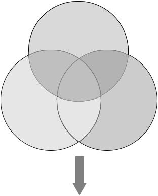
To offset the short supply of donor organs as well as the problems caused by the poor biocompatibility of the biomaterials used, a new hybridized method of ‘‘tissue engineering’’, which combines both cells and biomaterials has been introduced (8). To reconstruct new tissue by tissue engineering, a triad of components are requried: (1) harvested and dissociated cells from the donor tissue including nerve, liver, pancreas, cartilage, and bone as well as embryonic stem, adult stem, or precursor cell; (2) scaffolds
made of biomaterials on which cells are attached and cultured, then implanted at the desired site of the functioning tissue; (3) growth factors that promote and/or prevent cell adhesion, proliferation, migration, and differentiation by up-regulating or down-regulating the synthesis of protein, growth factors, and receptors (see Fig. 1). In a typical application for cartilage regeneration, donor cartilage is harvested from the patient and dissociated into individual chondrocyte cells using enzymes as collagenase, and then mass cultured in vitro. The chondrocyte cells are then seeded onto a porous and synthetic biodegradable scaffold. This cell–polymer structure is massively cultured in a bioreactor. The abnormal tissue is removed and the cell– polymer structure is then implanted in the patient. Finally, the synthetic biodegradable scaffold resorbs into the body and the chondrocyte cell produces collagen and glycosaminoglycan as its own natural extracellular matrix (ECM), which results in regenerated cartilage. This approach can theoretically be applied to the manufacture of almost all organs and tissues except for organs such as the brain (3).
In this section, a review is given of the biomaterials and procedures used in the development of tissue-engineered scaffolds, including: (1) natural and synthetic biomaterials, (2) natural–synthetic hybrid scaffolds, (3) the fabrication methods and techniques for scaffolds, (4) the required physicochemical properties for scaffolds, and (5) cytokinereleased scaffolds.
Cells
(e.g., chondrocytes, osteoblasts, stem cells)
|
Tissue |
|
Engineering |
Scaffolds |
Signaling |
(e.g., collagen, |
molecules |
gelatin, PGA, PLA, |
(e.g., growth |
PLGA) |
factors, |
|
morphogens, |
|
adhesins) |
Time
Appropriate
Environment
Regeneration of
tissues/organs
Figure 1. Tissue engineering triad. The combination of three key elements, cells, biomaterials, and signaling molecules, results in regenerated tissue-engineered neo-organs.
BIOMATERIALS FOR TISSUE ENGINEERING
The Importance of Scaffold Matrices in Tissue Engineering
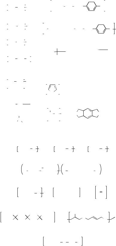
Scaffolds play a very critical role in tissue engineering. Scaffolds direct the growth (1) of cells seeded within the porous structure of the scaffold, or (2) of cells migrating from surrounding tissue. Most mammalian cell types are anchorage dependent; the cells die if an adhesion substrate is not provided. Scaffold matrices can be used to achieve cell delivery with high loading and efficiency to specific sites. Therefore, the scaffold must provide a suitable substrate for cell attachment, cell proliferation, differentiated function, and cell migration. The prerequisite physicochemical properties of scaffolds are (1) to support and deliver the cells; (2) to induce, differentiate, and promote conduit tissue growth; (3) to target the cell-adhesion substrate, (4) to stimulate cellular response; (5) to create a wound healing barrier; (6) to be biocompatible and biodegradable; (7) to have relatively easy processability and malleability into the desired shapes; (8) to be highly porous with large surface–volume; (9) to have mechanical strength and dimensional stability; and (10) to have sterilizability (9–16). Generally, three-dimensional (3D) porous scaffolds can be fabricated from natural and synthetic polymers (Fig. 2 shows these chemical structures), ceramics, metal, and in a very few cases, composite biomaterials and cyto- kine-releasing materials.
Natural Polymers
Many naturally occurring scaffolds can be used for tissue engineering purposes. One such example is the ECM, which is composed of very complex biomaterials and controls cell function. For the ECM used in tissue engineering, natural and synthetic scaffolds are designed to mimic specific function. The natural polymers used are alginate, proteins, collagens (gelatin), fibrins, albumin, gluten, elastin, fibroin, hyarulonic acid, cellulose, starch, chitosan (chitin), sclerolucan, elsinan, pectin (pectinic acid), galactan, curdlan, gellan, levan, emulsan, dextran, pullulan, heparin, silk, chondroitin 6-sulfate, polyhydroxyalkanoates, and others. Much of the interest in these natural polymers comes from their biocompatibility, relatively abundance and commercial availability, and ease of processing (17).
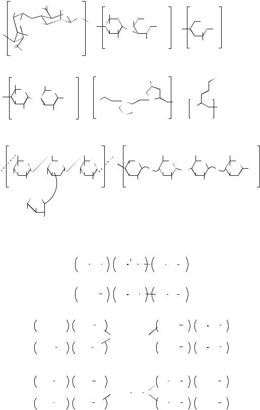
Alginate. Alginate (from seaweed) is composed of two repeating monosaccharides: L-guluronic acid and D-man- nuronic acid. Repeating strands of these monomers form linear, water-soluble polysaccharides. Gelation occurs by interaction of divalent cations (e.g., Ca2þ, Mg2þ) with blocks of guluronic acid from different polysaccharide chains (as shown in Fig. 3). From this gelation property, the encapsulation of calcium alginate beads impregnated with various pharmaceutics, cytokines, or cultured cells, has been extensively investigated. Varying the preparation conditions of the gelation can control structure and physicochemical properties. Calcium alginate scaffolds do not degrade by hydrolytic reaction, whereas they can be degraded by a chelating agent such as ethyleneaminetetraaceticacid (EDTA) or by an enzyme. Also, the diffusion
BIOMATERIALS: TISSUE-ENGINEERING AND SCAFFOLDS |
367 |
of calcium ions from an alginate gel can cause dissociation between alginate chains, which results in a decrease of mechanical strength over time. One of the disadvantages of an alginate matrix is a potential immune response and the lack of complete degradation, since alginate is produced in the human body (10). For these reasons, the chemical modification and incorporation of biological peptides, such as Arg-Gly-Asp cell adhesion peptides, have been used to improve the functionality and flexibility of natural scaffolds and their potential application (18).
Many researchers have studied the encapsulation of chondrocytes. Growth plate chondrocytes, fetal chondrocytes, and mesenchymal stem cells derived from bone marrow have been encapsulated in alginate (19). In each system, the chondrocytes demonstrated a differentiated phenotype, producing an ECM and retaining the cell morphology of typical chondrocytes. In addition, novel hybrid composites, such as alginate/agarose (a thermosensitive polysaccharide), alginate/fibrin, alginate/collagen and alginate/hyaruronic acid, and different gelling agents (water, sucrose, sodium chloride, and calcium sulfate) were investigated to optimize the advantages of each component material for tissue engineered cartilage (20–22). It was found that this hybrid material provides a reason why the microenvironments of composite materials affect chondrogenesis.
Collagen. At least 22 types of collagen exist in the human body. Among these, collagen types I, II, and III are the most abundant and ubiquitous. Conformation of the collagen chain consists of triple helices that are packed or processed into microfibrils. Molecularly, the three repeating amino acid sequences, such as glycine, proline, and hydroxyproline, form protein chains resulting in the formation of a triple helix arrangement. Type I collagen is the most abundant and is the major constituent of bone, skin, ligament, and tendon, whereas type II collagen is the collagen in cartilage. Collagen can promote cell adhesion as demonstrated by the Asp-Gly-Glu-Ala peptide in type I collagen, which functions as a cell-binding domain. Due to the abundance and ready accessibility of these tissues, they have been used frequently in the preparation of collagen (23).
The purified collagen materials obtained from either molecular or fibrillar technology are subjected to additional processing to fabricate the materials into useful scaffold types for specific tissue-engineered organs. Collagen can be processed into several types such as membrane (film and sheet), porous (sponge, felt, and fiber), gel, solution, filamentous, tubular (membrane and sponge), and composite matrix for the application of tissue repair, patches, bone and cartilage repair, nerve regeneration, and vascular and skin repair with/without cells (24). The Physicochemical properties of collagen can be improved by the addition of a variety of homogeneous and heterogeneous composites. Homogeneous composites can be formed between ions, peptides, proteins, and polysaccharides in a collagen matrix by means of ionic and covalent bonding, entrapment, entanglement, and coprecipitation. Heterogeneous composites, such as collagen–synthetic polymers, col- lagen–biological polymers, and collagen–ceramic hybrid
368 BIOMATERIALS: TISSUE-ENGINEERING AND SCAFFOLDS
|
|
|
|
|
|
|
|
|
|
|
|
|
|
|
|
|
|
|
|
|
|
|
|
|
|
|
|
|
|
|
|
|
|
|
|
|
|
|
|
|
|
|
|
|
|
|
|
|
|
|
|
O |
|
|
|
|
|
|
|
|
|
|
|
|
|
|
O |
|
|
|
|
|
|||||||||
|
|
|
|
|
|
|
|
|
|
|
|
|
|
|
|
|
|
|
|
|
|
|
|
|
|
|
|
|
|
|
|
|
|
O |
|
|
CH2 CH2 O |
|
|
|
|
|
|
|
|
|
|
|
|
|
|
|
|
|
|
|
|
|
|
|
|
|
|
|
|
|
|
|
|||||||||||||
|
|
|
|
CH2 |
CH2 |
|
|
n |
|
|
|
|
|
|
|
|
|
|
|
|
|
|
|
|
|
|
C |
|
|
|
|
|
|
|
|
|
|
|
|
|
|
|
C |
|
|
|
|
|
|
|
|
|
|||||||||||||||||||||||||||||
|
|
|
|
|
|
|
|
|
|
|
|
|
|
|
|
|
|
|
|
|
|
|
|
|
|
|
|
n |
|
|
|
|
|||||||||||||||||||||||||||||||||||||||||||||||||
|
|
|
|
|
|
|
|
|
|
|
|
|
|
|
|
|
|
|
|
|
|
||||||||||||||||||||||||||||||||||||||||||||||||||||||||||||
1 |
|
|
|
|
|
|
|
|
|
|
|
|
|
|
|
|
|
|
|
|
|
|
|
|
|
|
|
|
|
|
|
|
|
|
|
|
|
6 |
|
|
|
|
|
|
|
|
|
|
|
|
|
|
|
|
|
|
|
|
|
|
|
|
|
|
|
||||||||||||||||
|
|
|
|
|
|
|
|
|
|
|
|
|
|
|
|
|
|
|
|
|
|
|
|
|
|
|
|
|
|
|
|
|
|
|
|
|
|
|
|
|
|
|
|
|
|
|
|
|
|
|
|
|
|
|
|
|
|
|
|
|
|
|
|
|
|
||||||||||||||||
|
|
|
|
CH2 |
CF2 |
|
|
|
|
|
|
|
|
|
|
|
|
|
|
|
|
|
|
|
|
|
|
CH2 |
|
|
|
|
|
|
|
|
|
|
|
|
|
|
|
|
O |
|
|
|
|
|
|
|
|
|
|
|
|
|
|
|
|
O |
|||||||||||||||||||
|
|
|
|
|
|
|
|
|
|
|
|
|
|
|
|
|
|
|
|
|
|
|
|
|
|
|
|
|
|
|
|
|
|
|
|
|
|
|
|
|
|
|
|
|
|
|
|
|
|
|
|
|
|
|
|
|
|
||||||||||||||||||||||||
|
|
|
|
|
|
|
|
|
|
|
|
|
|
|
|
|
|
|
|
|
|
|
|
|
|
|
|
|
|
|
|
|
|
|
|
|
|
|
|
|
|
|
|
|
|
|
|
|
|
|
|
|
|
|
|
|
|
|
|
|
|
|
|
||||||||||||||||||
|
|
|
|
|
|
n |
|
|
|
|
|
|
|
O CH2 |
|
|
CH2 CH2 |
O |
|
|
C |
|
|
|
|
|
|
|
|
|
|
|
|
|
|
|
|
C |
|||||||||||||||||||||||||||||||||||||||||||
2 |
|
|
|
|
|
|
|
|
|
|
|
|
|
|
|
|
|
|
|
|
|
|
|
|
|
|
|
|
|
|
|
|
|
|
|
|
|||||||||||||||||||||||||||||||||||||||||||||
|
|
|
|
|
|
|
|
|
|
|
|
|
|
|
|
|
|
|
|
|
|
|
|
|
|
|
|
|
|
|
|
|
|
|
|
|
|
|
|
|
|
|
|
|
|
|
|
|
|
|
|
|
|
|
|
|
|
|
|
|
|
|
|
|
|
|
|
|
|
|
|
|
|
||||||||
|
|
|
|
|
|
|
|
|
|
|
|
|
|
|
|
|
|
|
|
|
|
|
|
|
|
|
|
|
|
|
|
|
|
|
|
|
|
|
|
|
|
|
|
|
7 |
|
|
|
|
|
|
|
|
|
|
|
|
|
|
|
|
|
|
|
|
|
|
|
|
|
|
|
|
|
|||||||
|
|
|
|
CF2 |
CF2 |
|
n |
|
|
|
|
|
|
|
|
|
|
|
|
|
|
|
|
|
|
|
|
|
|
|
|
|
|
|
|
|
|
|
|
|
|
|
|
|
|
|
|
|
|
|
|
|
|
|
|
|
|
|
|
|
|
|
|
|
|
|
|
|
|
||||||||||||
|
|
|
|
|
|
|
|
|
|
|
|
|
|
|
|
|
|
|
|
|
|
|
|
|
|
|
|
|
|
|
|
|
|
|
|
|
|
|
|
|
|
|
|
|
|
|
|
|
|
|
|
|
|
|
|
|
|
|
|
|
|
|
|
|
|
|
|||||||||||||||
|
|
|
|
|
|
|
|
|
|
|
|
|
|
|
|
|
|
|
|
|
|
|
|
|
|
|
|
|
|
|
|
|
|
|
|
|
|
|
|
|
|
|
|
|
|
|
|
|
|
|
|
|
|
|
|
|
|
|
|
|
|
|
|
|
|
||||||||||||||||
3 |
|
|
|
|
|
|
|
|
|
|
|
|
|
|
|
|
|
|
|
|
|
|
|
|
|
|
|
|
CH3 |
|
|
|
|
|
|
|
|
|
|
|
|
|
|
|
|
|
|
|
|
|
|
|
CH3 |
|
|
|
|
||||||||||||||||||||||||
|
|
|
|
|
|
|
|
|
|
|
|
|
|
|
|
|
|
|
|
|
|
|
|
|
|
|
|
|
|
|
|
|
|
|
|
|
|
|
|
|
|
|
|
|
CH2 |
|
|
|
|
|
|
|
|
|
|
|
|
||||||||||||||||||||||||
|
|
|
|
|
|
|
|
|
|
|
|
|
|
|
|
|
|
|
|
|
|
|
|
|
|
|
|
|
CH2 |
|
|
|
|
|
|
|
|
|
|
|
|
|
|
|
|
|
|
|
|
|
|
|
|
|
|
|
|
|
|
|
|
|
|
|
|
|
|
||||||||||||||
|
|
|
|
CH2 |
CH2 |
|
|
O |
|
|
|
|
|
|
|
|
|
|
C |
|
O |
|
|
|
|
|
|
|
|
|
|
|
|
|
|
|
|
|
|
|
|
|
|
|
C |
|
|
|
|
O |
|
|
|
|
|||||||||||||||||||||||||||
|
|
|
|
|
|
|
|
|
|
|
|
|
|
|
|
|
|
|
|
|
|
|
|
|
|
|
|
|
|
|
|
|
|
|
|
|
|
|
|
|
|
|
|
|
|
|
|
|
|
|
|
|
|
||||||||||||||||||||||||||||
|
|
|
|
|
|
|
|
|
|
|
|
|
|
|
|
|
|
|
|
|
|
|
|
|
|
|
|
|
|
|
|
|
|
|
|
|
|
|
|
|
|
|
|
|
|
|
|
|
|
|
|
|
|
||||||||||||||||||||||||||||
|
|
|
|
|
|
|
|
|
|
|
|
|
|
|
|
|
|
|
|
|
|
|
|
|
|
|
|
|
|
|
|
|
|
|
|
|
|
|
|
|
|
|
|
|
|
|
|
|
|
|
|
|
|
|
|
|
|
|
|
|
|||||||||||||||||||||
|
|
|
|
|
|
|
n |
|
|
|
|
|
|
|
|
|
|
|
|
|
|
|
|
|
|
|
|
|
|
|
|
|
|
|
|
|
|
|
|
|
|
|
|
|
|
|
|
|
|
|
O |
|
|
|
|
|
|
|
|
|
|
|
|
||||||||||||||||||
|
|
|
|
|
|
|
|
|
|
|
|
|
|
|
|
O |
|
|
|
|
|
|
|
|
|
|
|
|
|
|
|
|
|
|
|
|
|
|
|
|
|
|
|
|
|
|
|
|
|
|
|
|
|
|
|
|
|||||||||||||||||||||||||
|
|
|
|
|
|
|
|
4 |
|
|
|
|
|
|
|
|
|
|
|
|
|
|
|
|
|
|
|
|
|
|
|
|
|
|
|
|
|
|
|
|
|
|
|
|
|
|
|
|
|
|
|
|
|
|
|
|
|
|
|
CH2 |
|
|
|
|
|||||||||||||||||
|
|
|
|
|
|
|
|
|
|
|
|
|
|
|
|
|
|
|
|
|
|
|
|
|
|
|
|
|
|
|
|
|
|
|
|
|
|
|
|
|
|
|
|
|
|
|
|
|
|
|
|
|
|
|
|
|
|
|
|
|
|
|
|
|
|
|
|
|
|||||||||||||
|
|
|
|
|
|
|
|
|
|
|
|
|
|
|
|
|
|
|
|
|
|
|
|
|
|
|
|
|
|
|
|
|
CH3 |
|
n |
|
|
|
|
|
|
|
|
|
|
|
|
|
|
|
|
|
|
|
|
|
|
|
|
|
|
||||||||||||||||||||
|
|
|
|
|
|
|
|
|
|
|
|
|
|
|
|
|
|
|
|
|
|
|
|
|
|
|
|
|
|
|
|
|
|
|
|
|
|
|
|
|
|
|
|
|
|
|
|
|
|
|
|
|
|
|
|
|
|
|
|
|
|
|
|
|
|
|
|
|
|
|
|
|
|||||||||
|
|
|
|
|
|
|
|
|
|
|
|
|
|
|
|
|
|
|
|
|
|
|
|
|
|
|
|
|
|
|
|
|
|
|
|
|
|
|
|
|
|
|
|
|
|
|
|
|
|
|
|
|
|
|
|
|
|
CH2 |
|
|
|
|
|||||||||||||||||||
|
|
|
|
|
|
|
|
|
|
|
|
|
|
|
|
|
|
|
|
|
|
|
|
|
8 |
|
|
|
|
|
|
|
|
|
|
|
|
|
|
|
|
|
|
|
|
|
|
|
|
|
|
|
|
|
|
|
|
|
|||||||||||||||||||||||
|
|
|
|
|
|
|
|
|
|
|
|
|
|
|
|
|
|
|
|
|
|
|
|
|
|
|
|
|
|
|
|
|
|
|
|
|
|
|
|
|
|
|
|
|
|
|
|
|
|
|
|
|
|
|
|
|
|
|
|||||||||||||||||||||||
|
|
|
|
|
|
|
|
|
|
|
|
|
|
|
|
|
|
|
|
|
|
|
|
|
|
|
|
|
|
|
|
|
|
|
|
|
|
|
|
|
|
|
|
|
|
|
|
|
|
|
|
|
|
|
|
|
|
|
|
|
|
|
|
|
|
|
|
|
|
||||||||||||
|
|
|
|
|
|
|
|
|
|
|
|
|
|
|
|
|
|
|
|
|
|
|
|
|
|
|
|
|
|
|
|
|
|
|
|
|
|
|
|
|
|
|
|
|
|
|
|
|
|
|
|
|
|
|
OH |
n |
|||||||||||||||||||||||||
|
|
|
|
|
|
|
|
|
|
|
|
|
|
|
|
|
|
|
|
|
|
|
|
|
|
|
|
|
|
|
|
|
|
|
|
|
|
|
|
|
|
|
|
|
|
|
|
|
|
|
|
|
|
|
|
|
|
|
|
|
|
|
|
|
|
|
|||||||||||||||
|
|
|
|
CH |
CH2 |
|
|
|
|
|
|
|
|
|
|
|
|
|
|
|
|
|
|
|
|
|
|
|
|
|
|
|
|
|
|
|
|
|
|
|
|
|
|
|
|
|
|
|
|
|
|
|
|
9 |
|
|
|
|
|
|
|
|
|
|
|||||||||||||||||
|
|
|
|
|
|
|
|
|
|
|
|
|
|
|
|
|
|
|
|
|
|
|
|
|
|
|
|
|
|
|
|
|
|
|
|
|
|
|
|
|
|
|
|
|
|
|
|
|
|
|
|
|
|
|
|
|
|
|
|
|
|
|
|
|
|
||||||||||||||||
|
|
|
|
|
|
|
|
|
|
|
|
|
|
|
|
|
|
|
|
|
|
|
|
|
|
|
|
|
|
|
|
|
|
|
|
|
|
|
|
|
|
|
|
|
|
|
|
|
|
|
|
|
|
|
|
|
|
|
|
|
|
|
|
|
|
||||||||||||||||
|
|
|
|
|
|
|
|
|
|
|
|
|
|
|
|
|
|
|
|
|
|
|
|
|
|
|
|
|
|
|
|
|
|
|
|
|
|
|
|
|
|
|
|
|
|
|
|
|
|
|
|
|
|
|
|
|
|
|
|
|
|
|
|
|
|
|
|
|
|
|
|||||||||||
|
|
|
|
|
|
|
|
|
|
|
|
|
|
|
|
|
|
|
|
|
|
|
|
|
|
|
|
|
|
|
|
|
|
|
|
|
|
|
|
|
|
|
|
|
|
|
|
|
|
|
|
|
|
|
|
|
|
|
|
|
|
|
|
|
|
|
|
|
|
|
|
|
|
|
|
|
|
|
|
|
|
|
|
|
OH |
|
|
|
|
|
|
|
|
n |
|
|
|
|
|
|
|
|
|
|
|
|
|
|
|
|
|
|
|
|
|
|
|
|
|
|
|
|
|
|
|
|
|
|
|
|
|
|
|
|
|
|
|
|
|
|
|
|
|
|
|
|
|
|
|
|
|
|
|
|
|
|
|||||||
5 |
|
|
|
|
|
|
|
|
|
|
|
|
|
|
|
|
|
|
|
N |
|
|
|
|
|
|
|
|
|
|
|
|
|
|
|
|
|
|
|
|
|
|
|
|
|
|
|
|
|
|
|
|
|
|
|
|
|
|
|
|
|
|
|
|
|
|
|
||||||||||||||
|
|
|
|
|
|
|
|
|
|
|
|
|
|
|
|
|
|
|
|
|
|
|
|
|
|
|
|
|
|
|
|
|
|
|
|
|
|
|
|
|
|
|
|
|
|
|
|
|
|
|
|
|
|
|
|
|
|
|
|
|
|
|
|
|
|
|
|
|
|
||||||||||||
|
|
|
|
|
|
|
|
|
|
|
|
|
|
|
|
|
|
|
|
|
|
|
|
|
|
|
|
|
|
|
|
|
|
|
|
n |
|
|
|
|
|
|
|
|
|
|
|
|
|
|
|
|
|
|
|
|
|
|
|
|
|
|
|
|
|
|
|
|
|
|
|
|
|
|
|||||||
|
|
|
|
|
|
|
|
|
|
|
|
|
|
|
|
|
|
|
|
|
|
|
|
|
|
|
|
|
|
|
|
|
|
|
|
|
|
|
|
|
|
|
|
|
|
|
|
|
|
|
|
|
|
|
|
|
|
|
|
|
|
|
|
|
|
|
|
|
|
|
|
|
|
||||||||
|
|
|
|
|
|
|
|
|
|
|
|
|
|
|
|
|
|
|
|
|
|
|
|
|
11 |
|
|
|
|
|
|
|
|
|
|
|
|
|
|
|
|
|
|
|
|
|
|
|
|
|
|
|
|
|
|
|
|
|
|
|
|
|
|
|
|
|
|
|
|
|
|
|
|||||||||
|
|
|
|
|
|
CH |
|
|
|
|
|
|
|
|
|
|
|
|
|
|
|
|
CH3 |
|
|
|
|
|
|
|
|
|
|
|
|
|
|
|
O |
|
|
|
|
|
|
|
|
|
|
|
O |
|
|
|
|
|
|
|
|
|
|
|
|
||||||||||||||||||
|
|
|
|
|
CH2 |
|
|
|
|
|
|
|
|
|
|
|
|
|
|
|
|
|
|
|
|
|
|
|
|
|
|
|
|
|
|
|
|
|
|
|
|
|
|
|
|
|
|
|
|
|
|
|
|
|
|
|
|
|
|
||||||||||||||||||||||
|
|
|
|
|
|
|
|
|
|
|
|
|
|
|
|
|
|
|
|
|
|
|
|
|
|
|
|
|
|
|
|
|
|
|
|
|
|
|
|
|
|
|
|
|
|
|
|
|
|
|
|
|
|
|
|
||||||||||||||||||||||||||
|
|
|
|
|
|
|
|
|
|
|
|
|
|
|
|
|
|
|
|
|
|
|
|
|
|
|
|
|
|
|
|
|
|
|
|
|
|
|
|
|
|
|
|
|
|
|
|
|
|
|
|
|
|
|
|
|
|
|
|
|
|
|
|
|
|
|
|
|
|
|
|
|
|
|
|
|
|
||||
|
|
|
|
|
|
|
|
C |
|
|
O |
|
|
|
|
|
|
|
|
|
|
|
|
|
|
|
|
|
|
|
|
|
|
|
|
|
|
|
|
|
|
|
|
|
|
|
|
|
|
|
|
|
|
|
|
|
|
|
|
|
|
|
|
|
|
|
|
|
|
|
|||||||||||
|
|
|
|
|
|
|
|
|
|
|
|
|
|
|
|
|
|
|
|
|
|
|
|
|
|
|
|
|
Si |
O |
|
|
|
|
|
|
|
|
|
|
|
N |
|
|
|
|
|
|
|
|
|
|
|
|
N |
|
|
|
|
R |
|
|
|
|
|
||||||||||||||||
|
|
|
|
|
|
|
|
NH |
|
|
|
|
|
|
|
|
|
|
|
|
|
|
|
|
|
|
|
|
|
|
|
|
|
|
|
|
|
|
|
|
|
|
|
|
|
|
|
|
|
|
|
|
|
|
|
|
|||||||||||||||||||||||||
|
|
|
|
|
|
|
|
|
|
|
|
|
|
|
|
|
|
|
|
|
|
|
|
|
|
|
|
|
|
|
|
|
|
|
|
|
|
|
|
|
|
|
|
|
|
|
|
|
|||||||||||||||||||||||||||||||||
|
|
|
|
|
|
|
|
CH |
|
|
|
|
|
|
|
|
|
|
|
|
|
|
|
|
|
CH3 |
|
n |
|
|
|
|
|
|
|
|
|
|
|
|
|
|
|
|
|
|
|
|
|
|
|
|
|
|
|
|
|
|
|
|
|
|
|
|
|
|
|||||||||||||||
|
|
|
|
|
|
|
|
|
|
|
|
|
|
|
|
|
|
|
|
|
|
|
|
|
|
|
|
|
|
|
|
|
|
|
|
|
|
|
|
|
|
|
|
|
|
|
|
|
|
|
|
|
|
|
|
|
|
|
|
|
|
|
|
||||||||||||||||||
|
|
|
|
|
|
H3C |
|
|
|
|
|
CH3 |
|
n |
12 |
|
|
|
|
|
|
|
|
|
|
|
|
|
|
|
O |
13 |
|
|
|
|
O |
|
|
|
|
|
|
|
|
n |
|||||||||||||||||||||||||||||||||||
|
|
|
|
|
|
|
|
|
|
|
|
|
|
|
|
|
|
|
|
|
|
|
|
|
|
|
|
|
|
|
|
|
|
|
|
||||||||||||||||||||||||||||||||||||||||||||||
|
|
|
|
|
|
|
|
|
|
|
|
|
|
|
|
|
|
|
|
|
|
|
|
|
|
|
|
|
|
|
|
|
|
|
|
|
|
|
|
|
|
|
|
|
|
|
|
|
|
|
|
||||||||||||||||||||||||||||||
|
|
|
|
|
|
|
|
|
|
|
|
|
|
|
|
|
|
|
|
|
|
|
|
|
|
|
|
|
|
|
|
|
|
|
|
|
|
|
|
|
|
|
|
|
|
|
|
|
|
|
|
|
|
|
|
|
|
|
|
|
|
|
|
|
|
|
|
|
|
||||||||||||
10 |
|
|
|
|
|
|
|
|
|
|
|
|
|
|
|
|
|
|
|
|
|
|
|
|
|
|
|
|
|
|
|
|
|
|
|
|
|
|
|
|
|
|
|
|
|
|
|
|
|
|
|
|
|
|
|
|
|
|
|||||||||||||||||||||||
|
|
|
|
|
|
|
|
|
|
|
|
|
|
|
|
|
|
|
|
|
|
|
|
|
|
|
|
|
|
|
|
|
|
|
|
|
|
|
|
|
|
|
|
|
|
|
|
|
|
|
|
|
|
|
|
|
|
|
|
|
|
|
|
|
|
|
|
|
|
|
|||||||||||
(a) |
|
|
|
|
|
|
|
|
|
|
|
|
|
|
|
|
|
|
|
|
|
|
|
|
|
|
|
|
|
|
|
|
|
|
|
|
|
|
|
|
|
|
|
|
|
|
|
|
|
|
|
|
|
|
|
|
|
|
|
|
|
|
|
|
|
|
|
|
|
|
|
|
|
|
|||||||
|
|
|
|
|
|
|
|
|
|
|
|
|
|
|
|
|
|
|
|
|
O |
|
|
|
|
|
|
|
|
|
|
|
|
|
|
|
CH3 |
O |
|
|
|
|
|
|
|
|
|
R |
|
O |
|
|
|
|
|||||||||||||||||||||||||||
|
|
|
|
|
|
|
|
|
|
O |
|
|
|
CH2 |
|
|
|
|
|
|
|
|
|
|
|
|
|
|
|
|
O |
|
|
|
|
CH |
|
|
|
|
|
|
|
|
O |
|
|
|
|
CH |
|
|
|
|
|
|
|
|
|
||||||||||||||||||||||
|
|
|
|
|
|
|
|
|
|
|
|
|
|
|
C |
|
|
|
|
|
|
|
|
|
|
|
|
|
C |
|
|
|
|
|
|
C |
n |
||||||||||||||||||||||||||||||||||||||||||||
|
|
|
|
|
|
|
|
|
|
|
|
|
|
|
|
|
|
|
|
|
|
|
|
|
|
|
|
|
|
|
|
|
|
|
|
|
|
|
|
|
|
||||||||||||||||||||||||||||||||||||||||
|
|
|
|
|
|
|
|
|
|
|
|
|
|
|
|
|
|
|
|
|
|
|
|
|
|
|
|
|
|
|
|
|
|
||||||||||||||||||||||||||||||||||||||||||||||||
|
|
|
|
|
|
|
|
|
|
|
|
|
|
|
|
|
|
|
|
|
|
|
|
n |
|
|
|
|
|
|
|
|
|
|
|
|
|
|
|
|
|
|
|
|
|
|
|
|
|
n |
|
|
|
|
|
|
|
|
|
|
|
|
|
|
|
|
|
|
|
|
|
|
|
||||||||
Figure 2. Chemical structures of some commonly used biodegradable and nondegradable polymers in tissue engineering. (a) Synthetic nondegradable polymers: (1). polyethylene, (2). poly(vinylidene f- luoride), (3). polytetrafluoroethylene, (4). poly- (ethylene oxide), (5). poly(vinyl alcohol), (6). poly- (ethyleneterephthalate), (7). poly(butyleneterethphalate), (8). poly(methylmethacrylate), (9). poly- (hydroxymethylmetacrylate), (10). poly(N- isopropylacrylamide), (11). polypyrrole, (12). poly(dimethyl siloxane), and (13). polyimides. (b) Synthetic biodegradable polymers: (14). poly(glycolic acid), (15). poly(lactic acid), (16). poly(hydroxyalkanoate), (17). poly(lactide-co-glycolide), (18). poly(e-caprolactone), (19). polyanhydride, (20). polyphsphazene, (21). poly(orthoester), (22). poly- (propylene fumarate), and (23). poly(dioxanone).
(c) Natural polymers: (24). alginate, (25). chondroi- tin-6-sulfate, (26). chitosan, (27). hyarunonan, (28). collagen, (29). polylysine, (30). dextran, and (31). heparin. (d) PEO-based hydrogels: (32). Pluronic, (33). Pluronic R, (34). Tetronic, and (35). Tetronic R.
|
|
|
|
|
|
|
|
|
|
|
14 |
|
|
|
|
|
|
|
|
|
|
|
|
|
|
|
|
|
|
|
|
|
15 |
|
|
|
|
|
|
|
|
|
|
|
|
|
16 |
|
|
|
|
|
|
|||||||||||||||
|
|
|
|
|
|
|
|
O |
CH3 |
|
|
O |
|
|
CH3 |
|
|
|
|
|
|
|
O |
|
|
|
|
|
|
|
|
|
|
|
|
|
|
O |
|
|
|
|
|
|
|
|||||||||||||||||||||||
|
|
|
|
|
|
|
|
|
|
|
|
|
|
|
O C |
|
|
|
|
|
|
|
|
|
|
|
O |
|
|
|
|
|
|
|
|
|
O |
|
|
|
|
|
|
|
|
|
|
|
|
|
|
|
|
|
|
|||||||||||||
|
|
|
|
|
|
|
|
C |
CH |
|
|
|
|
CH |
|
C CH2 |
|
|
|
|
|
C |
|
CH2 O |
|
|
|
|
|
|
|
|||||||||||||||||||||||||||||||||||||
|
|
|
|
|
|
|
|
|
|
|
|
|
|
|
|
|
n |
|||||||||||||||||||||||||||||||||||||||||||||||||||
|
|
|
|
|
|
|
|
|
|
|
|
|
|
|
|
|
|
|
|
|
|
|
|
|
|
|
|
|
|
|
m |
|
|
|
|
|
|
|
|
|
|
|
|
|
|
|
|
|
|
|
|
|
|
|
|
|
|
|
|
|
|
|||||||
|
|
|
|
|
|
|
|
|
|
|
|
|
|
|
|
|
|
|
|
|
|
|
|
|
|
|
|
17 |
|
|
|
|
|
|
|
|
|
|
|
|
|
|
|
|
|
|
|
|
|
|
|
|
|
|
|
|
|
|
|
|
|
|
||||||
|
|
|
O |
|
|
|
|
|
|
|
|
|
|
|
|
|
|
|
|
|
|
|
|
|
|
|
|
|
O |
|
O |
|
|
|
|
|
|
|
|
|
|
|
|
|
|
|
|
|
|
R |
||||||||||||||||||
|
|
|
|
|
|
|
|
|
|
|
|
|
|
|
|
|
|
|
|
|
|
|
|
|
|
|
|
|
|
|
|
|
|
|
|
|
|
|
|
|
|
|
|
N |
|
|
|
|
|
|
|
|||||||||||||||||
|
|
|
|
|
|
|
|
|
|
|
|
|
|
|
|
|
|
|
|
|
|
|
|
|
|
|
|
|
|
|
|
|
|
|
|
|
|
|
|
|
|
|
|
|
|
|
|
|
|
|
|
|
|
|
|
|
|
|
|
|
|
|
|
|
|
|
|
|
|
|
|
C |
|
|
|
|
(CH2)5 |
|
O |
|
|
|
|
|
|
|
|
|
|
|
|
O |
|
|
C |
|
|
R |
|
|
C |
|
|
O |
|
|
|
|
|
|
|
|
|
|
|
|
|
|
P |
|
|
||||||||||||||||
|
|
|
|
|
|
|
|
n |
|
|
|
|
|
|
|
|
|
|
|
|
|
|
|
|
|
|
n |
|
|
|
|
|
|
|
||||||||||||||||||||||||||||||||||
|
|
|
|
|
|
|
|
|
|
|
18 |
|
|
|
|
|
|
|
|
|
|
|
|
|
|
|
|
|
|
19 |
|
|
|
|
|
|
|
|
|
|
|
|
|
|
R n |
|||||||||||||||||||||||
|
|
|
|
|
|
|
|
|
|
|
|
|
|
|
|
|
|
|
|
|
|
|
|
|
|
|
|
|
|
|
|
|
|
|
|
|
|
|
|
|
|
|
|
|
|
|
|
|
|
|
|
|
|
|
||||||||||||||
|
|
|
|
|
|
|
|
|
|
|
|
|
|
|
|
|
|
|
|
|
|
|
|
|
|
|
|
|
|
|
|
|
|
|
|
|
|
|
|
|
|
|
|
|
|
|
|
|
|
|
|
|
20 |
|
|
|
||||||||||||
|
|
|
|
|
|
|
|
|
|
|
|
|
|
|
|
|
|
|
|
|
|
|
|
|
|
|
|
|
|
|
|
|
|
|
|
|
|
|
|
|
|
|
|
|
|
|
|
|
|
|
|
|
|
|
|
|
|
|
|
|
|
|
|
|
||||
|
|
|
|
|
|
|
|
|
|
|
|
|
|
|
|
|
|
|
|
|
|
|
|
|
|
|
|
|
|
|
|
|
|
|
|
|
|
|
|
|
|
|
|
CH3 |
|
|
|
|
|
|
|
|
|
|
O |
|||||||||||||
H3CH2C |
O C C O CH2CH3 |
|
|
|
|
|
|
|
HO |
|
|
|
|
|
|
|
|
|
|
|
|
|
|
O |
|
|
|
|
O |
|||||||||||||||||||||||||||||||||||||||
|
O |
O |
|
|
|
C |
|
C |
|
|
O |
O |
|
|
R |
|
|
|
|
|
|
|
|
|
|
|
|
|
|
|
|
|
|
|
|
|
|
|
|
|
|
|
|
|
||||||||||||||||||||||||
|
|
|
|
|
|
|
|
|
|
|
|
|
|
|
|
|
|
|
|
|
|
|
|
|
|
|
|
|
|
|
|
|
|
|
|
|
|
|
|
|
|
|
|
|
|
|||||||||||||||||||||||
|
|
|
|
|
|
|
|
|
n |
|
|
|
|
|
|
|
|
|
|
|
|
|
|
|
|
|
|
|
|
|
|
|
|
|
|
|
|
|
|
|
|
|
|
|
n |
|||||||||||||||||||||||
|
|
|
|
|
|
|
|
|
|
|
|
|
|
|
|
|
|
|
|
|
|
|
|
|
|
|
|
|
|
|
|
|
|
|
|
|
|
|
|
|
|
|
|
|
|
|
|
|
|
|
|
|
|
|
|
|
O |
|
|
|
|
|
|
|
||||
|
|
|
|
|
|
|
|
|
|
|
21 |
|
|
|
|
|
|
|
|
|
|
|
|
|
|
|
|
|
|
|
|
|
|
|
|
|
|
|
|
|
|
|
|
|
|
|
|
|
|
|
|
22 |
|
|
|
|
|
|
|
|
||||||||
|
|
|
|
|
|
|
|
|
|
|
|
|
|
|
|
|
|
|
|
|
|
|
|
|
|
|
|
|
|
|
|
|
|
|
|
|
|
|
|
|
|
|
|
|
|
|
O |
|
|
|
|
|
|
|
|
|
|
|
|
|
|
|
|
|
||||
|
|
|
|
|
|
|
|
|
|
|
|
|
|
|
|
|
|
|
O |
|
|
|
(CH2)2 |
|
|
|
O |
CH2 |
|
|
|
|
|
|
|
|
|
|
|
|
|
|
|
|
|
|
|
|
|
|||||||||||||||||||
|
|
|
|
|
|
|
|
|
|
|
|
|
|
|
|
|
|
|
|
|
|
|
|
|
|
C |
|
|
|
|
|
|
|
|
|
|
|
|
|
|
|
|
|
|||||||||||||||||||||||||
|
|
|
|
|
|
|
|
|
|
|
|
|
|
|
|
|
|
|
|
|
|
|
|
|
|
|
n |
|
|
|
|
|
|
|
||||||||||||||||||||||||||||||||||
|
|
|
|
|
|
|
|
|
|
|
|
|
|
|
|
|
|
|
|
|
|
|
|
|
|
|
|
|
|
|
|
|
|
|
|
|
|
|
|
|
|
|
|
|
|
|
|
|
|
|
|
|
|
|
|
|
|
|
|
|
||||||||
(b) |
|
|
|
|
|
|
|
|
|
|
|
|
|
|
|
|
|
|
|
|
|
|
|
|
|
|
|
|
|
|
|
23 |
|
|
|
|
|
|
|
|
|
|
|
|
|
|
|
|
|
|
|
|
|
|
|
|
|
|
|
|
|
|
||||||
n
OH
 CH3
CH3
|
|
|
|
|
|
|
|
|
|
|
|
|
|
BIOMATERIALS: TISSUE-ENGINEERING AND SCAFFOLDS |
369 |
||||||||||||||||||
|
|
–O |
O |
|
|
|
|
|
|
|
|
|
|
|
|
|
|
|
|
|
|
|
|
|
|
|
|
|
|
|
|
|
|
OH |
|
OH |
|
|
|
|
|
COOH |
|
CH2OSO3H |
|
|
|
CH2OH |
|
|
|
|
|
|
|
||||||||||||
|
|
O |
|
|
|
|
|
|
|
|
|
|
|
|
|
|
|
|
|||||||||||||||
|
|
|
|
O |
|
|
|
HO |
|
|
|
|
|
|
|
|
|
|
|||||||||||||||
|
O HO |
|
|
|
|
|
|
|
|
|
|
O |
|
|
|
|
O |
|
|
|
|
|
|
|
|||||||||
|
|
|
|
|
|
|
|
OH |
|
|
|
O |
|
|
|
|
|
|
|
|
|
|
|
|
|
|
|||||||
|
|
|
|
|
|
|
|
|
|
|
|
|
|
|
|
|
|
|
|
|
|
|
|
|
|
|
|
|
|
||||
|
|
|
|
|
|
|
|
|
|
O |
|
|
|
|
|
|
OH |
O |
|
|
|
|
|||||||||||
|
|
|
|
|
|
|
|
|
|
|
|
|
|
|
|
|
|
|
|
|
|
|
|
|
|
||||||||
|
OH |
|
|
|
|
|
|
|
|
|
|
|
|
|
HNCOCH3 |
|
|
|
|
|
|
|
|
|
|
|
|
|
|
||||
O |
|
|
|
|
|
|
|
|
|
|
|
|
|
|
|
n |
|
NH2 |
|
n |
|
|
|
|
|||||||||
O– |
|
|
|
|
|
|
|
|
|
|
|
|
25 |
|
|
|
|
26 |
|
|
|
|
|
|
|
||||||||
|
|
|
|
|
|
n |
|
|
|
|
|
|
|
|
|
|
|
|
|
|
|
|
|
|
|
|
|||||||
|
|
|
24 |
|
|
|
|
|
|
|
|
|
|
|
|
|
|
|
|
|
|
|
|
|
|
|
|||||||
|
|
|
|
|
|
|
|
|
|
|
|
|
|
|
|
|
|
|
|
|
|
|
|
|
|
|
|
|
|
|
|
|
|
|
|
|
|
|
|
|
|
|
|
|
|
|
|
|
|
|
|
|
|
|
|
|
|
|
|
|
NH2 |
|
|
|
|
||
COOH |
|
CH2OH |
|
|
|
|
|
|
|
|
|
|
|
HO |
|
|
|
|
|
|
|
|
|
|
|
|
|
|
|||||
|
|
|
|
|
|
|
|
|
|
|
|
|
|
|
|
|
|
|
|
|
|
|
|
|
|
|
|
|
|
||||
|
O |
|
|
O |
|
|
|
|
|
|
|
|
|
O |
|
|
|
|
|
|
|
|
|
|
|
|
|
|
|||||
OH |
O |
|
O |
|
|
H |
|
N |
|
|
N |
|
|
|
N |
|
|
OH |
|
|
|
|
|
|
|
|
|
|
|
||||
|
|
|
|
|
|
|
|
|
|
|
|
|
|
|
|
|
|
|
|
|
|
|
|
|
|||||||||
|
|
HO |
|
|
|
|
|
H |
|
|
|
|
|
|
|
|
|
|
|
|
|
N |
|
|
|
|
|
|
|
|
|
|
|
|
OH |
HNCOCH3 |
|
|
|
O |
|
|
|
|
O |
|
|
|
H |
|
|
|
|
|
|
|
|
|
|
||||||||
|
|
|
|
|
|
|
|
|
|
|
|
|
|
|
|
n |
O n |
|
|
|
|
||||||||||||
|
|
|
|
|
n |
|
|
|
|
|
|
|
|
|
|
|
|
|
|
|
|
|
|
||||||||||
|
|
|
27 |
|
|
|
|
|
|
|
|
|
28 |
|
|
|
|
|
|
|
|
|
29 |
|
|
|
|
|
|
|
|
|
|
|
CH2 |
|
|
CH2 |
|
|
CH2 |
|
|
|
|
|
|
CH2OSO3H |
COOH |
|
|
CH2OSO3H |
|
||||||||||||||
|
O |
|
|
O |
|
|
O |
|
|
|
|
O |
O |
|
O |
|
|
|
O |
|
|||||||||||||
|
|
|
|
|
|
|
|
|
|
|
|
|
COOH |
|
|
|
|
|
|
|
|
|
|
O |
|
|
|
||||||
|
OH |
|
|
O |
|
|
OH |
|
|
|
OH |
|
|
O OH |
|
O |
OHH |
O OH |
|
|
|
||||||||||||
HO |
O |
|
HO |
O |
HO |
|
|
O |
|
|
|
|
|
|
|
|
|
|
|
|
|
|
|
|
|
|
|
|
|
|
|
|
|
OH |
|
OH |
NH2 |
|
|
|
|
OSO3H |
HNSO3H |
|
OH |
|
|
|
HNSO3H |
|
|||||||||||||||||
|
|
|
|
|
n |
|
|
|
|
|
|
||||||||||||||||||||||
|
CH2OH |
|
|
|
|
|
|
|
|
|
|
|
|
|
|
|
|
|
|
|
|
|
|
|
|
n |
|
||||||
|
|
|
|
|
|
|
|
|
|
|
|
|
|
|
|
|
|
|
|
|
|
|
|
|
|
|
|
|
|
|
|||
O |
31 |
|
|
OH |
|
HO |
|
OH
30
(c)
|
|
|
|
|
|
|
|
|
|
|
|
|
|
|
|
|
|
|
|
|
|
|
CH3 |
|
|
|
|
|
|
|
|
|
|
|
|
|
|
|
|
|
|
|
|
|
HO |
|
|
|
CH2 CH2 O |
|
CH2 CH O |
CH2 CH2 |
O |
|
|
H |
|
|
|
|
|
|
|||||||||||||||
|
|
|
|
|
|
|
|
|
|
|
|
|
|
|
|
|
|
|||||||||||||||||||||
|
|
|
|
|
|
|
|
|
|
|
|
|
|
|
|
|
|
|
|
|
n |
|
m |
|
|
|
|
|
|
n |
|
|
|
|
|
|
|
|
|
|
|
|
|
|
|
|
|
|
|
|
|
|
|
|
|
|
|
|
|
|
32 |
|
|
|
|
|
|
|
|
|
|
|
|
|
|
|
|
|
|
|
|
|
|
HO |
|
|
|
CH2 |
|
CH3 |
CH2 CH2 O |
|
CH3 |
|
|
|
|
|
|
|
|
|
|
|
||||||||||||
|
|
|
|
|
|
|
|
|
|
|
|
|
|
|
|
|
|
|
|
|
|
|
|
|
|
|
||||||||||||
|
|
|
|
|
|
|
|
|
|
CH O |
|
CH2 |
|
CH O |
|
|
H |
|
|
|
|
|
|
|||||||||||||||
|
|
|
|
|
|
|
|
|
|
|
|
|
|
|
|
|
|
|||||||||||||||||||||
|
|
|
|
|
|
|
|
|
|
|
|
|
|
|
|
|
|
|
|
|
n |
|
m |
|
|
|
|
|
|
n |
|
|
|
|
|
|
|
|
|
|
|
|
|
|
|
|
|
|
|
|
|
|
|
|
|
|
|
|
|
|
33 |
|
|
|
|
|
|
|
|
|
|
|
|
|
|
|
|
|
|
|
|
|
|
|
|
|
|
|
|
|
|
|
|
CH3 |
|
|
|
|
CH3 |
|
|
|
|
|
|
|
|
|||||||||
H |
|
O |
|
CH2 |
CH2 |
|
|
|
O |
|
|
|
|
CH |
|
CH2 |
|
|
CH2 |
|
|
|
O |
|
|
CH2 |
CH2 |
O |
|
H |
||||||||
|
|
|
|
|
|
|
|
|
|
|
|
CH |
||||||||||||||||||||||||||
|
|
m |
|
|
|
|
|
|
|
|
n |
|
|
|||||||||||||||||||||||||
|
|
|
|
|
|
|
|
|
|
|
|
|
|
|
|
|
|
|
n |
|
|
|
|
|
|
|
|
|
|
|
|
|
|
m |
||||
|
|
|
|
|
|
|
|
|
|
|
|
|
|
|
|
|
|
|
|
|
N |
|
CH2 CH2 N |
CH2 |
|
CH |
O |
|
|
CH2 |
CH2 |
O |
|
H |
||||
|
|
|
|
|
|
|
|
|
|
|
|
|
|
|
|
|
|
|
|
|
|
|
|
|
|
|||||||||||||
H |
|
O |
|
CH2 |
CH2 |
|
|
|
O |
|
|
CH CH2 |
|
|
|
|
|
|
||||||||||||||||||||
|
|
|
|
|
|
|
|
|
|
|
|
|
||||||||||||||||||||||||||
|
|
|
|
|
|
|
|
m |
|
|
|
|
|
|
|
|
|
|
|
n |
|
|
|
|
|
|
|
|
n |
|
|
|
|
|
|
m |
||
|
|
|
|
|
|
|
|
|
|
|
|
|
|
|
|
|
|
|
|
|
|
|
|
|
|
|
|
|
|
|
|
|
||||||
|
|
|
|
|
|
|
|
|
|
|
|
|
|
|
|
CH3 |
34 |
|
|
CH3 |
|
|
|
|
|
|
|
|
||||||||||
|
|
|
|
|
|
|
|
|
|
|
|
|
|
|
|
|
|
|
|
|
|
|
|
|
|
|
|
|
|
|
|
|
|
|
|
|
||
|
|
|
|
CH3 |
|
|
|
|
|
|
|
|
|
|
|
|
|
|
|
|
|
|
|
|
|
|
|
|
|
|
|
|
CH3 |
|
|
|
||
H |
|
O |
|
|
CH CH2 |
|
|
|
O |
|
|
CH2 |
|
CH2 |
|
|
CH2 CH2 |
O |
|
|
CH2 |
|
|
CH |
O |
|
H |
|||||||||||
|
|
|
|
|
|
|
|
n |
|
|
|
|||||||||||||||||||||||||||
|
|
|
|
|
|
|
|
m |
|
|
|
|
|
|
|
|
|
|
|
n |
|
|
|
|
|
|
|
|
|
|
|
|
|
|
m |
|||
|
|
|
|
|
|
|
|
|
|
|
|
|
|
|
|
|
|
|
|
|
N |
|
CH2 CH2 N |
CH2 CH2 |
O |
|
|
CH2 |
|
|
CH |
O |
|
H |
||||
|
|
|
|
|
|
|
|
|
|
|
|
|
|
|
|
|
|
|
|
|
|
|
|
|
|
|
||||||||||||
H |
|
O |
|
CH CH2 |
|
|
O |
|
CH2 CH2 |
|
|
|
|
|
|
|
||||||||||||||||||||||
|
|
|
|
|
|
|
|
|
|
|
||||||||||||||||||||||||||||
|
|
|
|
|
|
|
|
m |
|
|
|
|
|
|
|
|
|
|
|
n |
|
|
|
|
|
|
|
|
n |
|
|
|
|
|
|
m |
||
|
|
|
|
CH3 |
|
|
|
|
|
|
|
|
|
|
|
|
|
35 |
|
|
|
|
|
|
|
|
|
CH3 |
|
|||||||||
|
|
|
|
|
|
|
|
|
|
|
|
|
|
|
|
|
|
|
|
|
|
|
|
|
|
|
|
|
|
|
|
|
||||||
|
|
|
|
|
|
|
|
|
|
|
|
|
|
|
|
|
|
|
|
|
|
|
|
|
|
|
|
|
|
|
|
|
|
|
|
|
||
(d)
Figure 2. (continued )

370 BIOMATERIALS: TISSUE-ENGINEERING AND SCAFFOLDS
O |
O |
-OOC |
COO- |
O |
O |
O OH OH Ca2+ |
HO HO O |
COO- |
-OOC |
|
O |
HO HO |
OH OH |
O |
O |
|
|
Figure 3. Schematic representation of the guluronate junction zone in alginate; eggbox model. The circles represent calcium ions.
polymers (collagen–nano-hydroxyapatite and collagen– calcium phosphate) have been investigated for use in tis- sue-engineered products (10).
Fibrin. Fibrin plays a major role during wound healing as a hemostatic barrier to prevent bleeding and to support a natural scaffold for fibroblasts. Actual polymerization is triggered by the conversion of fibrinogen to fibrin monomer by thrombin, and gelation occurs within 30–60 s. One advantage of using fibrin in this manner is its ability to completely fill the defect by gelling in situ. Fibrin sealant composed of fibrinogen and thrombin in addition to antifibronolytic agents has been used already in such surgical applications as sealing lung tears, cerebral spinal fluid leaks, and bleeding ulcers, because of its natural role in wound healing. Fibrin sealant might be made from autologous blood or from recombinant proteins (22). Fibrin gels can degrade either through hydrolytic or proteolytic means. Fibrinogen is commercially available from several manufacturers, so the cost of the fabrication of fibrin gels is relatively low. Recently, much work has been done to develop fibrin as a potential tissue-engineered scaffold matrix, especially for cartilage, which is formed from a fibrin/chondrocyte construct. Biochemical and mechanical analysis has demonstrated its cartilage-like properties. In neural tissue engineering, fibrin modified the incorporation of bioactive peptide in fibrin gels (25). Also, fibrin/ hydroxyapatite hybrid composites have been investigated to optimize the mechanical strength of tissue-engineered subchondral bone substitutes.
Hyaluronan. Hyaluronic acid, a natural glycosaminoglycans polymer, can be found in abundance within cartilaginous ECM. It has some disadvantages in its natural form, such as high water solubility, fast resorption, and fast tissue clearance times, which are not conducive to biomaterials. To overcome these undesirable characteristics,
chemical modifications were made to increase biocompatibility, tailor the degradation rate, control water solubility, and to fit the mechanical property. To increase hydrophobicity, esterification was carried out to increase the hydrocarbon content of the added alcohol, which resulted in tailored degradation rates since hydrophobicity directly influences hydration and the deesterification reaction (10). Another approach, the condensation reaction between the carboxylic group of unmodified hyaluronan molecules with the hydroxyl group of other hyalunonic acid molecules, was used to fabricate the sponge form. Then, bone marrow-derived mesenchymal progenitor cells were seeded to induce chondrogenesis and osteogenesis on this scaffold. Results from animal studies indicate that modified hyaluronic acid can successfully support mesenchymal stem cell proliferation and differentiation for osteochondral application (15). Also, a sulfate reaction on a hyaluronan gel created a variety of sulfate derivatives, ranging from one-to-four sulfate groups per disaccharide subunit. A crosslinking network hydrogel can be formed by using diamines from individual hyaluronic acid chains. Chondrocytes seeded on sulfated hyaluronic acid hydrogels appear to have good cell compatibility with the tissueengineered cartilage. The benzyl ester hyaluronan products HYAFF-11 and LaserSkin (Fidia Advanced Biopolymers, FAB, Abano Terme, Italy) have been introduced to engineer skin bilayers in vitro (26).
Chitosan. Chitosan, a polysaccharide derived from chitin, is composed of a simple glucosamine monomer and has physicochemical properties similar to many glycosaminoglycans. Chitosan is relatively biocompatible and biodegradable; it does not evoke a strong immune response. It is relatively cheap due to its abundance and good reactivity with diverse methods of chemical processing. Chitin is typically extracted from arthropod shells by means of acid–alkali treatment to hydrolyze acetamido groups from the N-acetylglucosamine resulting in the production chitosan. It has a molecular weight of 800,000–1,500,000 g mol 1 and dissolves easier than the native chitin polymer (27). For its use in the tissue-engineered cartilage, a 3D composite, such as a chondroitin sulfate A/chitosan hydrogel scaffold, was prepared. This hydrogel supported a differentiated phenotype of seeded articular chondrocytes and type II collagen and proteoglycan production (28). Also, the organic–inorganic hybrid scaffold, used as a chitosan/tricalcium phosphate scaffold, was fabricated for tissue-engineered bone. When osteoblast cells collected from rat fetal calvary were seeded onto a chitosan/tricalcium phosphate scaffold, the cells proliferated in a multiplayer manner and deposited a mineralized matrix (29).
Agarose. Agarose is another type of marine source polysaccharide purified extract from sea creatures, such as agar or agar-bearing algae. One of the unique properties of agarose is the formation of a thermally reversible gel, which starts to set at a concentration in excess of 0.1% at a temperature 40 8C and a gel melting temperature of 90 8C. Agarose gel is widely used in the electrophoresis of proteins and nucleic acid. Its good gelling behavior could make it a suitable injectable bone substitute and cell
carrier matrix (17). Allogenic chondrocyte-seeded agarose gels have been used as a model to repair osteochondral defects in vivo. The repaired tissues were scored histologically based on the intensity and extent of the proteoglycan and the type II collagen immunoassay, the structural features of the various cartilaginous zones, integration with host cartilage, and the morphological features and arrangement of chondrocytic cells. The allogenic chondro- cyte–agarose-grafted repairs had a higher semiquantitative score than control grafts. These results showed a good potential for use in tissue engineering (30). More detailed studies, such as the in vivo mechanical properties, biocompatibility and toxicity, and the balance degradation and synthesis kinetics of agarose-based tissue-engineered products, must be undertaken to further successful agarose applications (31).
Small Intestine Submucosa. Porcine small intestine submucosa (SIS) is an important material for natural ECM scaffolds (15). Many experiments have shown systematically that an acellular resorbable scaffold material, derived from SIS, is rapidly resorbed, supports early and abundant new blood vessel growth, and serves as a template for the constructive remodeling of several body tissues including musculoskeletal structures, skin, body wall, dura mater, urinary bladder, and blood vessels (32). The SIS material consists of a naturally occurring ECM, rich in components that support angiogenesis, including fibronectin, glycosaminoglycans including heparin, several collagens (including types I, III, IV, V, and VI), and angiogenic growth factors such as basic fibroblast growth factor and vascular endothelial cell growth factor (33). For these reasons, SIS scaffolds have been successfully used to reconstruct the urinary bladder, for vascular grafts, to reconstruct cartilage and bone alone or as a composite with synthetic polymers and inorganic biomaterials (34).
Acellular Dermis. Acellular human skin, that is skin removed of all cellular components, may be one of the most significant ECMs. An acellular dermis can be seeded with fibroblasts and keratinocytes to fabricate a dermal–epider- mal composite for the regeneration of skin. AlloDerm (LifeCell, Branchburgh, NJ) is a typical commercialized product, a split-thickness acellular allograft prepared from human cadaver skin and cryopreserved for off-shelf use (35). Alloderm has been successful in the treatment of burn patients because of its nonantigenic dermal scaffold that includes elastin, proteoglycan, and basement membrane.
Poly(hydroxyalkanoates). Poly(hydroxyalkanoates) are entirely natural and are obtained from the microorganism Alcaligen eutrophus as Gram-negative bacteria. The physical properties of polyhydroxybutyrate (PHB) are similar to nondegradable polypropylene. Its copolymers with hydroxyvalerate [poly(hydroxybutylate-co-hydrovalerate); PHBV] have a modest range of mechanical properties and a correspondingly modest range of chemical compositions for monomers and processing conditions. Due to their good processability, these polymers can be manufactured into many forms, such as fibers, meshes, sponges, films, tubes, and matrices through standard processing techniques.
BIOMATERIALS: TISSUE-ENGINEERING AND SCAFFOLDS |
371 |
The family of poly(hydroxyalkanoates) does not appear to cause any acute inflammation, abscess formation, or tissue necrosis in whethers in the form of nonporous disks or cylinders, adjacent tissues (36). To optimize the mechanical property of PHBV, organic–inorganic hybrid composites such as PHBV–hydroxyapatite were developed for the tissue engineering of bone; hydroxyapatite promotes osteoconductive activity (13). Also, Schwann cell-seeded PHB was applied to regenerate a nerve in the shape of a conduit to guide and induce neonerve tissue at the nerve ends. Good nerve regeneration in PHB conduits as compared to nerve grafts was observed. The shape, mechanical strength, porosity, thickness, and degradation rate of PHB and its copolymers can be engineered.
Other Natural Polymers. Excluding those polymers discussed in the Natural Polymers section above, other natural polymers, are proteins, albumin, gluten, elastin, fibroin, cellulose, starch, sclerolucan, elsinan, pectin (pectinic acid), galactan, curdlan, gellan, levan, emulsan, dextran, pullulan, heparin, silk, and chondroitin 6-sulfate. Although they are not discussed here, these biopolymers are of interest because of their unusual and useful functional properties as well as their abundance. This group of natural polymers are (1) biocompatible and nontoxic, (2) easily processed as film and gel, (3) heat stable and thermal processable over a broad temperature range, and (4) water soluble (17). In vivo and in vitro experiments, and physicochemical modifications should be performed in the near future to promote the use of these natural polymers in tissue-engineered scaffolds.
Synthetic Polymers
Natural polymers are not used more extensively because they are expensive, differ from batch to batch, and there is a possibility of cross-contamination from unknown viruses or unwanted diseases due to their isolation from plant, animal, and human tissue. Alternatively, synthetic polymeric biomaterials have easily controlled physicochemical properties and quality, and no immunogenecity. Also, they can be processed by various techniques and supplied consistently in large quantities. To adjust the physical and mechanical properties of a tissue-engineered scaffold at a desired place in the human body, the molecular structure, and molecular weight are adjusted during the synthetic process. Synthetic polymers are largely divided two categories: biodegradable, and nonbiodegradable. Some nondegradable polymers include poly(vinylalcohol) (PVA), poly(hydroxylethylmethacryalte), and poly(N-isopropyla- cryamide). Some synthetic degradable polymers are in the family of poly(a-hydroxy ester)s, such as polyglycolide (PGA), polylactide (PLA) and its copolymer poly(lactide-co- glycolide) (PLGA), polyphosphazene, polyanhydride, poly(- propylene fumarate), polycyanoacrylate, polycaprolactone, polydioxanone and biodegradable polyurethanes.
Between these two polymers, synthetic biodegradable polymers are preferred for use in tissue-engineered scaffolds because they have minimal chronic foreign body reactions and they promote the formation of completely natural tissue. That is, they can form a temporary scaffold
372 BIOMATERIALS: TISSUE-ENGINEERING AND SCAFFOLDS
for mechanical and biochemical support. More detailed polymer fabrication methods are discussed in the section, Scaffold Fabrication and Characterization.
Poly(a-Hydroxy Ester)s. The family of poly(a-hydroxy acid)s, such as PGA, PLA, and its copolymer PLGA, are among the few synthetic polymers approved for human clinical use by the U.S. Food and Drug Administration (FDA). These polymers are extensively used or tested as scaffold materials, because they are as bioerodible with good biocompatibility, have controllable biodegradability, and relatively good processability (37). This family of poly(a-hydroxy e´ster)s has been used for three decades: PGA as a suture; PLA in bone plate, screw and reinforced materials; and PLGA in surgical and drug delivery devices. The safety of these materials has been prove for many medical applications (38–47).
These polymers degrade by nonspecific hydrolytic scission of their ester bonds. Polyglycolide biodegrades by a combination of hydrolytic scission and enzymatic (esterase) action producing glycolic acid, which can either enter the tricarboxylic acid (TCA) cycle or be excreted in urine and eliminated as carbon dioxide and water. The hydrolysis of PLA yields lactic acid, which is a normal byproduct of anaerobic metabolism in the human body and is incorporated in the TCA cycle to be excreted finally by the body as carbon dioxide and water. With the addition of a methyl group to glycolide, PLA is much more hydrophobic than the highly crystalline PGA. As a result, PLA has a slower degradation rate over a year’s time. The degradation time of PLGA as a copolymer can be controlled from weeks to over a year by varying the ratio of monomers, its molecular weight, and the processing conditions. The synthetic methods and physicochemical properties, such as melting temperature, glass transition temperature, tensile strength, Young’s modulus, and elongation, were reviewed elsewhere (48).
The mechanism of biodegradation of poly(a-hydroxy acid)s is bulk degradation, which is characterized by a loss in polymer molecular weight, while its mass is maintained. Mass maintenance is useful for tissue-engineering applications that require a specific shape. However, a loss in molecular weight causes a significant decrease in mechanical properties. Degradation depends on its chemical history, porosity, crystallinity, steric hindrance, molecular weight, water uptake, and pH. Degradable products, such as lactic acid and glycolic acid, decrease the pH in the surrounding tissue resulting in inflammation and potentially poor tissue development. The PGA, PLA, and PLGA scaffolds are applied for the regeneration of all tissue, including skin, cartilage, blood vessel, nerve, liver, dura mater, bone, and other tissue (10,12,17). For the application of these polymers as scaffolds, the development of fabrication methods for porous structures is also important.
The hybrid structure of chondrocytes and fibroblast/ PGA fiber felts was successfully tested in the regeneration of cartilage and skin, respectively (49). Also, porous PLGA scaffolds with an average pore sizes of 150–300 or 500– 710 mm were seeded with osteoblast cells, which resulted in good bone generation. Composites of PLA/tricalcium phos-
phate and PLA/hydroxyapatite were attempted to induce bone formation both in vitro and in vivo (13,50). Porous PLA tubes with an inside diameter of 1.6 mm, are outside diameter of 3.2 mm, and lengths of 12 mm, were implanted into 12 mm gaps in the rat sciatic nerve model. Compared to control grafts, both the number and density of axons were significantly less for the tabulated implants. The PGA tube was also tested for the regeneration of vascular grafts, and showed good in vivo results.
To improve the physicochemical properties of poly(a- hydroxy acid)s for use as scaffold materials, the chemical modification of both end groups of PLA and PGA was undertaken; the additional reaction of the moieties helps to control the biological and/or physical properties of biomaterials (17). For example, poly(lactic acid-co-lysine-co- aspartic acid) (PLAL-ASP) was synthesized to add a cell adhesion property. Similarly, a copolymer of lactide and e-caprolactone was synthesized to improve the elastic property of PLA. The PLA-poly(ethylene oxide) (PEO) copolymers were synthesized to have the degradative and mechanical properties of PLA and the biological control offered by PEO and its functionalization (51). One of the unique characteristics of PLA-PEO block copolymers is its temperature sensitivity. Because of the hydrophobicity of PLA and hydrophilicity of PEO, the sol–gel property can be applied to injectable cell carriers. Also, a nano-hybrid composite with other materials has been developed for application to all organs in the body.
Poly(vinyl Alcohol). Poly(vinyl alcohol) is synthesized from poly(vinyl acetate) by saponification. The result is a hydrogel that contains some water, which is similar to cartilage. It is relatively biocompatible, swells with a large amount of water, easily sterilized, and easily fabricated and molded into desired shapes. It has a reactive pendant alcohol group that can be modified by chemical crosslinking, physical cross-linking, or by incorporating an acrylate group, which results in improvement of its mechanical properties. A typical commercialized PVA gel is Salubria (Salumedica, Atlanta, GA), which was created by completing a series of freeze–thaw cycles with PVA polymers and 0.9% saline solution. By changing the ratio of PVA and H2O, the molecular weight of PVA, and the quantity and duration of the freeze–thaw cycles, the physical properties of the PVA hydrogel can be controlled. Poly(vinyl alcohol) has been used in cartilage regeneration; it has similar mechanical properties needed in breast augmentation, diaphragm replacement, and bone replacement (10). One significant drawback is that it is not fully biodegradable because of the lack of labile bonds within the polymer backbone. So, it is recommended that low molecular weight PVA, 15,000 g mol 1, which can be absorbed through the kidney, might be applied to tissue-engineered scaffolds.
Polyanhydride. Polyanhydride is synthesized by the reaction of diacids with anhydride to form acetyl anhydride prepolymers. High molecular weight anhydrides are synthesized from the anhydride prepolymer in a melt condensation. Polyanhydrides are modified to increase their physical properties by a reaction with imides (17). A typical example of this is copolymerization with an
aromatic imide monomer that results in the polyanhy- dride-co-imide used in hard tissue engineering. To control degradability and to enhance mechanical properties, photo-crosslinkable functional groups were introduced by the substituted methacrylate groups on polyanhydrides for orthopedic tissue engineering (48,50). The degradation mechanism of polyanhydrides is a highly predictable and controlled, surface erosion whereas that of poly(a-hydroxy ester) is bulk erosion. To optimize the degradation behavior of anhydride-based copolymers, the polymer backbone chemistry needs to be controlled to achieve a ratio of monomer and molecular weight.
Poly(Propylene Fumarate). Poly(propylene fumarate) and its copolymer, a biodegradable and unsaturated linear polyester, were synthesized as potential scaffold biomaterials. The degradation mechanism is a hydrolytic chain scission similar to poly(a-hydroxy ester). The mechanical strength and degradable behaviors were controlled by crosslinking with a vinyl monomer at the unsaturated double bonds. The physical properties are enhanced by a composite with degradable bioceramic b-tricalcium phosphate, which is used as injectable bone (52). Copolymerization of propylene fumarate with ethylene glycol can be made elastic with poly(propylene fumarate) and used as a cardiovascular stent. New materials for propylene fumarate polymers are continually being investigated through copolymer synthesis, hybrid composites, and blends.
PEO and Its Derivatives. Poly(ethylene oxide) is one of the most important and widely used polymers in biomedical applications because of its excellent biocompatibility (51,53,54). It can be produced by anionic or cationic polymerization from ethylene oxide by initiators. Poly(ethylene oxide) is used to coat materials used in medical devices to prevent tissue and cell adhesion, as well as in the preparation of biologically relevant conjugates, and in induction cell membrane fusion. These PEO hydrogels can be fabricated by crosslinking reactions which gamma rays, electron beam irradiation, or chemical reactions. This hydrogel can be used for drug delivery and tissue engineering. Vigilon (C.R. Bard, Inc., Murray Hill, NJ) is a radiated crosslinked, high molecular weight PEO, which swells with water and is used as a wound-covering material. The hydroxyl in the glycol end group is very active, making it appropriate for chemical modification. The attachment of bioactive molecules, such as cytokines and peptides to PEO or poly(ethylene glycol) (PEG) promotes the efficient delivery of bioactive molecules. See the section, Cytokine Release System for Tissue Engineering, for a more detailed explanation.
To synthesize biodegradable PEO, block copolymerization with PGA or PLA degradable units has been carried out. The hydrogel can be polymerized into twoor threeblock copolymers such as PEO-PLA, PEO-PLA-PEO, and PLA-PEO-PLA. For the biodegradable block, e-caprolac- tone, d-valerolactone, and PLGA can be used (50). A characteristic of this series of hydrogels is a temperaturesensitive phenomena. A solid state at room temperature changes to a gel state at body temperature. Hence, biodegradable hydrogels are very useful in injectable cell loading
BIOMATERIALS: TISSUE-ENGINEERING AND SCAFFOLDS |
373 |
scaffolds (55). After injection of the chondrocyte cell hybrid structure and biodegradable hydrogels, the hydrogels degrade in vivo and neocartilage tissue remains.
Also, the copolymers of PEO and poly(propylene oxide) (PPO), including PPO-PEO-PPO or PEO-PPO-PEO block copolymers, are the basis for the commercially available Pluronics and Tetronics. Pluronics form a thermosensitive gel by shrinking hydrophobic segments of the copolymer PPO (54). The physicochemical property of the hydrogel can be varied with the composition and structure of the ratio of PPO and PEO. Some have been approved by the FDA and EPA for use as food additives, pharmaceutical ingredients, and agricultural products. Although the polymer is not degraded by the body, the gels dissolve slowly and the polymer is eventually cleared. Chondrocytesloaded Pluronics, when directly injected at the injured site containing tissue-engineered cartilage, maintained its original shape in the developing neocartilage (56). Also, these polymers are used in the treatment of burn patients and for protein delivery. The advantages of these injectable hydrogels include: (1) no need for surgical intervention, (2) easy pore-size manipulation, and (3) no need for complex shape fobrication.
Polyphosphazene. Polyphossphazene consists of an inorganic backbone of alternating single and double bonds between phosphorous and nitrogen atoms, while most of the polymer is made up of a carbon–carbon organic backbone (10,12,17). It has side groups that can react with other functional groups which result in block or star polymers. Biological and physical properties can be controlled by the substitution of functional side groups. For example, the rate of degradation can be varied by controlling the proportion of hydrolytically labile side groups. The wettability such as hydrophilicity, hydrophobicity, and amphiphilicity, of polyphosphazene might be dependent on the properties of the side group. It can be made into films, membranes, and hydrogels for scaffold applications by cross-linking or grafting modifications (48). The cytocompatibility of highly porous polyphosphazene scaffolds offers possibilities for skeletal tissue engineering. Also, the blend of polyphosphazene with PLGA may be modified and its miscibility and degradability determined (57).
Biodegradable Polyurethane. Polyurethane is one of the most widely used polymeric biomaterials in biomedical fields due to its unique physical properties, such as durability, elasticity, elastomer-like character, fatigue resistance, compliance, and tolerance. Moreover, the reactivity of the functional group of the polyurethane backbone can be achieved by the attachment of biologically active biomolecules and the adjustment of their hydrophilicity– hydrophobicity (58). Recently, the synthesis of a new generation of nontoxic biodegradable peptide-based polyurethanes was achieved. Typical biodegradable polyurethane is composed of an amino acid-based hard segment (such as lysine diisocyanate), a polyol soft segment (such as a hydroxyl doner-like polyester), and sugar (59). Hence, the degradation products of these nontoxic lysine diisocyanatebased urethane polymers are nontoxic lysine and the polyol. If the covalent bonding of various proteins, such
374 BIOMATERIALS: TISSUE-ENGINEERING AND SCAFFOLDS
as cytokines, growth factors, and peptides, are introduced in the polymer backbone, the controlled release of the bioactive molecules can be achieved in a degradable manner using polyurethane scaffolds. The mechanisms of degradation are hydrolysis, oxidation—both thermal, and enzymatic. Both the chemistry and the composition of soft and hard segments play an important role in the degradability of polyurethane. Poly(urethane-urea) matrices with lysine diisocyanate as the hard segment and glucose, glycerol, or PEG as the soft segments have been studied. In the application of biodegradable polyurethane as a scaffold various types of cells, such as chondrocytes, bone marrow stromal cells, endothelial cells, and osteoblast cells, were successfully adhered and proliferated. Also, toxicity, induction of a foreign body reaction, and antibody formation were not observed in in vivo experiments. The long-term safety and biocompatibility of biodegradable polyurethane must be continuously monitored for use in tissue-engineered scaffold substrates.
Other Synthetic Polymers. Besides the synthetic polymers already introduced in the above sections, many other synthetic polymers, either degradable or nondegradable, are being developed and tested to mimic the natural tissue and wound-healing environment. Examples are poly(2-hydro- xyethylmethacrylate) hydrogel, injectable poly(N-isopropy- lacrylamide) hydrogel, and polyethylene for neocartilage; poly(iminocarbonates) and tyrosine-based poly(iminocarbonates) for bone and cornea; crosslinked collagen–PVA films and an injectable biphasic calcium phosphate–methylhy- droxypropylcellulose composite for bone regeneration materials; a polyethylene oxide-co-polybutylene terephthalate for bone bonding; poly(orthoester) and its composites with ceramics for tissue-engineered bone; synthesized conducting polymer polypyrrole–hyaruronic acid composite films for the stimulation of nerve regeneration; and pep- tide-modified synthetic polymers for the stimulation of cell and tissue.
It is very important for the design and synthesis of more biodegradable and biocompatible scaffold biomaterials to mimic the natural ECM in terms of bioactivity, mechanical properties, and structures. The more biocompatible biomaterials tend to elicit less of an immune response and reduce an inflammatory response at the implantation site.
Bioceramic Scaffolds
Bioceramic is a term used for biomaterials that are produced by sintering or melting inorganic raw materials to create an amorphous or a crystalline solid body that can be used as an implant. Porous final products have been used mainly as scaffolds. The components of ceramics are calcium, silica, phosphorous, magnesium, potassium, and sodium. Bioceramics used in tissue engineering might be classified as nonresorbable (relatively inert), bioactive, or surface active (semi-inert), and biodegradable or resorbable (non-inert). Alumina, zirconia, silicone nitride, and carbons are inert bioceramics. Certain glass ceramics are dense hydroxyapatites [9CaO Ca(OH)2 3P2O5] and semi-inert (bioactive). Calcium phosphates, aluminum–calcium–phosphates, coralline, tricalcium phosphates (3CaO P2O5), zinc-calcium-
phosphorous oxides, zinc-sulfate-calcium-phosphates, ferric–calcium–phosphorous–oxides, and calcium aluminates are resorbable ceramics (60). Among these bioceramics, synthetic apatite and calcium phosphate minerals, coral-derived apatite, bioactive glass, and demineralized bone particles are widely used in the hard tissue engineering area, hence, they will be discussed in this section.
Synthetic crystalline calcium phosphate can be crystallized into salts such as hydroxyapatite and b-whitlockite, depending on the Ca / P ratio. These salts are very tissue compatible and are used as bone substitutes in a granular, sponge form or as a solid block. The apatite formed with calcium phosphate is considered to be closely related to the mineral phase of bone and teeth. The chemical composition of crystalline calcium phosphate is a mixture of 3CaO P2O5, 9CaO Ca(OH)2 3P2O5 and calcium pyrophosphate (4CaO P2O5). The active exchange of ions occurs on the surface and leads to the exchange composition of minerals (9,61). When porous ceramic scaffolds were implanted in the body, both with or without cells for tissue-engineered bone, the delivery of some elements to the new bone was at the interface between the materials and the osteogenic cells.
Tricalcium phosphate is the rapidly resorbable calcium phosphate ceramic resulting in resorption 10–20 times faster than hydroxyapatite (13). Porous tricalcium phosphate may stimulate local osteoblasts for new bone formation. Injectable calcium phosphate cement containing b- tricalcium phosphate, dibasic dicalcium phosphate, and tricalcium phosphate monoxide, was investigated for the treatment of distal radius fractures. Calcium sulfate hemihydrate (plaster of Paris), as a synthetic graft material, was also tested for tissue-engineered bone.
Coral-derived apatite (Interpore; Interpore international, Irvine, CA) is a natural substance made by marine vertebrate (62). The porous structure of coral, which is structurally similar to bone, is a unique physicochemical property that promotes its use as a scaffold matrix for bone. The main component of natural coral is calcium carbonate or aragonite, the metastable form of calcium carbonate. This compound can be converted to hydroxyapatite by a hydrothermal exchange process, which results in a mixture of hydroxyapatites, 9CaO Ca(OH)2 3P2O5, and fluoroapatite, Ca5(PO4)3F. For tissue-engineered bone, the hybrid structure of porous coral-derived scaffolds and mesenchymal stem cells were demonstrated in vitro. The results showed the differentiation of bone marrow derived from stem cells to osteoblasts; successive mineralizations were successfully accomplished (63).
Glass ceramics are polycrystalline materials manufactured by controlled crystallization of glasses using nucleating agents, such as small amounts of metallic agent Pt groups, TiO2, ZrO2, and P2O5, which result in a finegrained ceramic that possesses excellent mechanical and thermal properties (60,61). Typical bioglass ceramics developed for implantations are SiO2-CaO-Na2O-P2O5 and Li2O-ZnO-SiO2 systems. These bioglass scaffolds are suitable for inducing direct bonding with bone. Bonding to bone is related to the composition of each component.
One significant natural bioactive material is the demineralized bone particle, which is a powerful inducer of new
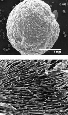
bone growth (38,41). Demineralized bone particles contain many kinds of osteogenic and chondrogenic cytokines such as bone morphogenetic protein, and are widely used as filling agent for bony defects. Because of their improved availability through the tissue bank industry, demineralized bone particles are widely used in clinical settings. To achieve more optimal results in the application of demineralized bone particles to tissue engineering, nanohybridization with synthetic (PLGA/demineralized bone particle hybrid scaffolds) and with natural organic compounds (collagen/demineralized bone particle hybrid scaffolds), has been carried out.
Porosity—the size of the mean diameter and the surface area—is a critical factor for the growth and migration of tissue into bioceramic scaffolds (60). Several methods have been introduced to optimize the fabrication of porous ceramics, such as dip casting, starch consolidation, the polymeric sponge method, the foaming method, organic additives, gel casting, slip casting, direct coagulation consolidation, hydrolysis-assisted solidification, and freezing methods. Therefore, it is very important to choose an appropriate method of preparation based on the physical properties of the desired organs.
A CYTOKINE-RELEASE SYSTEM FOR TISSUE ENGINEERING
Growth factors, a type of cytokine, are polypeptides that transmit signals to modulate cellular activity and tissue development including cell patterning, motility, proliferation, aggregation, and gene expression. As in the development of tissue-engineered organs, regeneration of functional tissue requires maintenance of cell viability and differentiated function, encouragement of cell proliferation, modulation of the direction and speed of cell migration, and regulation of cellular adhesion. For example, transforming growth factor-b1 (TGF-b1) might be required to induce osteogenesis and chondrogenesis from bone marrow derived mesenchymal stem cells. Also, brainderived neurotrophic factor (BDNF) can be enhanced to regenerate the spinal cord after injury. The easiest method for the delivery of growth factor is injection near the site of cell differentiation and proliferation (4). The most significant problems associated with the direct injection method are that the growth factors have a relatively short half-life, have a relatively high molecular weight and size, display very low tissue penetration, and have potential toxicity at systemic levels (4,10,11,16).
A promising technique for the improvement of their efficacy is to locally control the release of bioactive molecules for a specified release period to promote impregnation into a biomaterial scaffold. Through impregnation into the scaffold carrier, protein structure and biological activity can be stabilized to a certain extent, resulting in prolonging the release time at the local site. The duration of cytokine release from a scaffold can be controlled by the types of biomaterials used, the loading amount of cytokine, the formulation factors, and the fabrication process. The release mechanisms are largely divided into three categories: (1) diffusion controlled, (2) degradation controlled, and (3) solvent controlled. The mechanism of biodegrad-
BIOMATERIALS: TISSUE-ENGINEERING AND SCAFFOLDS |
375 |
Figure 4. (a) Bone marrow-derived mesenchymal stem cells impregnated TGF-b1 loaded alginate beads (original magnifications 40 ), and (b) inner structure of alginate beads (original magnifications 100 ).
able scaffold materials was regulated by degradation control, whereas that of the nondegradable material was regulated by diffusion and/or solvent control. The desired release pattern, such as a constant, pulsatile, and time programed behavior over the specific site and injury can be achieved by the appropriate combination of these mechanisms. Also, the cytokine-release system’s geometries and configurations can be altered to produce the necessary scaffold, tube, microsphere, injectable form or fiber (46,51,54).
Figures 4–6 show the TGF-b1 loaded alginate bead and the release pattern of TGF-b1 from alginate beads for the chondrogenesis from bone marrow-derived mesenchymal stem cells (64). The pore structure of 10 mm width and 100 mm length, was well suited to promote cell proliferation (Fig. 4); TGF-b1 released at a near zero-order rate for 35 days (Fig. 5). By using the alginate bead with TGF-b1 delivery system, chondrogenesis was successfully attained, as shown in Fig. 6.
To fabricate a new sustained delivery device for nerve growth factor (NGF), we developed NGF-loaded biodegradable PLGA films by a novel and simple sandwich solvent casting method for possible applications in the central nervous system (45). The release of NGF from the NGFloaded PLGA films was prolonged > 35 days with a zeroorder rate, without initial burst, and controlled by variation of different molecular weights and different NGF loading amounts as shown in Fig. 7. After 7 days, NGF
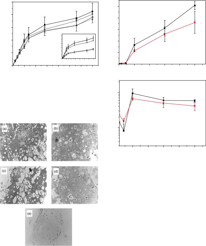
376 BIOMATERIALS: TISSUE-ENGINEERING AND SCAFFOLDS
1000
(pg/mL) |
800 |
|
|
|
|
|
|
|
|
|
|
|
|
|
|
|
|
1 |
|
|
|
|
|
|
|
|
-β |
|
|
|
(%) |
|
|
|
|
TGFof |
600 |
|
|
|
|
|
|
|
|
|
|
|
|
|
|
|
|
amount |
|
|
|
1 |
0.20 |
|
|
|
|
|
|
TGFof -β |
|
|
|
||
|
|
|
|
|
0.30 |
|
|
|
Released |
400 |
|
|
Releasedamount |
0.25 |
|
|
|
|
|
|
0 |
5 10 |
15 20 25 30 |
35 |
||
|
|
|
|
|
0.15 |
|
|
|
|
200 |
|
|
|
0.10 |
|
|
|
|
|
|
|
0.05 |
|
|
|
|
|
|
|
|
|
|
|
|
|
|
|
|
|
|
0.00 |
|
|
|
|
0 |
|
|
|
|
Release time (day) |
|
|
|
|
|
|
|
|
|
|
|
|
0 |
5 |
10 |
15 |
20 |
25 |
30 |
35 |
Release time (day)
Figure 5. Release pattern of TGF-b1 from TGF-b1 loaded alginate beads; (&) 0.5 mg TGF-b1, ( ) 0.5 mg TGF-b1 with heparin, (~) 1.0 mg TGF-b1, and (!) 1.0 mg TGF-b1 with heparin.
was released in a phosphate buffered saline solution (PBS; pH 7.0) and rat pheochromocytoma (PC-12) cells were cultured on the NGF-loaded PLGA film for 3 days. The released NGF stimulated neurite sprouting in the cultured
Figure 6. Safranin-O staining of chondrogenesis cells from bone marrow-derived mesenchymal stemcells in alginate beads. We can observe typical chondrocyte cells in alginate beads; (a) 0.5 mg mL 1 TGF-b1, (b) 1.0 mg mL 1 TGF-b1, (c) 0.5 mg mL 1 TGF-b1 with heparin (d) 1.0 mg mL 1 TGF-b1 with heparin, and
(e) control (without TGF-b1) (Original magnification 100 ).
(ng/mL) |
10 |
(a) |
|
||
NGF |
8 |
|
released |
6 |
|
amount |
|
|
4 |
|
|
Cumulative |
|
|
2 |
|
|
|
|
|
|
0 |
|
0 |
7 |
14 |
21 |
28 |
35 |
|
|
Release time (days) |
|
|
|
|
(b) |
|
|
|
|
(Release rate (ng/day)) |
1 |
|
|
|
|
. |
|
|
|
|
|
10 |
|
|
|
|
|
01 |
|
|
|
|
|
Log |
|
|
|
|
|
|
7 |
14 |
21 |
28 |
35 |
|
|
Release time (day) |
|
|
|
Figure 7. (a) Release profiles and (b) logarithmic plot of release rate for NGF from NGF-loaded PLGA films of 43,000 g/mol. ( ) 25.4 ng, and (&) 50.9 ng NGF/cm2 PLGA.
PC-12 cells; the remaining NGF in the NGF/PLGA film at 378C for 7 days was still bioactive, as shown in Fig. 8. These studies suggest that NGF-loaded PLGA sandwich film can be released in the delivery system over the desired time period, thus, it can be a useful neuronal growth culture serving as a nerve contact guidance tube for applications in neural tissue engineering.
One serious problem during the fabrication of cytokineloaded scaffolds is the denaturation and deactivation of cytokines, which result in loss of biological activity (65,66). Hence, the optimized method must be developed for stabilized cytokine-release scaffolds. For example, the release of NGF from a PLGA matrix was investigated using codispersants, such as polysaccharides (dextran) and proteins (albumin and b-lactoglobulin), with different molecular weights and charges. Negatively charged codispersants stabilized NGF in the PLGA system. Similarly, albumin stabilized epidermal growth factor (EGF) and heparin stabilized other growth factors.
Another available emerging technology is the ‘‘tethering of protein’’, that is, immobilization of protein on the surface
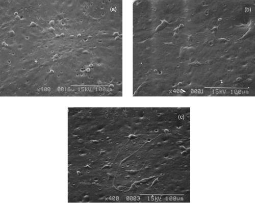
BIOMATERIALS: TISSUE-ENGINEERING AND SCAFFOLDS |
377 |
Figure 8. Effect of NGF released on neurites formation of PC-12 cells for 3-day cultivation on control (a) PLGA, (b) 25.4 ng, and (c) 50.9 ng NGF/cm2 PLGA just after 7 days. There were total medium changes (Molecular weight of PLGA; 83,000 g/mol, original magnification; 400 ).
of a scaffold matrix. Immobilization of insulin and transferrin to the poly(methylmetacrylate) films stimulates the growth of fibroblast cells compared to the same concentrations of soluble or physically adsorbed proteins (67). For the enhancement of cytokine activity, the PEO chain was applied as a short spacer between the surface of the scaffold and the cytokine. Tethered EGF, immobilized to the scaffold through the PEO chain, showed better DNA synthesis or cell rounding compared to the physically adsorbed EGF surface (68).
Conjugation of cytokine with an inert carrier prolongs the short half-life of protein molecules. Inert carriers are albumin, gelatin, dextran, and PEG. In PEGylation, PEG conjugated cytokine is most widely used for the release. This carrier appears to decrease the rate of cytokine degradation, attenuate the immunological response, and reduce clearance by the kidneys (69). Also, this PEGylated cytokine can be impregnated into scaffold materials by physical entrapment for sustained release. For example, the NGF-conjugated dextran (70,000 g mol 1) impregnated polymeric device was implanted directly into the brain of adult rats. Conjugated NGF could penetrate into the brain tissue 8 times faster than the unconjugated NGF. This conjugation method can be applied to the delivery of proteins and peptides. Immobilized RGD (arginin- glycine-aspartic acid) and YIGSR (tyrosin-leucineglycine- serine-arginine), which are typical ECM proteins, can enhance cell viability, function, and recombinant products in the cell (70).
Gene-activating scaffolds are being designed to deliver the targeted gene that results in the stimulation of specific cellular responses at the molecular level (4,3,11). Modification of bioactive molecules with resorbable biomaterial systems obtain specific interactions with cell integrins resulting in cell activation. These bioactive bioglasses and macroporous scaffolds also can be designed to activate genes that stimulate regeneration of living tissue (9). Gene delivery would be accomplished by complexation with positively charged polymers, encapsulation, and gel by means of the scaffold structure (51). Methods of gene delivery for gene-activating scaffolds are almost the same methods as for those with protein, drug, and peptides.
SCAFFOLD FABRICATION AND CHARACTERIZATION
Scaffold Fabrication Methods
Engineered scaffolds may enhance the functionalities of cells and tissues to support the adhesion and growth of a large number of cells because they provide a large surface area and pore structure within a 3D structure. The pore structure needs to provide enough space, permit cell suspension, and allow penetration of the 3D structure. Also, these porous structures help to promote ECM production, transport nutrients from nutrient media, and excrete waste products (10,12,15). Therefore, an adequate pore size and a uniformly distributed, and an interconnected pore structure, which allow for easy distribution of cells
378 BIOMATERIALS: TISSUE-ENGINEERING AND SCAFFOLDS
throughout the scaffold structure, are very important. Scaffold structures are directly related to their fabrication methods; over 20 methods have been proposed (10,71).
The most common and commercialized scaffold is the PGA nonwoven sheet (Albany International Research Co., Mansfield, MA; porosity 97%, 1–5 mm thick); it is one of the most tested scaffolds for tissue-engineered organs. To stabilize dimensionally and provide mechanical integrity, fiber-bonding technology was developed using heat and PLGA or PLA solution spray coating methods (72).
Porogen leaching methods have been combined with polymerization, solvent casting, gas foaming, or compression molding of natural and synthetic scaffolds biomaterials. The leaching of pore-generating particles such as sodium chloride crystal, sodium tartrate, and sodium citrate were sieved using a molecular sieve (10,71). PLGA, PLA, collagen, poly(orthoester), or SIS-impregnated PLGA scaffolds were successfully fabricated into a biodegradable sponge structure by this method with >93% porosity and a desired pore size of 1000 mm. By using the solvent casting/particulate leaching method, complex geometries, such as tube, nose, and specific organ types (e.g., nano-composite hybrid scaffolds), could be fabricated by means of conventional polymer-processing techniques, such as calendaring, extrusion, and injection. Complex geometry can be fabricated from porous film lamination (33,39,42,47). The advantage of this method is its easy control of porosity and geometry. However, the disadvantages include: (1) the loss of watersoluble biomolecules or cytokines during the leaching porogen process, (2) the possibility that the remaining porogen as a salt can be harmful to the cell culture, and (3) the different geometry surface and cross-section that results.
The gas-foaming method consists of a solid scaffold matrix exposured to a sudden expansion of CO2 gas under high pressure, which results in the formation of a sponge structure due to nucleation and expansion in a dissolved CO2 scaffold matrix. The PLGA scaffolds with >93% porosity and 100 mm median pore size were developed by this method (71). A significant advantage is that there is no loss of bioactive molecules in the scaffold matrix, since there is no more need for the leaching process and there is no residual organic solvent. The disadvantage is the presence of a skimming film layer on the scaffold surface, which results in a need for an additional process to remove the skin layer.
The phase-separation method is divided into the freeze– drying, freeze–thaw, freeze–immersion precipitation, and emulsion freeze–drying techniques (37,72,73). Phase separation by freeze–drying can be induced by the appropriate concentration of polymer solution obtained by rapid freezing. Then, the used solvent is removed by freeze– drying, leaving in porous structure made up of a portion of the solvent. These can be collagen scaffolds with pores50–150 mm; collagen–glycosaminoglycan blend scaffolds with an average pore size 90–120 mm; or chitosan scaffolds with a pore size 1–250 mm, dependent on the freezing conditions (71). Also, scaffold structures of synthetic polymers, such as PLA or PLGA, have been successfully made much >90% porosity and 15–250 mm size by this method. The freeze–thaw technique induces phase separation between a solvent and a hydrophilic monomer upon freezing, followed by the polymerization of the hydro-
philic monomer by means of ultraviolet (UV) irradiation and removal of the solvent by thawing. This technique leads to the formation of a macroporous hydrogel. A similar method is the freeze–immersion precipitation technique. The polymer solution is cooled, immersed in a nonsolvent, and then the vaporized solvent leads to a porous scaffold structure. Also, the emulsion freeze–drying method is used to fabricate a porous structure. Mixtures of polymer solution and nonsolvent are thoroughly sonicated, freezed quickly in liquid nitrogen at 198 8C, and then freeze– dried, resulting in a sponge structure. The advantage of these techniques is that they result in the loading of hydrophilic or hydrophobic bioactive molecules, whereas the disadvantages are relatively small pore sized scaffolds with precise pore structures that are hard to control (73).
Nano-electrospinning of PGA, PLA, PLGA, caprolactone copolymers, collagen, and elastin, has been extensively developed (74). For example, electrostatic processing can consistently produce PGA fiber diameters 41 mm. By controlling the pick-up of these fibers, the orientation and mechanical properties can be tailored to the specific needs of the injured site. Also, collagen electrospinning was performed utilizing type I collagen dissolved in 1,1,1,3,3,3-hexafluoro-2-propanol with a concentration of 0.083 g mL 1. The optimally electrospun type I collagen nonwoven fabric appeared with an average diameter of 100 40 nm, which resulted in biomimicking fibrous scaffolds.
Injectable gel scaffolds have also been reported (10,16,51,54). An injectable, gelforming scaffold offers several advantages: (1) it can fill any space based on its ability to flow; (2) it can load various types of bioactive molecules and cells by simple mixing; (3) it does not contain residual solvents that may be present in a preformed scaffold; and (4) it does not require a surgical procedure for placement. Typical examples are thermosensitive gels such as Pluronics and PEG-PLGA- PEG triblock copolymer, pH sensitive gels such as chitosan and its derivates, an ionically cross-linked gel such as alginate, and fibrin and hyaluronan gels, as well as others previously introduced in the Natural Polymers section. In the near future, multifunctional gels which are tissue-spe- cific, have a very fast sol–gel transition, are fully degradable over the necessary time period will be available.
Newly hybridized fabrication techniques such as organic–inorganic and synthetic–natural techniques at the nanosize level that biomimic, are also being developed for use in engineered scaffolds.
Physicochemical Characterization of Scaffolds
For the successful achievement of 3D scaffolds, several characterization methods are needed. These methods can be divided into four categories. (1) Morphology—porosity, pore size,andsurface area;(2)mechanicalproperties—compressive and tensile strength; (3) bulk properties—degradation and its relevant mechanical properties; and (4) surface properties—surface energy, chemistry, and charge.
Porosity is defined as the fraction of the total volume occupied by voids that appear as percentages. The most widely used methods for the measurement of porosity are mercury porosimetry, scanning electron microscopy (SEM), and confocal laser microscopy.
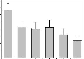
Mechanical properties are extremely important when designing tissue-engineered products. Conventional testing instruments can be used to determine the mechanical properties of a porous structure. Mechanical tests can be divided into (1) creep tests, (2) stress–relaxation tests, (3) stress– strain tests, and (4) dynamic mechanical tests. These test methods are similar to those used for conventional biomaterials.
The rate of degradation of manufactured scaffolds is a very important factor in the design of tissue-engineered products. Ideally, the scaffold constructs provide mechanical and biochemical supports until the entire tissue regenerates, then the scaffold completely biodegrades at a rate consistent with tissue generation. Immersion studies are commonly conducted to track the degradation of the biodegradable matrix. Changes in weight loss and molecular weight can be evaluated by the chemical balance of the matrix, by SEM, and by gel permeation chromatography. These results produce the mechanism of biodegradation.
It is generally recognized that the adhesion and proliferation of different types of cells on polymeric materials depend largely on the materials’ surface characteristics, such as wettability (hydrophilicity/hydrophobicity of surface free energy), chemistry, charge, roughness, and rigidity (37,40,41,44,45). The 3D aspects of tissue engineering are more important for cell migration, proliferation, DNA/ RNA synthesis, and phenotype presentation on the scaffold materials. Surface chemistry and charge can be analyzed by electron scanning chemical analysis and streaming potential, respectively. Also, wettability of the scaffold surface can be measured by the contact angle using static and dynamic methods.
SURFACE MODIFICATION OF SCAFFOLDS FOR THE IMPROVEMENT OF BIOCOMPATIBILITY
As explained above, the surface properties of scaffold materials are very important. For example, the hydrophobic surfaces of PLA, PGA, and PLGA possess high interfacial free energy in aqueous solutions, which tends to unfavorably influence their cell, tissue, and blood compatibility in the initial stage of contact. Moreover, it does not allow the nutrient media to permeate into the center of the scaffolds. For these reasons, a surface treatment is applied by several methods: (1) chemical treatment using oxidants, (2) physical treatment using glow discharge, and (3) a blend with hydrophilic biomaterials or bioactive molecules.
The physicochemical treatment has been demonstrated to improve the wetting property and hydrophilicity of PLGA porous scaffolds fabricated by the emulsion freeze–drying method (37,45). The chemical treatments were 70% perchloric acid, 50% sulfuric acid, and 0.5 N sodium hydroxide solution. The physical methods included corona and plasma treatments generated by a radiofrequency glow discharge. After treatment, water contact angles decreased (Fig. 9). The wetting property of chemically treated PLGA scaffolds also ranked in the order of perchloric acid, sulfuric acid, and sodium hydroxide solution by blue dye intrusion experiment, whereas phy-
BIOMATERIALS: TISSUE-ENGINEERING AND SCAFFOLDS |
379 |
|
80 |
|
angle (degrees) |
70 |
|
60 |
||
contact |
||
|
||
Water |
50 |
|
|
||
|
40 |
Control |
Sulfuric acid |
Chloric acid |
Sodium hydroxide |
Corona |
Plasma |
Figure 9. Changes of water contact angles after physicochemical treatment. The significant decreasing of water contact angle, that is, increased hydrophilicity, was observed.
sical methods had no effect, as shown in Fig. 10. Thus, the chemical treatment method may be useful in uniform cell seeding into porous biodegradable PLGA scaffolds. Wettability plays an important role in cell adhesion, spreading, and growth on the PLGA surface, and the intrusion of nutrient media into the PLGA scaffold.
Scaffolds impregnated with bioactive and hydrophilic material might be better for cell proliferation, differentiation, and migration due to cell stimulation. To give scaffolds new bioactive functionality from SIS powder as a natural source, scaffolds consisting of porous SIS/PLA and SIS/PLGA as a natural–synthetic composite, were prepared by the solvent casting–salt leaching method for use in tissue-engineered bone. A uniform distribution of good interconnected pores from the surface-to-core region was observed (pore size 40–500 mm), independent of the SIS amount, by using the solvent casting–salt leaching method. Porosities, specific pore areas as well as pore size distribution were also similar. After the fabrication of SIS/ PLGA hybrid scaffolds, the wetting properties were greatly improved resulting in more uniform cell seeding and distribution, as shown in Fig. 11. Five different scaffolds, a PGA nonwoven mesh scaffold without glutaraldehyde (GA) treatment, PLA scaffolds without and with GA treatment, PLA/SIS scaffolds without and with GA treatment, were implanted into the back of nude mouse to observe the effect of SIS on the induction of cell proliferation by hematoxylin and eosin using von Kossa staining, for 8 weeks. It was observed that the effect of PLA/SIS scaffolds with GA treatment on bone induction is stronger than PLA scaffolds, that is the effects of PLA/SIS scaffolds with GA treatment > PLA/SIS scaffolds without GA treatment > PGA nonwoven > PLA scaffolds only with GA treatment ¼ PLA scaffolds only without GA treatment for osteoinduction activity (Fig. 12).
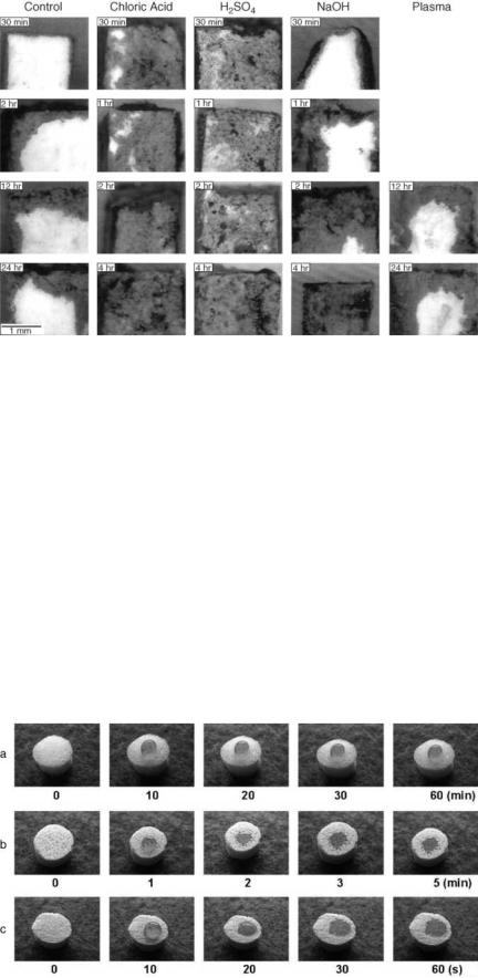
380 BIOMATERIALS: TISSUE-ENGINEERING AND SCAFFOLDS
Figure 10. Wetting properties of physicochemically treated porous PLGA scaffolds by blue dye intrusion methods for 0.5, 1, 2, 4, 12, and 24 h.
STERILIZATION METHODS FOR SCAFFOLDS
The sterilizability of polymeric scaffold biomaterials is an important property, since polymers have lower thermal and chemical stability than other materials, such as ceramics and metals. Consequently, polymers are more difficult to sterilize using conventional techniques. Commonly used sterilization techniques are dry heat, autoclaving, radiation, and ethylene oxide gas (EOG). In addition, plasma glow discharge and electron beam sterilization recently were proposed due to their convenience (6,75).
In dry heat sterilization, the temperature varies between 160 and 190 8C. This temperature is above the melting and softening temperatures of many linear polymers, such as PLGA, resulting in the shrinking of the scaffold dimension. The PLA scaffolds were sterilized at 129 8C for 60 s, resulting in a minimal change in tensile properties. One of the significant problems was a decrease in molecular weight, which might have an affect on the
degradation kinetics of the polymers. In the case of polyamide (Nylon) used as a nonbiodegradable polymer, oxidation occurs at the dry sterilization temperature, even though this is below its melting temperature. The only polymers that can safely be dry sterilized are polytetrafluoroethylene (PTFE) and silicone rubber. However, ceramic and metallic scaffolds were safe in this temperature range.
Steam sterilization (autoclaving) is performed under high steam pressure at a relatively low temperature (125–130 8C). However, if the polymer is subjected to attack by water vapor, this method cannot be employed. The PVC, polyacetals, PE (low density variety), and polyamides belong to this category. In the poly(a-hydroxy ester) family, a trace of water can deteriorate the PLGA backbone.
Chemical agents such as EOG and propylene oxide gases, and phenolic and hypochloride solutions are used widely for sterilizing all biomaterials, since they can be used at relatively low temperatures. Chemical agents
Figure 11. Wetting properties of SIS impregnated PLGA scaffolds by red dye intrusion methods. We observed the rapid penetration of water into SIS/PLGA scaffolds compared to the control PLGA scaffolds; (a) control PLGA,
(b) 40% SIS/PLGA, and (c) 160% SIS/PLGA scaffolds.
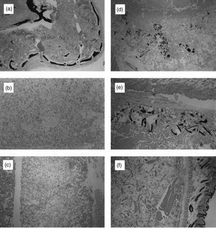
BIOMATERIALS: TISSUE-ENGINEERING AND SCAFFOLDS |
381 |
sometimes cause polymer deterioration even when sterilization takes place at room temperature. However, the time of exposure is relatively short (overnight), and most scaffolds can be sterilized with this method. The cold EOG sterilization method is the most widely used, with conditions of 358C and 95% humidity. While the hot EOG method, which uses 608C and 95% humidity, can cause shrinkage of the PLGA scaffold. One significant problem is residual EOG, which is harmful on the surface and within the polymer. Therefore, it is important that the scaffolds are subjected to adequate degassing or aeration subsequent to EOG sterilization, so that the concentration of residual EOG can be reduced to acceptable levels.
Radiation sterilization using isotopic 60Co can also deteriorate polymers, since at high dosages the polymer chains can be dissociated or crosslinked according to the characteristics of the chemical structures. At a 2.5 Mrad dose, the tensile strength and molecular weight of PLGA decreases. Also, there is a rapid decrease in the molecular weight of the PGA nonwoven felt with increasing doses of radiation. It is important to remember that the properties and useful lifetime of the PLGA implant can be significantly affected by irradiation. In the case of polyethylene, it becomes a brittle and hard material at doses as high as 25 Mrad; This is due to a combination of random chain scission crosslinking. Polypropylene will often discolor during irradiation giving the product an undesirable tint, but a more severe problem is the embrittlement resulting in flange breakage, luer crack-
Figure 12. Photomicrographs of von Kossa and H&E histological sections of implanted
(a) PGA nonwoven, (b) PLA scaffold only without GA treatment, (c) PLA scaffold only with GA treatment, (d) SIS/PLA scaffold without GA treatment, (e) SIS/PLA scaffold with GA treatment, and (f) SIS/PLA scaffold with GA treatment (H&E) (Original magnification 100 ).
ing, and tip breakage. The physical properties continued to deteriorate with time following irradiation.
Sterilization methods might significantly affect the physicochemical properties of the scaffold matrix. The specific effects with various methods are determined by the kinds of scaffold materials themselves, the scaffold preparation methods, and the sterilization factors. It is essential that a new standard for sterilizing scaffold devices be designed and established.
CONCLUSIONS
Tissue engineering, including regenerative medicine in recognition of its tremendous potential, has received a revolutionary ‘‘research push.’’ As a result, there have been many reports on the successful regeneration of tissues and organs including skin, bone, cartilage, the peripheral and central nerves, tendon, muscle, cornea, bladder and urethra, and liver as well as composite systems like the human phalanx and joint, using scaffold biomaterials from polymers, ceramic, metal, composites and its hybrids. As previously emphasized, scaffold materials must contain a site of cellular and molecular induction and adhesion, and must allow for the migration and proliferation of cells through porosity. They must also maintain strength, flexibility, biostability, and biocompatibility to mimic a more natural, 3D environment. From this standpoint, control over a precise biochemical signal must be fostered by the combination
382 BIOMATERIALS: TISSUE-ENGINEERING AND SCAFFOLDS
of a scaffold matrix and bioactive molecules including genes, peptide molecules, and cytokines. Moreover, the combination of cells and redesigned bioactive scaffolds should expand to a tissue level of hierarchy. To achieve this goal, novel scaffold biomaterials, scaffold fabrication methods, and characterization methods must be developed.
ACKNOWLEDGMENTS
This work was supported by grants from the Korea Ministry of Wealth and Health (0405-BO01-0204-0006) and stem cell Research Center (SC3100).
BIBLIOGRAPHY
Cited References
1.Langer R, Vacanti J. Tissue engineering. Science 1993;260: 920–926.
2.Nerem RM , Sambanis A. Tissue engineering: from biology to biological substitutes. Tissue Eng 1995;1:3–13.
3.Griffith LG, Naughton G. Tissue engineering—Current challenges and expanding opportunity. Science 2002;295:1009– 1014.
4.Baldwin SP, Saltzman WM. Materials for protein delivery in tissue engineering. Adv Drug Deliv Rev 1998;33:71–86.
5.Mann BK, West JL. Tissue engineering in the cardiovascular system: Progress toward a tissue engineered heart. Anat Record 2001;263:367–371.
6.Lee HB, Khang G, Lee JH. Chapter 3, Polymeric biomaterials. In: Park JB, Bronzino JD, editors. Biomaterials: Principles and Applications. Boca Raton: CRC Press; 2003.
7.Patrick CW, Jr. Tissue engineering strategies for adipose tissue repair. Anat Record 2001;263:361–376.
8.Petit-Zeman S. Regenerative medicine. Nature Biotech 2001;19:201–206.
9.Hench LL, Polak JM. Third-generation biomedical materials. Science 2002;295:1014–1017.
10.Seal BL, Otero TC, Panitch A. Polymeric biomaterials for tissue and organ generation. Mater Sci Eng 2001;R34:147–230.
11.Babensee JE, McIntire LV, Mikos AG. Growth factor delivery for tissue engineering. Pharm Res 2000;17:497–504.
12.Chaignaud BE, Langer R , Vacanti JP. Chapter 1, The history of tissue engineering using synthetic biodegradable polymer scaffolds and cells. In: Atala A, Mooney DJ, editors. Synthetic Biodegradable Polymer Scaffolds. Boston: Birkhauser; 1996.
13.Rose FRA, Oreffo ROC. Bone tissue engineering: Hope vs Hype. Biochem Biophys Res Commun 2002;292:1–7.
14.Freyman TM, Yannas IV, Gibson LJ. Cellular materials as porous scaffolds for tissue engineering. Prog Mater Sci 2001;46:273–282.
15.Woolverton CJ, Fulton JA, Lopina ST, Landis WJ. Chapter 3, Mimicking the natural tissue environment. In: Lewandrowski K-U, Wise DL, Trantolo DJ, Gresser JD, Yasemski MJ, Altobeli DE, editors. Tissue Engineering and Biodegradable Equivalents: Scientific and Clinical Applications. New York: Marcel Dekker; 2002.
16.Tabata Y. The importance of drug delivery systems in tissue engineering. PSTT 2000;3:80–89.
17.Wong WH, Mooney DJ. Chapter 4, Synthesis of properties of biodegradable polymers used as synthetic matrices for tissue engineering. In: Atala A, Mooney DJ, editors. Synthetic Biodegradable Polymer Scaffolds. Boston: Birkhauser; 1996.
18.Rwoley JA, Madlambayan G, Mooney DJ. Alginate hydrogels as synthetic extracellular matrix. Biomaterials 1999;20:45–53.
19.Shakibaei M, De Souza P. Differentiation of mesenchymal limb bud cells to chondrocytes in alginate bead. Cell Biol Int 1997;21:75–86.
20.Madihally SV, Matthew HW. Porous chitosan scaffolds for tissue engineering. Biomaterials 1999;20:1133–1142.
21.Kang HW, Tabata Y, Ikada Y. Fabrication of porous gelatin scaffolds for tissue engineering. Biomaterials 1999;20:1339– 1344.
22.Dunn CJ, Goa KL. Fibrin sealant: A review of its use in surgery and endoscopy. Drugs 1999;58:863–886.
23.Mayne R, Burgeson RE. Structure and function of collagen types. In: Mecham RP, editor. Biology of extracellular matrix: A Series. Orlando: Academic Press; 1987.
24.Li S-T. Chapter 6, Biologic biomaterials: Tissue-derived biomaterials (Collagen). In: Park JB, Bronzino JD, editors. Biomaterials: Principles and Applications. Boca Raton (FL): CRC Press; 2003.
25.Schense JC, Bloch J, Aebischer P, Hubbell JA. Enzymatic incorporation of bioactive peptides into fibrin matrices enhances neurite extension. Nature Biotech 2000;18:415–419.
26.Zacchi V, Soranzo C, Cortivo R, Radice M, Brun P, Abatangelo G. In vitro engineering of human skin-like tissue. J Biomed Mater Res 1998;40:187–194.
27.Malette WG, Quigley HJ, Gaines RD, Johnson ND, Rainer WG. Chitosan: a new hemostatic. Ann Thorac Surg 1983;36: 55–58.
28.Sechriest VF, Miao YJ, Niyibizi C, Westerhauzen-Larson A, Matthew HW, Evans CF, Fu FH, Suh J-K. GAG-augmented polysaccharide hydrogel: a novel biocompatible and biodegradable material to support chondrogenesis. J Biomed Mater Res 2000;49:534–541.
29.Lee YM, Park YJ, Lee SJ, Ku Y, Han SB, Choi SM, Klokkevoid PR, Chung CP. Tissue engineered bone formation using chitosan/tricalcium phosphate sponges. J Periodontol 2000; 71:410–417.
30.Lee DA, Noguchi T, Knight MM, O’Donnell L, Bently G, Bader DL. Response of chondrocyte subpopulations cultured within unloaded and loaded agarose. J Orthop Res 1998;16: 726–733.
31.Lee DA, Frean SP, Lee P, Bader DL. Dynamic mechanical compression influences nitric oxide production by articular chondrocytes seeded in agarose. Biochem Biophys Res Commun 1998;251:580–585.
32.Badylak SF, Record R, Lindberg K, Hodde J, Park K. Small intestine submucosa: a substrate for in vitro cell growth. J Biomater Sci, Polymer Ed 1998;9:863–878.
33.Khang G, Shin P, Kim I, Lee B, Lee SJ, Lee YM, Lee HB, Lee I. Preparation and characterization of small intestine submucosa particle impregnated PLA scaffolds: The application of tissue engineered bone and cartilage. Macromol Res 2002;10:158–167.
34.Badylak SF. The extracellular matrix as a scaffolds for tissue reconstruction. Cell Develop Biol 2002;13:377–383.
35.Gustafson C-J, Katz G. Cultured autologous keratinocytes on a cell-free dermis in the treatment of full-thickness wounds. Burns 1999;25:331–335.
36. Williams SF, Martin DP, Horowitz DM, Peoples OP. PHA applications: Addressing the price performance issue, I. Tissue Engineering. Int J Biolog Macromol 1999;25:111–121.
37.Khang G, Lee HB. Chapter 67. Cell-synthetic surface interaction: Physicochemical surface modifications. In: Atala A, Lanza R, editors. Orlando: Academic Press; 2001.
38.Khang G, Seong H, Lee HB. Sustained delivery of drugs with biodegradable. In: Hsuie GH, Okano T, Kim YU, Sung W-W, Yui N, Park KD, editors. Taipei, Taiwan: Princeton International Publishing Co.; 2002.
39.Khang G, Lee SJ, Han CW, Rhee JM, Lee HB. Preparation and characterization of natural/synthetic hybrid scaffolds.
In: Elcin M, editor. London, England: Kluwer-Plenum Press; 2003.
40.Khang G, Lee JH, Lee I, Rhee JM, Lee HB. Interaction of different types of cells on PLGA surfaces with wettability chemogradient. Macromol Res 2000;8:276–284.
41.Khang G, Choi MK, Rhee JM, Rhee SJ, Lee HB, Iwasaki Y, Nakabayashi N, Ishihara K. Biocompatibility of poly(MPC- co-EHMA)/PLGA blends. Macromol Res 2001;9:107–115.
42.Khang G, Park CS, Rhee JM, Lee SJ, Lee YM, Lee I, Choi MK, Lee HB. Preparation and characterization of demineralized bone particle impregnated PLA scaffolds. Macromol Res 2001;9:267–276.
43.Choi HS, Khang G, Shin H-C, Rhee JM, Lee HB. Preparation and characterization of fentanyl-loaded PLGA microspheres; In vitro release profiles. Int J Pharm 2002;234:195–203.
44.Lee SJ, Khang G, Lee YM, Lee HB. Interaction of human chondrocyte and fibroblast cell onto chloric acid treated poly(a-hydroxy acid) surface. J Biomater Sci, Polym Ed 2002;13:197–212.
45.Khang G, Choi CW, Rhee JM, Lee HB. Interaction of different types of cells on physicochemically treated PLGA surfaces. J Appl Polym Sci 2002;85:1253–1262.
46.Khang G, Jeon EK, Rhee JM, Lee I, Lee SJ, Lee HB. Controlled release of NGF from sandwiched PLGA films for the application of neural tissue engineering. Macromol Res 2003; 11:334–340.
47.Jang JW, Lee B, Han CW, Lee I, Lee HB, Khang G. Preparation and characterization of ipriflavone-loaded PLGA scaffolds for tissue engineered bone. Polymer(Korea) 2003;27:226–234.
48.Khon J, Langer R. Chapter 2.5, Bioresorbable and bioerodible materials. In: Ratner BD, Hoffman AS, Scheon FJ, Lemons JE, editors. Biomaterials Science: An Introduction to Materials in Medicine, San Diego: Academic Press; 1996.
49.Vacanti CA, Langer R, Schloo B, Vacanti JP. Synthetic polymers seeded with chondrocytes provide a template for new cartilage formation. Plast Reconstr Surg 1991;88:753–759.
50.Burg KJL, Porter S, Kellam JF. Biomaterials developments for tissue engineering. Biomaterials 2000;21:2347–2359.
51.Gutowska A, Jeong B, Jasionowski M. Injectable gel for tissue engineering. Anat Record 2001;263:342–349.
52.Suggs LJ, Krishna RS, Garcia CA, Peter SJ, Anderson JM, Mikos AG. In vitro and in vivo degradation of poly(propylene fumarate-co-ethylene glycol) hydrogel. J Biomed Mater Res 1998;42:312–320.
53.Harris JM, editor. Poly(ethylene glycol) Chemistry: Biotechnical and Biomedical Applications. New York: Plenum Publish. Co.; 1997.
54.Qui Y, Park K. Environment-sensitive hydrogels for drug delivery. Adv Drug Deliv Rev 2001;53:321–339.
55.Webb D, An YH, Gutowska A, Mironov VA, Friedman RJ. Propagation of chondrocytes using thermosensitive polymer gel culture. Orthoped J Musc Orthoped Surg 2000;3:18– 22.
56.Sims CD, Butler P, Casanova R, Lee BT, Randolph MA, Lee A, Vacanti CA, Yaremchuk MJ. Injectable cartilage using polyethylene oxide polymer substrate. Plast Reconstruct Surg 1996;95:843–850.
57.Laurencin CT, El-Amin SF, Ibim SE, Willoughby DA, Attawia M, Allcock HR, Ambrosio AA. A highly porous 3-dimensional polyphosphazene polymer matrix for skeletal tissue engineering. J Biomed Mater Res 1996;30:133–138.
58.Agarwal S, Gassner R, Piesco NP, Ganta SR. Chapter 7, Biodegradable urethanes for biomedical applications. In: Lewandrowski K-U, Wise DL, Trantolo DJ, Gresser JD, Yasemski MJ, Altobeli DE, editors. Tissue Engineering and Biodegradable Equivalents: Scientific and Clinical Applications. New York: Marcel Dekker; 2002.
BIOMATERIALS: TISSUE-ENGINEERING AND SCAFFOLDS |
383 |
59.Zhang JY, Beckman EJ, Piesco NP, Agarwal S. A new peptide based urethane polymer: synthesis, degradation, and potential to support cell growth in vitro. Biomaterials 2000;21:1247– 1258.
60.Billotte WG. Chapt. 2, Ceramic biomaterials. In: Park JB, Bronzino JD, editors. Biomaterials: Principles and Applications. Boca Raton (FL): CRC Press; 2003.
61.Hench LL. Bioactive ceramics. Ann NY Acad Sci 1988; 523:54–71.
62.Frician JC, Bareille R, Rouais F. In vitro dissolution of coral in periodontal or fibroblast cell culture. J Dent Res 1998;77:406–411.
63.Yoshikawa T, Oghushi H, Uemura T. Human marrow cells derived cultured bone in porous ceramics. Bio-Med Mater Eng 1998;8:311–320.
64.Unpublished data.
65.Krewson C, Dause R, Mak M, Saltzman WM. Stabilization of nerve growth factor in controlled release polymers and in tissue. J Biomater Sci, Polym Ed 1996;8:103–117.
66.Haller MF, Saltzman WM. Localized delivery of proteins in the brain. Pharm Res 1998;15:377–385.
67.Ito Y, Lui SQ, Imanishi Y. Enhancement of cell growth on growth factor-immobilized polymer films. Biomaterials 1991; 12:449–453.
68.Khul PR, Grriffith-Cima LG. Tethered epidermal growth factor as a paradigm for growth factor-induced stimulation from the solid phase. Nature Med 1996;2:1022–1027.
69.Duncan R, Spreafico F. Polymer conjugates. Pharmacokinetic considerations for design and development. Clin Pharmacokinet 1994;27:290–306.
70.Massia SP, Hubbell JA. Covalent surface immobilization of Arg-Gly-Asp- and Tyr-Ile-Gly-Ser-Arg-containing peptides to obtain well-defined cell-adhesive substrate. Anal Biochem 1990;187:292–301.
71.Leibmann-Vinson A, Hemperly JJ, Guarino RD, Spargo CA, Heidaran MA. Chapter 36, Bioactive extracellular matrices: Biological and biochemical evaluation. In: Lewandrowski KU, Wise DL, Trantolo DJ, Gresser JD, Yasemski MJ, Altobeli DE, editors. Tissue Engineering and Biodegradable Equivalents: Scientific and Clinical Applications. New York: Marcel Dekker; 2002.
72.Thompson RC, Wake MC, Yasemski MJ, Mikos AG. Biodegradable polymer scaffolds to regenerate organs. Adv Polym Sci 1995;122:245–274.
73.Khang G, Jeon JH, Cho JC, Lee HB. Fabrication of tubular porous PLGA scaffolds by emulsion freeze drying methods. Polymer(Korea) 1999;23:471–177.
74.Bowlin GL, Pawlowski KJ, Boland ED, Simpson DG, Fenn JB, Wnek GE, Stitzel JD. Chapter 9, Electrospinning of polymer scaffolds for tissue engineering. In: Lewandrowski K-U, Wise DL, Trantolo DJ, Gresser JD, Yasemski MJ, Altobeli DE, editors. Tissue Engineering and Biodegradable Equivalents: Scientific and Clinical Applications. New York: Marcel Dekker; 2002.
75.Athanasious KA, Neiderauer GG, Agrawal CM. Sterilization, toxicity, biocompatibility and clinical applications of polylactic acid/polyglycolic acid copolymers. Biomaterials 1996;17:93–102.
Reading List
Jeon EK, Khang G, Lee I, Rhee JM, Lee HB. Preparation and release profile of NGF-loaded PLA scaffolds for tissue engineered nerve regeneration. Polymer(Korea) 2001;25:893–901.
See also ENGINEERED TISSUE; STERILIZATION OF BIOLOGIC SCAFFOLD MATERIALS.
