

- •VOLUME 1
- •CONTRIBUTOR LIST
- •PREFACE
- •LIST OF ARTICLES
- •ABBREVIATIONS AND ACRONYMS
- •CONVERSION FACTORS AND UNIT SYMBOLS
- •ABLATION.
- •ABSORBABLE BIOMATERIALS.
- •ACRYLIC BONE CEMENT.
- •ACTINOTHERAPY.
- •ADOPTIVE IMMUNOTHERAPY.
- •AFFINITY CHROMATOGRAPHY.
- •ALLOYS, SHAPE MEMORY
- •AMBULATORY MONITORING
- •ANALYTICAL METHODS, AUTOMATED
- •ANALYZER, OXYGEN.
- •ANESTHESIA MACHINES
- •ANESTHESIA MONITORING.
- •ANESTHESIA, COMPUTERS IN
- •ANGER CAMERA
- •ANGIOPLASTY.
- •ANORECTAL MANOMETRY
- •ANTIBODIES, MONOCLONAL.
- •APNEA DETECTION.
- •ARRHYTHMIA, TREATMENT.
- •ARRHYTHMIA ANALYSIS, AUTOMATED
- •ARTERIAL TONOMETRY.
- •ARTIFICIAL BLOOD.
- •ARTIFICIAL HEART.
- •ARTIFICIAL HEART VALVE.
- •ARTIFICIAL HIP JOINTS.
- •ARTIFICIAL LARYNX.
- •ARTIFICIAL PANCREAS.
- •ARTERIES, ELASTIC PROPERTIES OF
- •ASSISTIVE DEVICES FOR THE DISABLED.
- •ATOMIC ABSORPTION SPECTROMETRY.
- •AUDIOMETRY
- •BACTERIAL DETECTION SYSTEMS.
- •BALLOON PUMP.
- •BANKED BLOOD.
- •BAROTRAUMA.
- •BARRIER CONTRACEPTIVE DEVICES.
- •BIOCERAMICS.
- •BIOCOMPATIBILITY OF MATERIALS
- •BIOELECTRODES
- •BIOFEEDBACK
- •BIOHEAT TRANSFER
- •BIOIMPEDANCE IN CARDIOVASCULAR MEDICINE
- •BIOINFORMATICS
- •BIOLOGIC THERAPY.
- •BIOMAGNETISM
- •BIOMATERIALS, ABSORBABLE
- •BIOMATERIALS: AN OVERVIEW
- •BIOMATERIALS: BIOCERAMICS
- •BIOMATERIALS: CARBON
- •BIOMATERIALS CORROSION AND WEAR OF
- •BIOMATERIALS FOR DENTISTRY
- •BIOMATERIALS, POLYMERS
- •BIOMATERIALS, SURFACE PROPERTIES OF
- •BIOMATERIALS, TESTING AND STRUCTURAL PROPERTIES OF
- •BIOMATERIALS: TISSUE-ENGINEERING AND SCAFFOLDS
- •BIOMECHANICS OF EXERCISE FITNESS
- •BIOMECHANICS OF JOINTS.
- •BIOMECHANICS OF SCOLIOSIS.
- •BIOMECHANICS OF SKIN.
- •BIOMECHANICS OF THE HUMAN SPINE.
- •BIOMECHANICS OF TOOTH AND JAW.
- •BIOMEDICAL ENGINEERING EDUCATION
- •BIOSURFACE ENGINEERING
- •BIOSENSORS.
- •BIOTELEMETRY
- •BIRTH CONTROL.
- •BLEEDING, GASTROINTESTINAL.
- •BLADDER DYSFUNCTION, NEUROSTIMULATION OF
- •BLIND AND VISUALLY IMPAIRED, ASSISTIVE TECHNOLOGY FOR
- •BLOOD BANKING.
- •BLOOD CELL COUNTERS.
- •BLOOD COLLECTION AND PROCESSING
- •BLOOD FLOW.
- •BLOOD GAS MEASUREMENTS
- •BLOOD PRESSURE MEASUREMENT
- •BLOOD PRESSURE, AUTOMATIC CONTROL OF
- •BLOOD RHEOLOGY
- •BLOOD, ARTIFICIAL
- •BONDING, ENAMEL.
- •BONE AND TEETH, PROPERTIES OF
- •BONE CEMENT, ACRYLIC
- •BONE DENSITY MEASUREMENT
- •BORON NEUTRON CAPTURE THERAPY
- •BRACHYTHERAPY, HIGH DOSAGE RATE
- •BRACHYTHERAPY, INTRAVASCULAR
- •BRAIN ELECTRICAL ACTIVITY.
- •BURN WOUND COVERINGS.
- •BYPASS, CORONARY.
- •BYPASS, CARDIOPULMONARY.
342BIOMATERIALS, SURFACE PROPERTIES OF
14.Kurtz SM, Muratoglu OK, Evans M, Edidin AA. Advances in the processing, sterilization of ultra-high molecular weight polyethylene for total joint arthroplasty. Biomaterials 1999;20:1659–1688.
15.Teoh SH, Tang ZG, Hastings GW. Thermoplastic polymers in biomedical applications: Structures, properties and and processing. In: Black J, Hastings G, editors. Handbook of Biomaterial Properties. London: Chapman & Hall; 1998.
16.(a) Ramshaw JAM, Werkmeister JA, Edwards GA. Tissuepolymer composite vascular prostheses. (b) Kowligi RR, Edwin TJ, Banas C, Calcote RW. Vascular grafts: materials, methods, and clinical applications. (c) Planell JA, Vila MM, Gil FJ, Driessens FCM. Acrylic bone cements. (d) Felder G III, Donachy JH, Sr. Fabrication techniques and polymer considerations for the blood contacting components of the Penn State circu- latory–assist devices. In: Wise DL, Trantolo DJ, Altobelli DE, Yaszemski MJ, Gresser JD, Schwartz ER, editors. Encyclopedic Handbook of Biomaterials and Bioengineering, Part B: Applications Vol. 2. New York: Marcel Dekker; 1995.
17.(a) Tomita N, Fujita H, Nagata K. Polymers for artificial joints. (b) El-Zaim HS, Heggers JP. Silicones for pharmaceutical and biomedical applications. (c) Kishida A, Ikada Y. Hydrogels for biomedical and pharmaceutical applications.
(d) Rokkanen PU. Bioabsorbable polymers for medical applications with an emphasis on orthopedic surgery. (e) Domb AJ, Kumar N, Sheskin T, Bentolila A, Slager J, Teomim D. Biodegradable polymers as drug carrier systems. (f) Miyata T, Uragami T. Biological stimulus-responsive hydrogels. (g) Dumitriu S. Polysaccharides as biomaterials. In: Dumitriu S, editor. Polymeric Biomaterials, 2nd ed. New York: Marcel Dekker; 2002.
18.Lamba NMK, Woodhouse KA, Cooper SL. Polyurethanes in Biomedical Applications, Boca Raton (FL): CRC Press; 1998. Ch. 2.
19.(a) Szycher M, Reed AM. Biodurable polyurethane elastomers. (b) Tiffany JS, Petraitis DJ. Silicone biomaterials. In: Wise DL, Trantolo DJ, Altobelli DE, Yaszemski MJ, Gresser JD, Schwartz ER, editors. Encyclopedic Handbook of Biomaterials and Bioengineering, Part A: Materials (Vol. 2). New York: Marcel Dekker; 1995.
20.(a) Pappas MA, Schmidt CC, Shanbhag AS, Whiteside TA, Rubash HE, Herndon JH. Biological response to particulate debris from nonmetallic orthopedic implants. (b) Gresser JD, Trantolo DJ, Lyons CH, Nagaoka H, Shuster L, Swift RM, Wise DL. In vitro and in vivo release of naltrexone from two types of poly(lactide-co-glycolide) matrices. In: Wise DL, Trantolo DJ, Altobelli DE, Yaszemski MJ, Gresser JD, editors. Human Biomaterials Applications. Totowa (NJ): Humana Press; 1996.
21.Minovic A, Milosev I, Pisot V, Cor A, Antolic V. Isolation of polyacetal wear particles from periprosthetic tissue of isoelastic femoral stems. J Bone Joint Surg 2001; 83B:1182– 1190.
22.Hoffman AS, Stayton PS, Bulmus V, Chen G. Really smart bioconjugates of smart polymers and receptor proteins. J Biomed Mater Res 2000;52:577–586.
23.Chandaroy P, Sen A, Hui SW. Temperature-controlled release from liposomes encapsulating Pluronic F127. J Controlled Release 2001;76:27–37.
24.Klemm D, Philipp B, Heinze T, Heinze U, Wagenknecht W. Comprehensive Cellulose Chemistry Vol. 2: Functionalization of Cellulose. Weinheim, Germany: Wiley-VCH; 1998.
25.Laurent TC. The Chemistry, Biology and Medical Applications of Hyaluronan and its Derivative. London: Portland Press; 1998.
26.Kennedy JF, Philips GO, Williams PA. Hyaluronan 2000. Cambridge (England): Woodhead Publishing Limited; 2002.
27.Wainwright SA, Biggs WD, Currey JD, Gosline JM. Mechanical Design in Organism. Princeton (NJ): Princeton University Press; 1982. Chapt 3.
28.Zhang M. Biocompatibility of materials. In: Shi D, editor. Biomaterials and Tissue Engineering, Heidelberg: Springers; 2003. p 83–137.
See also BONE CEMENT, ACRYLIC; CONTACT LENSES; LENSES, INTRAOCULAR;
POLYMERIC MATERIALS.
BIOMATERIALS, SURFACE PROPERTIES OF
SALLY L. MCARTHUR
ALEXANDER G. SHARD
University of Sheffield
INTRODUCTION
In the broadest of definitions, biomaterials are nonliving materials that come into contact with biological systems. The point of contact between the two different phases is at the interface, or surface, of the material. It is quite common for the surface of a material to have properties that are not trivially related to the bulk of the material. These differences can arise because of a number of processes, such as surface segregation, surface reactions, contamination, scratching, and phase separation. It should therefore be recognized that the interactions between a biomaterial and the biological medium and in turn, the physical and chemical activity or stability of a medical device, can depend critically upon the properties of the surface.
In general, materials selection for biomedical devices and applications is based on a combination of physical properties, manufacturability, and availability. In many cases, materials are chosen because they have been used previously in medical devices and as such, detailed records of in vivo behavior and performance already exist. Due to the costs and time involved with the testing of new materials to meet regulatory standards for safety and efficacy, relatively few materials are currently used in the manufacture of biomedical devices. The most common of these are titanium-based alloys, 316L stainless steels, ultrahigh molecular weight polyethylene (UHMWPE), expanded poly(tetrafluoroethylene) (e-PFTE), poly(ethylene terephthalate) (PET), poly(hydroxyethyl methacrylate) (pHEMA), polyglycolic and lactic acids (PGA and PLA), polystyrene, polyurethanes, hydroxyapatite, alumina, and zirconia.
Of course, mechanical properties are only one of a number of materials characteristics that may be required for biomedical applications. Each biomedical application may desire a range of properties that are directly influenced by the nature of the surface. Specific characteristics and modifications made to biomaterial surfaces include:
1. Orthopedic Devices
Improved wear resistance and frictional properties for joints and bearing surfaces via cross-linking of UHMWPE and the introduction of carbide, nitride and crystalline structures on metallic components.
Bone conductive coatings for improved osseointegration via implantation of specific chemical species (e.g., Ca, P) into metals and deposition of hydroxyapatite coatings
2. Cardiovascular Devices
Improved hemocompatibility via diamond-like carbon (DLC) coatings (e.g., mechanical heart valve leaflets) and via the immobilization of biomolecules to promote epithelialization
Shortand long-term drug delivery via degradable polymeric coatings (e.g., stents).
Polymeric barrier coatings to prevent transmission of electrical signals and improve corrosion resistance (e.g., pacemaker cases and leads).
3. Diagnostics, Sensors and in vitro Applications,
Reflective coatings for optical sensors. Nonfouling coatings to prevent protein and cell
attachment and reduce background signal in biological assays and sensors.
Oriented biomolecule immobilization for DNA, protein, and antibody arrays.
Topographical and chemical patterning of microfluidic devices and sensors for the control of fluid flow and chemical mixing.
4. Tissue Engineering
Improved cell proliferation and growth in culture via oxidation of polystyrene to produce a hydrophilic substrate (tissue culture polystyrene, TCPS).
Immobilization of biological ligands for controlled cell adhesion (e.g., RGD and other cell receptor binding domains).
In this article, we intend to provide a broad overview of the basic properties of surfaces, their interactions with biological systems, and how surfaces can be changed to suit particular biomaterial applications. Of particular importance is the requirement for surface characterization. As stated earlier, differences between surface and bulk properties can arise via a number of different processes. However they arise, it is important to ensure that the surface properties of the material are verified before ascribing any biological effect to the material. To complete this article, we provide an outline of the most commonly employed surface characterization techniques and include references to more detailed texts to aid the interested reader.
PROPERTIES OF SURFACES
One of the most important properties of a surface or interface is that it exhibits free energy. This means that if the surface was extended in some way so that it had a larger
BIOMATERIALS, SURFACE PROPERTIES OF |
343 |
area then work would have to be done. If this was not the case, then for fluid interfaces at least, the surface could grow without limit, eventually resulting in a homogenous mixture. The existence of surface energy leads to a tendency for surfaces to contract resulting in a higher pressure on the inside of curved surfaces. Measuring the interfacial energy between liquids and air is relatively trivial, as the surface may be extended without producing a bulk strain. Thus the energy required to extend an area of surface or, more usually, the force of contraction normal to a length of surface can be directly obtained. Liquid surface tensions scale with the strength of intermolecular interactions in the bulk of the liquid, so hydrocarbons typically have surface energies of25 mN m 1, water has 72 mN m 1 due to hydrogen bonding and the metallic bonding in mercury results in a surface energy of over 470 mN m 1 (1).
In contrast, the surface energy of solids cannot be obtained directly. There are, however, a variety of methods of estimating it from a series of contact angle experiments and it is found that the surface energies between solids and air are very similar to those of analogous liquids. However, for most biomaterial applications it is the solid–water interfacial energy that is important. One should note that ‘‘low energy’’ hydrophobic surfaces typically have interfacial energies with both air and water of 30 mN m 1. Hydrophilic surfaces, such as clean glass or aluminium, can have high surface energies in air, higher than 80 mN m 1, but have negligibly small interfacial energies with water. The difference between the air and water interfacial energies for glass and aluminium is greater than the surface energy of water, and hence water does not form drops, but completely wets these materials. Protein adsorption, described in detail later, can be thought of as being driven by the minimization of surface energy. A comparison of the surface energies for hydrophilic and hydrophobic materials gives an appreciation of the strength and importance of the hydrophobic interaction, described later, during this process.
Other properties of surfaces that are important are chemistry, mechanical properties, and topology. In the context of a biomaterial, the chemistry of a surface will determine the initial interactions with proteins through ionic, hydrogen bonding, and hydrophobic interactions as well as the promotion of specific interactions by the presence of surface bound ligands. It is important to realize that the surface chemistry of a material may bear little or no resemblance to the bulk chemistry. In many cases, this is due to the presence of thin layers of contaminants that naturally accumulate on the surfaces of all materials. The deliberate alteration of biomaterial surface chemistry is carried out to enhance or inhibit certain properties, usually the alteration of protein adsorption and cell attachment. Whether the surface chemistry is a result of contamination or modification, it is important to specifically characterize the surface to ensure that correlations between biomaterial chemistry and performance are correctly obtained. The mechanical properties of a biomaterial surface are also of some importance, particularly for cellular attachment. It is generally found that cells attach more strongly to rigid substrates and will migrate from soft-to-hard materials. Note that the mechanical properties of some materials, in
344 BIOMATERIALS, SURFACE PROPERTIES OF
particular polymers, may be somewhat different at the surface compared to the bulk. It has been observed, for example, that the glass transition temperature of polymers is reduced close to an interface. The topology of a surface is also important as it defines the surface area of the interface, and has been shown to influence cell behavior (2).
The Adsorption of Proteins at Surfaces
One of the most important events that occur in biomaterial applications is the sequestration of proteins from solution to the surface of the material. Proteins are polyamino acids in which, for each protein, there is a predetermined and specific sequence of amino acids. This sequence is termed the primary structure of the protein. The secondary structure consists of a variety of common folding motifs, such as a-helices and b-sheets. The tertiary structure of the protein comprises the folding and packing of the secondary structure into a particular three-dimensional (3D) shape. For most proteins, the tertiary structure creates unique, and often rather small sites of activity that allow the protein to function (e.g., cell-binding domains). In contrast, synthetic macromolecules form random coils because they lack the well-defined structure that allows the strong bonding that occurs between different parts of the protein chain.
When one considers protein adsorption at interfaces, it is common to draw analogies to the adsorption of synthetic macromolecules. While these comparisons are extremely useful, it is important to remember that proteins are capable of site specific and highly selective binding, whereas synthetic macromolecules in general are not. Examples of such selective binding include the much utilized affinity of avidin for biotin and the binding of antigens to antibodies. Protein adsorption occurs primarily due to a number of intermolecular forces. These include ionic and hydrogen bonding and the hydrophobic interaction (3). Although the ionic interaction is rather strong in solid materials, in aqueous media it is diminished due to strong ion–dipole interactions with water, the high dielectric constant of water and the presence of other solvated ions that cause a decrease in the effective range of ionic interactions. Nevertheless, ionic interactions are important at short ranges and can have a strong effect on the rate of adsorption of proteins at surfaces. It is commonly observed, for example, that a protein adsorbs most rapidly to an uncharged surface when it is at its isoelectric point, that is, when it is itself uncharged. The presence of a charged interface can decrease or increase the rate of adsorption depending on whether the protein has a like or an unlike total charge. Furthermore, if the protein has a dipole moment, then it may be possible to influence the orientation of the protein upon adsorption at a charged surface.
Hydrogen bonding is a particularly strong example of a dipole–dipole interaction. A hydrogen atom bound to an electronegative element such as oxygen or nitrogen forms a strong association with a lone pair of electrons on another electronegative atom, which may be part of another molecule. There is no great driving force for the formation of hydrogen bonds in the presence of water, since water very effectively makes such bonds. Without the generation of highly specific geometries of complementary hydrogen donors and acceptors, hydrogen bonding is almost certainly
not a major driving force for adsorption of proteins at interfaces.
The ‘‘hydrophobic interaction’’ is something of a misnomer, since the driving force is in fact the formation of hydrogen bonds in water and not the attraction between two hydrophobic species. Water cannot form hydrogen bonds with regions of predominantly hydrocarbon species, whether these are part of a protein or on a surface. The result is that at such an interface water is in a state of higher free energy than if the interface was not present. Hydrocarbons thus tend to aggregate together to minimize the area of contact between themselves and water and lower the free energy of the system as a whole. These interactions are critical to the folding of proteins, with the interior of the protein generally consisting of hydrophobic amino acids and the exterior of hydrophilic amino acids. It is undoubtedly also an important interaction in the adsorption of proteins at interfaces. While the exterior surface of most globular proteins contain few hydrophobic sites, if the protein can unfold upon the surface (denature) then many more such sites become available.
When a surface is exposed to a solution of a single protein it is generally found that adsorption occurs rapidly and in many cases is diffusion limited. It is usual for adsorption to reach a maximum at a single layer with close contact between adsorbed proteins. Following adsorption, the rate of desorption from the surface is extremely slow. Proteins cannot commonly be removed from surfaces simply by changing the protein solution for pure solvent. However, if other proteins are present there may be exchange between adsorbed and solvated proteins. This includes self-exchange, as has been demonstrated by the exchange of unlabelled proteins with their radiolabeled analogues (4). Different proteins can have different affinities for surfaces, so that one protein may adsorb initially because it is in a high solution concentration, but at later times be displaced by other proteins that have higher affinity, but are in low concentrations. This effect is named after Leo Vroman and the classic example is the adsorption of proteins from serum that occurs in the order albumin, fibrinogen, and high molecular weight kininogen. It is also thought that immunoglobulin G adsorbs transiently between albumin and fibrinogen (5). This exchange can take place in a matter of seconds in pure serum, but may take minutes or hours in diluted serum. It is also noted that the amount of protein that can be exchanged in this manner diminishes the longer the protein is in contact with the surface. This indicates that the initial state of adsorption is metastable and that some activation energy barrier needs to be overcome for an adsorbed protein to reach a free energy minimum. It is possible that this energy barrier relates to the unfolding of tertiary or secondary structure and represents a denaturation of the protein.
Although the precise details of protein adsorption are unclear, it is generally agreed that the stability of adsorbed protein layers derives from the large number of contact points possible between a single protein molecule and a surface. Although each individual contact may be weak and temporarily displaced by smaller molecules the probability of breaking enough bonds for the protein to actually desorb is extremely small. The stability of an adsorbed layer is
therefore related to both the strength of individual interactions with the surface and the number of interactions. One should expect on this basis that, neglecting the detailed interactions and protein conformation, a high molecular weight protein should displace a low molecular weight protein because it is able to form more bonds to the surface. It is instructive to note that this trend is at least partially followed in the Vroman effect, the exception being high molecular weight kininogen that has a slightly lower molecular weight than fibrinogen.
Cell Behavior at Surfaces
In comparison with protein adsorption, the adhesion of cells to a biomaterial surface is a rather slow process. In standard cell culture, the adsorption and equilibration of proteins at the surface will occur much more rapidly than cellular attachment. The behavior of cells at a surface is thought to be governed by the initial layer of protein. Cells with surfaces via interactions of their transmembrane proteins (e.g., integrins) with proteins in the extracellular matrix. One approach to encourage cell adhesion is to incorporate such specific sequences at the surface of the biomaterial. A variety of suitable peptide sequences have been reported. From fibronectin, the RGD sequence mentioned above and also REDV, which targets integrins found in endothelial cells, but not blood platelets. Laminin contains sequences such as YIGSR and SIKVAV, which may be employed to encourage nerve cell growth (6).
If cell attachment and growth is to be discouraged, then the biomaterial surface should adsorb as few proteins as possible or only adsorb proteins that are not implicated in cellular adhesion. In the first alternative, this is typically achieved by using a hydrogel-like polymer layer, such as grafted chains of polyethylene glycol. These highly hydrated films provide few sites for protein attachment and cell attachment is also strongly discouraged. It is commonly observed that cells attach poorly to hydrophobic surfaces; this may indicate that there is a selective adsorption of proteins that do not contain binding domains for cells. The modification of surfaces to promote and inhibit cell attachment is discussed later in this article.
Once a cell has formed attachment points at a surface it will strengthen these by accumulating integrin receptors in the vicinity of each site. These eventually form a focal adhesion that acts as a connection between the actin cytoskeleton of the cell and the surface. As these adhesive contacts are made the cell spreads upon the surface and will then enter the normal cell cycle. The formation of focal adhesions is critical to the survival of the cell, without sufficient spreading a cell will normally die. There are proteins that trigger signals from the focal adhesion to the cellular interior such as focal adhesion kinase, which may be implicated in this decision making process.
The movement of mammalian cells is achieved by crawling. This involves the myosin driven contraction of actin filaments in the cell to supply the mechanical power, the detachment of focal adhesions at the trailing edge of the cell and the formation of new adhesions at the leading edge
(7). Cells will generally move in the direction in which they can make the largest number of focal adhesions. The sur-
BIOMATERIALS, SURFACE PROPERTIES OF |
345 |
face of a biomaterial can thus be tailored to concentrate cells in particular locations.
SURFACE MODIFICATION
In many cases, surface characteristics can be modified by designing the chemical constituents of the materials, for example, surface segregating components in polymer blends to alter frictional properties; or induced during the manufacturing process, for example, the introduction of topography via die and mould design. However, it is not always possible or practical to use these approaches and secondary processing capable of inducing specific surface properties without detrimentally affecting the bulk characteristics is often required.
In broad terms, surface modification techniques can be divided into two categories: those that treat the existing surface and those that result in the addition of a surface coating. As shown in Table 1, there are a number of different surface modification techniques that are currently used in industry or applied to bioengineering research. In this section, we give a brief overview of a number of these techniques, discuss their advantages, and limitations and give some specific examples of their application.
Plasma Treatment and Polymerization
Plasma-based modifications have been applied, with varying degrees of success, to biomaterials and biomedical devices since the early 1960s. Also termed radio frequency glow discharge (rfgd), the process involves the volatilization of a liquid or gaseous ‘‘monomer’’ into an evacuated process chamber. An electric field at rf is applied across the vapor, ionizing a fraction of the molecules and generating electrons, ions, free radicals, photons, and molecules in both ground and excited states, within the gas plasma. When the resultant reactive species impinge on a surface within the plasma zone, they create reactive sites resulting in alteration of the surface chemistry and properties.
There are two classes of glow discharge plasma modification, treatment and Polymerization. Plasma treatment results in the introduction of chemical species or physical changes to the surface of the material. Plasma treatments are often used to etch polymeric, metallic and ceramic surfaces, remove contaminants, and improve adhesion and hydrophilicity (8). Chemical modifications resulting from plasma treatments can also be used as an activation step for graft polymerization. Plasma generated radicals can be used to initiate polymerization of monomers in the liquid or gas phase, resulting in surface -grafted polymer layers. Typically ‘‘monomers’’ used for plasma treatment include oxygen, argon, ammonia, air, and water.
Plasma Polymerization occurs when a plasma is struck in an organic vapor and results in the deposition of a polymeric film from the vapor phase. Excitation of the monomer results in reactive species impinging on a surface within the plasma zone creating reactive sites that are then used for the covalent attachment of other species and subsequent growth of a coating of controllable thickness (typically tens of nanometers). A wide range of monomers can be used to produce plasma polymer coatings suitable

346 |
BIOMATERIALS, SURFACE PROPERTIES OF |
|
|
Table 1. Methods and Applications of Surface Modification Commonly Used in Biomedical Devices |
|
||
|
|
|
|
Method |
|
Application |
References |
|
|
|
|
Plasma polymerization |
Organic and inorganic coatings for use as barrier coatings (thermal and chemical). |
8,9 |
|
|
|
Improved abrasion resistance, electrical and optical properties. Control of chemical |
|
|
|
functionality, cell and protein adhesion |
|
Plasma treatment |
Introduction of chemical functionality, crosslinking of polymers for improved wear and |
9,10 |
|
|
|
frictional properties |
|
Plasma immersion or source |
Wear resistance and improved friction properties for metals ceramics and polymers. |
11–13 |
|
ion implantation (PIII) |
Improved biocompatibility |
|
|
Radiation techniques |
Polymer grafting, introduction of topographical features and chemical functionality |
14–17 |
|
[ultraviolet (UV), gamma |
|
|
|
and laser irradiation] |
|
|
|
Ion implantation |
Improved wear and friction properties. Implantation of specific elements can improve |
18–20 |
|
|
|
cellular integration on polymers and metals |
|
IBAD |
|
Enhanced cell and tissue compatibility, antimicrobial properties, friction, wear, |
20–22 |
|
|
and chemical stability. |
|
Polymer grafting |
Nonfouling and biomimetic surfaces. Control of hydrophilicity, introduction of |
23–25 |
|
|
|
chemical functionality. Chemical, thermal, and biologically responsive coatings |
|
Biomolecule immobilization |
Biomimetic surfaces, introduction of specific biological function and activity. |
26–29 |
|
|
|
|
|
for biomaterials applications. Table 2 lists some of the most common monomers and their applications. In general, plasma polymers tend to be highly cross-linked and do not reproduce the chemistry of the monomer. In the last 10 years, there has been increasing interest in the production of plasma polymers with the functionality and specific characteristics of their parent monomer. This can range from simple systems for retaining more amine or acid functionality in coatings (30) to more complex cases such as optimizing the protein resistance of poly(ethylene oxide)- like plasma polymers (31) or the production of thermally responsive N-isopropylacrylamide (NIPPAm) surfaces (32).
A range of deposition parameters can be used to manipulate the characteristics of a plasma polymer and encourage coating properties that are commensurate with those of a traditionally synthesized polymer. Lower deposition powers, pulsing of the power supply, and copolymerization have all been used to modify the coating properties (30,33,34). The resulting materials have been shown to retain higher monomer functionality and in some cases specific physicochemical properties normally associated with multi-step polymer grafted surfaces (32).
Ion Implantation and Ion-Beam Assisted Deposition
As is the case with plasma techniques discussed previously, the key difference between these two ion-based
surface modification techniques is that ion implantation is a surface treatment while ion-beam assisted deposition (IBAD), as the name suggests, results in a surface coating. In both cases ionized species are produced via an ion source and accelerated in an electric field to reach the surface with kiloelectronvolt energies. Parameters affecting the process include beam energy, dose, and current density as well as the nature of the ion species (20).
In polymers, ion impacts and interactions induce modifications of the macromolecular structure through gas evolution, formation of double bonds, chain scissions, and cross-linking over a thickness corresponding to the penetration depth of the ions. Factors such as chain scission and cross-linking obviously have diametrically opposing effects on the properties of the polymeric surface. Generally, manufacturers utilize ion-beam implantation to increase cross-link density, a factor that can improve wear properties at load bearing interfaces and create polymers with improved chemical resistance. In metals and ceramics, ion implantation can be used to induce the formation of new surface phases, surface disorder. The formation of hard-phase nitride, carbide, and oxide precipitates via ion implantation has been used to harden the surfaces of Ti and Ti alloy orthopedic implants, improving their wear resistance (35). The application of specific ion species such as nitrogen and calcium enables the generation of specific chemical
Table 2. Common Plasma Polymerization Monomers and the Coating Properties They Produce
Monomer |
Coating Properties and Applications |
|
|
Organosilanes (silanes and disiloxanes) |
Thermal and chemical resistance |
|
Specific electrical and optical properties |
Fluorine (e.g.) and hydrocarbon (octadiene) containing |
Hydrophobic coatings |
|
Chemical barrier coatings, non cell adhesive. |
Acid containing (acrylic acid) |
Hydrophilic coatings |
Amine containing (heptylamine, allylamine) |
Acid and amine functionality used for polymer and |
|
biomolecule immobilization |
|
Controlled cell attachment and growth |
Ethylene oxide containing (glymes, diethylene glycol vinyl ether) |
Nonfouling coatings |
|
Controlled protein and cell adhesion |
|
|
changes at the surface. The bone conductivity, corrosion and wear resistance of Ti alloys have all been shown to improve after calcium and phosphorous ion implantation (35). On polymeric materials, nitrogen ion implantation has been used to induce complex crosslinked surfaces with increased solvent and wear resistance (36), while the incorporation of silver (Ag) ions has been used to impart antimicrobial properties on indwelling catheters (21). Ion-beam assisted deposition, combines ion-beam implantation with physical vapor deposition (PVD), producing a low stress, uniform and adherent coating via interactions of the ions from the beam with the coating atoms (20). Ion-beam assisted deposition (IBAD) has been used to produce a variety of metallic and inorganic coatings on Co–Cr and Ti alloys, alumina, and UHMWPE (37). Titanium alloy coatings have been produced on Co– Cr components to improved cellular integration in orthopedic applications (38) and bone conductivity has been improved on a variety of metallic substrates with the deposition of adherent hydroxyapatite coatings. Commercially, IBAD is used to produce DLC coatings that are chemically inert, optically transparent, have a low friction coefficient, and are extremely hard. These DLC coatings are used to treat the bearing surfaces of orthopedic implants to improve wear and friction properties and reduce the incidence of wear debris (39). On polymeric substrates, IBAD is used to produce adhesive silver coatings for antimicrobial applications (40).
Plasma Immersion Ion Implantation
Plasma immersion ion implantation (PIII) has a critical advantage over the standard ion implantation methods discussed in the previous section: it is not a line of sight technique, a factor that enables the modification of complex shapes commonly found in biomedical applications. Unlike ion implantation, PIII samples are pulse-biased to a high negative potential relative to the chamber wall and surrounded by high density plasma. Ions generated in the plasma are accelerated across the sheath formed around the samples and are implanted into the surface. Gaseous plasmas can be induced using a variety of sources including rf and microwave, and combining these gas plasmas with a metallic plasma allows interface mixing that result in metallic coatings with low intrinsic stress, significantly reducing the risk of coating delamination (12). Plasma immersion ion implantation has been used to surface modify skeletal prosthetic implants with Ti alloy coatings (for cell recruitment) while maintaining the mechanical properties of the Co–Cr substrate (41) and to deposit carbon and Ti–N coatings on both metals and polymers for improved wear and scratch resistance (12).
Wet Chemical Techniques
There is a vast array of wet chemical techniques that can be used to modify the surfaces of biomaterials. In their simplest incarnation, wet chemical routes for surface modification can involve the immersion of a device in a chemical bath to adsorb polymer to the surface. The complexity of the modification increases incrementally through the
BIOMATERIALS, SURFACE PROPERTIES OF |
347 |
grafting of polymers to form nonfouling and bioresponsive coatings toward the construction of biomimetic surfaces that attempt to imitate the outer surface of a cell and can contain a range of lipids, proteins, and sugars.
Grafted polymer layers can be used to manipulate both the physical and the chemical characteristics of a surface. Grafting can be achieved via a number of routes including covalent coupling, surface graft polymerization; surface segregation and interpenetration of a substrate. One of the most popular current applications is in the generation of nonfouling surfaces via the immobilization of watersoluble polymers like polyethylene oxide (PEO). While there is considerable debate on the efficacy of these coating, recent reviews on surface modification for nonfouling behavior have detailed the critical roles of polymer molecular weight, graft density, and residual charge on the performance of these types of grafted polymer layers (23,42).
An alternative approach to polymer grafting is the selfassembly of molecules to form monolayers. Self-assembled monolayers (SAMs) can be formed spontaneously via a range of specific molecule–surface interactions. Common systems include alkane thiol on gold or silver and chlorosilanes on hydroxyl-terminated surfaces. By tailoring the headgroup chemistry of the immobilized molecules, surface can be designed with a range of properties. Commercially, these systems are currently used as platforms for biosensors and bioarray technologies (43). The well-defined nature of these systems has resulted in their extensive application in research as model systems for protein and cell–surface interaction studies (42).
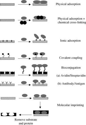
Increasing focus on the development of coatings capable of eliciting specific biological responses has seen a significant research focus on the incorporation of peptide sequences, particularly from the receptor-binding domains of adhesion proteins, in order to promote cell adhesion (6,27). In more general terms, there is significant interest in the immobilization of a range of biomolecules. Array and sensor technologies require antibody, protein, and DNA immobilization, while the immobilized proteoglycans such as heparin have been used to the modulate hemocompatibility of biomedical devices (44). As illustrated in Fig. 1, immobilization strategies for biomolecules can be as simple as nonspecific adsorption or as complex as molecular imprinting. The adsorption of biomolecules tends to result in coatings with limited functionality as the molecules are randomly oriented and tend to be desorbed from the surface over time. Covalent immobilization can eliminate problems associated with desorption, but there is often little control over conformation or orientation and thus activity of the biomolecule can be limited. The use of spacer polymer chains or amino acid sequences between the surface and the protein can reduce the denaturation of the molecule. Examples of this type of approach are the site specific modification of proteins with cysteine that enable immobilization of the proteins in specific orientations on gold (45). The success of strategies designed to present biological ligands can also be maximized if the immobilized molecule is coupled to a surface capable of preventing nonspecific adsorption. In general terms, biological response is influenced by the presentation, average density and the spatial distribution of the immobilized molecule
348 BIOMATERIALS, SURFACE PROPERTIES OF
Figure 1. A range of immobilization strategies for biomolecules at interfaces.
(1). One of the most challenging applications for wet chemical surface modification lies in the development of surfaces that borrow from structures observed in Nature. Surfaces that mimic the structure of a cell wall are increasingly sought for use in biosensors and as model systems to further investigate cell, protein, and pharmaceutical interactions. These systems generally consist of a lipid bilayer that may containing a range of different lipids, transmembrane and membrane proteins, and in some cases oligosaccharides. Critically, these structures need to retain their fluidity; molecules need to be able to move within and through the structure in order to maintain their activity and function. The most simple cell mimetic surfaces discussed in the literature have been based on transferring lipid bilayers onto glass substrates. Under these conditions, a thin film of water lubricates the interface between the glass and bilayer and allows free lateral diffusion. These types of bilayers generally have poor long-term stability, show limited transmembrane protein activity, and cannot be transferred through the air–water interface without disrupting the structure (46). An alternative approach lies in either the formation of hybrid bilayers, where the inner leaflet is formed from alkane thiol on gold (47), or the deposition of the lipids on a polymer support (46). Hydrogel layers can also be used and act as a hydrated cushion that is both a self-lubricating and a spacer, creating an area for protein insertion without affecting protein function. At present, there are a number
of commercial biosensors that utilize this type of technology for disease diagnosis (48).
SURFACE ANALYSIS
The study of surfaces and coatings is an advanced field and ranges from the investigation of elementary chemical processes on single crystals in ultrahigh vacuum (UHV) to the analysis of rather more ‘‘dirty’’ and real surfaces in engineering applications. The development of techniques suitable for surface science has a long history, the driving force for which has only recently included attempting to understand and control biomaterial surface interactions. Table 3 details some of the more common surface analysis techniques used today in the characterization of biomaterials. The physical principles of all the techniques commonly employed today are well understood and have been for many decades. Many of the approaches described here have their origin in the study of elementary chemical and physical processes, the semiconductor industry, and from engineering disciplines.
When considering the choice of surface analytical tools, it is important to appreciate the questions that require answering. A single technique cannot generally provide a complete picture of the surface characteristics. In this section, a description of the some of the most commonly used techniques is provided with reference to more detailed and extensive reviews. It is important to note that the techniques fall into two classes, those that operate inside a vacuum and those that can directly probe the biomaterial– water interface. While it is obviously preferable to use those techniques that can perform under ambient conditions, in general these techniques are either not as informative or not as surface sensitive as the vacuum techniques. For this reason, the vacuum techniques are commonly utilized to provide a detailed characterization, but in doing so it must always be under the assumption that the surface is the same in vacuum as it is under water. This is a rather large assumption, particularly if the material is able to reorganize itself relatively easily. The surface energy change following immersion in water can be rather large, as indicated above, and in mixed biomaterial phases components that are absent at the surface in vacuum may dominate when the material is immersed in an aqueous environment. There is evidence for this kind of surface reorientation in the contact angle hysteresis of water on some polymers.
Hysteresis is the difference in contact angle between a water contact line advancing or receding across a surface. For some polymers, the advancing angle is high and the receding angle is low, indicating that at the polymer–air interface the polymer is hydrophobic and at the polymer– water interface it is hydrophilic. As long as the surface is flat and homogenous, this is evidence of surface reorganization. For some materials it is possible to reduce the rate of reorganization by cooling. This is typically achieved by hydrating the sample in air, and then freezing the sample in liquid nitrogen prior to entry into the vacuum chamber. The sample needs to be held at low temperature while the ice on the surface sublimes and then vacuum techniques
|
BIOMATERIALS, SURFACE PROPERTIES OF |
349 |
||
Table 3. Surface Analysis Techniques Used in the Characterization of Biomaterials |
|
|
||
|
|
|
|
|
Technique |
Sampling Depth/Height (Spatial Resolution) |
Information Obtained |
References |
|
|
|
|
|
|
Ultrahigh Vacuum |
˚ |
|
|
|
Static secondary ion mass spectrometry |
Chemical |
|
49,50 |
|
<5 nm (500 A) |
|
|||
|
|
Spectroscopy and Imaging |
|
|
X-ray photoelectron spectroscopy |
2–10 nm (5 mm) |
Elemental, chemical |
|
51 |
|
|
Spectroscopy and Imaging |
|
|
Ambient Techniques |
|
|
|
|
Attenuated total reflectance |
> 100 nm (1 mm) |
Chemical |
|
52 |
Fourier transform |
|
|
|
|
Infrared (ATR/FTIR) spectrometry |
|
|
|
|
Contact angle measurement |
<1 nm (1 mm) |
Surface free energy, wettability |
|
53 |
Atomic force microscopy (AFM) |
Atomic ¼ 20 mm |
|
|
54 |
Imaging |
Topography, coverage, atomic structure |
|
|
|
Surface force measurement |
˚ |
|
|
|
A ¼ mm |
Chemical, conformational, structural |
|
|
|
|
˚ |
|
|
|
Ellipsometry |
Layer thickness, adsorption kinetics |
|
55,56 |
|
A ¼ 300 nm |
|
|||
Streaming potential |
Not applicable |
Electrokinetics |
|
57 |
measurements/electroosmosis |
300 nm |
|
|
|
Surface plasmon resonance |
Adsorbed mass and adsorption kinetics |
|
58 |
|
can be applied to the surface, which should not have reorganized from the hydrated state (59).
Vacuum Techniques
Traditional surface analysis techniques are usually based on ultrahigh vacuum instrumentation. One of the reasons for this was that much of the initial interest in the field was concentrated upon extremely clean and often highly reactive surfaces. To maintain the surface in this state during the analysis it is important to prevent undesired gas or vapor molecules sticking to the surface and changing its characteristics. This is only possible in ultrahigh vacuum (<10 9 mbar or so). A second important reason is that to study just the surface and eliminate contributions from the bulk of the material it is necessary to use probe species that strongly interact with matter, such as ions and electrons. These cannot penetrate through more than a few atomic layers, and hence provide highly surface sensitive information. However, the detection of such species normally requires that they travel a considerable distance from the surface. At atmospheric pressure the average distance traveled prior to interaction with gaseous species is too short for the detection systems to work. A vacuum of, typically, 10 7 mbar or better is required for the techniques described here to operate. In addition, many of the components necessary for the production and detection of probe species can only be operated in vacuum; at atmospheric pressure, they would be irreparably damaged. We will now briefly describe some of the key surface analysis techniques used in the characterization of biomaterials. More detailed information and discussion on the interpretation of these techniques can be found in books by Vickerman (54) and Watts (51).
X-Ray Photoelectron Spectroscopy
During X-ray photoelectron spectroscopy (XPS) analysis, the sample is illuminated with X rays of a particular energy
that causes electrons to be emitted from the sample. This phenomenon is called the photoelectric effect and it is generally found that an X-ray photon imparts all of its energy to a single electron during the process. Since the electrons are bound in orbitals of well-defined energy (binding energy) that are characteristic of the material, the outgoing electrons have a kinetic energy that is essentially the difference between the photon and binding energies. For core level electrons, this binding energy is characteristic of the nucleus to which the electron is closely bound. Only those electrons that are generated close to the surface can escape from the sample without loss of energy due to inelastic collisions with other atoms. Thus, by analyzing the number of electrons emitted from the surface as a function of electron kinetic energy it becomes possible to identify the elements present on, or near to, the material surface. As long as the photon energy is significantly larger than the binding energy of the electron, the probability of generating a photoelectron from a core level is independent of the chemical situation of the element. This means that it is possible not only to identify the elements present but, with appropriate sensitivity factors, quantify the relative amounts of each element.
The chemical situation of the element may, however, have an influence on the binding energy of the core level electron. Chemical bonds to different elements may cause some charge to accumulate on the element of interest, this will directly affect the binding energy of the core electrons. This change in the binding energy is termed the ‘‘chemical shift’’ by analogy to nuclear magnetic resonance (NMR) spectroscopy. The appearance of a chemical shift is extremely useful in surface analysis as it can provide information on how the various elements in the surface are bonded to each other. Where the same element is in a range of chemical environments it is often possible to deduce the fraction of atoms in each environment by careful curve fitting of the spectrum. An example of these types of chemical shifts is illustrated in Fig. 2. In this case, there are chemical shifts evident in a high resolution carbon 1s
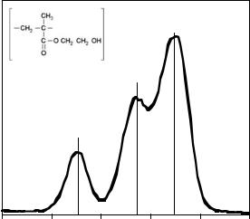
350 BIOMATERIALS, SURFACE PROPERTIES OF
|
|
|
|
285 eV |
|
|
|
|
|
CHx |
|
|
|
n |
286.5 eV |
|
|
|
|
|
|
|
|
|
|
|
C-O |
|
|
Intensity |
|
289 eV |
|
|
|
|
|
O-C=O |
|
|
|
292 |
290 |
288 |
286 |
284 |
282 |
Binding Energy (eV)
Figure 2. The XPS high resolution C1s spectrum of a HEMA contact lens. The figure illustrates the various chemical shifts associated with the chemical bonds in the polymer chain.
(C1s) spectrum due to the various chemical bonds present in poly(hydroxyethylmethacrylate), pHEMA, a common soft contact lens material.
The surface sensitivity of XPS is dependent on the angle at which electrons are emitted from the sample. For smooth, flat samples it is possible to enhance the surface sensitivity by analyzing electrons which are emitted at a grazing incidence from the sample. By collecting at a number of different angles it is possible to obtain information on the depth distribution of components close to the material surface. This depth profiling capability is particularly important when thin films and coatings of < 10 nm thickness are being studied.
The XPS has been utilized to chemically characterize biomaterials in four principal areas: identification and characterization of the surface chemistry of bulk polymers; characterization of surface specific modifications; characterization of coatings; detection of biomolecules. Factors that are commonly investigated using XPS include: surface oxidation and reorientation of polymer segments; surface segregation (blooming) of plasticizers, additives, and low molecular weight fragments; adventitious contaminants such as silicones and protein adsorption.
Secondary Ion Mass Spectrometry
The impact of a high kinetic energy (typically 1–100 keV) ion, atom, or molecule causes material to be sputtered from a surface. The origin of the vast majority of ejected species is from the topmost layer of the sample. Therefore analysis of the sputtered fragments can provide information on the composition of the material surface. A small proportion of the ejected atomic and molecular fragments are ionized, these are called the secondary ions. Secondary ion mass spectrometry (SIMS) is the application of mass spectrometry to the secondary ions. Note that SIMS is an ablative, destructive technique and this can be used to advantage in
generating a depth profile of layered surfaces. The use of SIMS for depth profiling is termed ‘‘dynamic’’ SIMS and is most often employed in the study of layered materials in which the elemental composition of the layers is of interest, for example, doping levels in semiconductors. It is not possible to obtain more detailed chemical information using dynamic SIMS because of the damage induced by the high energy primary ions. In contrast, ‘‘static’’ SIMS employs a low density, low dose ion bombardment such that the probability of two ion impacts occurring at the same place on the sample is negligibly small (< 1013 ions cm 2). The mass spectrum then contains information that is characteristic of the undamaged surface. This information is particularly useful in the analysis of organic materials, when the normal rules of organic mass spectrometry can be applied to the interpretation of SIMS data. Most modern static SIMS instruments are based on time-of- flight mass analyzers (TOF–SIMS), which have a far greater combined sensitivity and mass resolution than quadrupole or magnetic sector detectors. The probability of ion generation is influenced by a daunting range of factors and thus SIMS is regarded as a nonquantitative technique. However, it is commonly found that in a range of similar materials the characteristic ion intensities are approximately proportional to the concentration of species from which they are generated. With a suitable set of calibration data it is then possible to use SIMS in a quantitative manner. The application of TOF–SIMS in the analysis of biomaterials and biological interfaces has historically revolved around the characterization of polymeric interfaces. This has included the study of degradation pathways for biodegradable polymers, the monitoring of coating chemistries, detection of surface contamination and surface chemical characterization of copolymer systems. The surface sensitivity of TOF–SIMS has lead to its application in the detection and identification of biomolecules adsorbed at interfaces. The process is not without its problems though as the largest ions detected from any protein are the immonium ions (þNH2¼CHR) from each amino acid (MW<200). As a result of this fragmentation, the identification of proteins is often more like a jigsaw puzzle, where the amino acid fragments have to be pieced together using pattern recognition or multivariate analysis techniques, to identify and quantify the parent molecules (60). These types of statistical analysis are being increasingly used to analyze, compare and reconstruct data collected in TOF–SIMS.
In addition to spectroscopy, TOF–SIMS can be used in an imaging mode to chemically map the surface of a material. There is always a trade off between high special resolution and high mass resolution, but with the advent of liquid metal ion sources (e.g., Gaþ and Inþ), systems are typically capable of spatial resolution of < 10 mm, while retaining atomic mass resolution. As a result there is increasing application of TOF–SIMS for the chemical imaging of a range of biomaterial surfaces. Significantly, developments in ion sources have shown that polyatomic (e.g., Au3) and cluster ion (C60) sources can significantly improve the molecular ion yield of both biological and polymeric materials. With the development of integrated freeze hydration stages for sample preparation, this has
lead to increased activity in the application of TOF–SIMS in the analysis of cell membranes and other hydrated biological systems (49).
AMBIENT TECHNIQUES
Atomic Force Microscopy
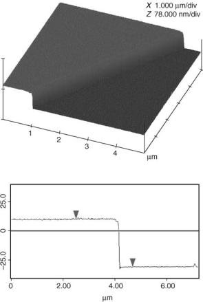
Atomic force microscopy (AFM) can be utilized to characterize surfaces via either an imaging or a spectroscopic mode. There are two common methods of imaging utilized in AFM, contact, and tapping mode. Both can be performed in either air or liquid, a factor that makes AFM particularly attractive in biomaterial research. In contact mode, the tip is scraped across the surface, while in tapping mode, the tip is in intermittent contact with the surface and as such, there is limited substrate disturbance. As a result, tapping mode AFM is more common for the characterization of biomaterials and biological surfaces. Common applications of AFM to biomaterial surfaces include: surface topography and coating continuity assessment, measurement and monitoring of coating thickness (see Fig. 3) and phase imaging. The last application is an extension of tapping mode imaging that gives nanometer-scale two-dimensional (2D) information about surface structure. It can be used to locate and characterize the distribution of discrete phases within polymer blends such as polyurethane–urea, and a range of other polymers of biological importance. There is also considerable interest in using AFM to image the surfaces of cells and biomolecules on surfaces (61,62).
Surface Force Measurements
The forces that act between particles and surfaces determine a wide range of interactions. They control the stability of dispersions and emulsions; determine the adhesion of colloids onto surfaces and the adsorption properties of proteins and cells at surfaces. Surface force measurements are used in variety of industries and increasingly there is interest in the application of surface force analysis to biomaterials. with the aim of characterizing protein–pro- tein, cell–protein, protein–surface and cell–surface interactions.
There are a number of techniques that can be used to measure force interactions between surfaces. They are divided into two classes based on the method used to determine surface separation. Absolute surface separation can only be determined using an interferometric technique such as the surface force apparatus (SFA). These techniques are limited by the need for a transparent substrate and specific geometric configuration. In response, noninterferometric techniques have been developed that can employ a wider variety of substrates, and that rely on indirect determination of the surface separation rather than interferometry. One of these is AFM surface force measurement.
Force measurements can be made with an AFM using both a bare tip and a tip modified with a probe particle. If the results from these measurements are to be quantitative, knowledge of the radius of curvature for the probe or tip is critical. While it is possible to measure the nominal
BIOMATERIALS, SURFACE PROPERTIES OF |
351 |
Figure 3. The AFM tapping mode image of a 40 nm step between coated and uncoated regions on a silicon wafer. The step is produced by masking a section of the sample surface prior to plasma polymerization. Once the coating is deposited, the mask is removed and surface imaged. This enables the plasma polymer thickness to be measured with nm resolution. (Image courtesy of Dr. P.G. Hartley, CSIRO Molecular Science, Australia.)
radius of a bare AFM tip, the indirect nature of the measurement adds to the error associated with the resulting force calculations (63). The Derjaguin approximation is only valid when the radius is much larger than the surface separation, which is not necessarily the case if an unmodified tip is used. If a colloid probe is utilized, the radius of curvature can be measured easily and accurately using scanning electron microscope (SEM) or optical microscopy.
The zero surface separation for noninterferometric techniques are set at the hard wall encountered when the two surfaces are forced together. Termed the constant compliance regime, this is the region of the force curve in which the displacement of the colloid probe is linear with respect to the surface motion. This assumption of hard wall contact is an inherent limitation of these techniques, particularly if the surface deforms or compresses under pressure, as is often the case with polymer surfaces. The compression of a polymer layer can have a number of effects on the force curve. In the first instance, compression of a dense polymer layer can result in a compliance line mimicking hard wall contact, with a layer of compressed polymer between the probe and the substrate. If the polymer is less densely
352 BIOMATERIALS, SURFACE PROPERTIES OF
packed, the probe may displace the material, squeezing the polymer out of the gap between the probe and the surface. This results in discontinuities in the compliance region as the force increases and the probe pushes through the polymer layers (63). As a result, conclusions about absolute layer thickness cannot be inferred from this data.
While XPS is able to characterize the chemistry of a surface in the dry state, surface force measurements are well suited to characterizing the intermolecular forces and stability in a variety of environments. A number of studies have investigated the surface force characteristics of rfgd films, grafted polymer layers, and adsorbed protein films in a variety of media (64). Other studies have used surface force measurements to characterize the interactions of polymer modified surfaces in an attempt to elucidate parameters that control the structure of the polymer layer (64). Surface force techniques have been used to investigate the effects of molecular weight, ionic strength, charge density, and polymer concentration on the interaction forces of adsorbed and grafted polymers layers. Increasingly, strategies to eliminate protein adsorption are based on the characterization and modification of the surfaces and thus the interaction forces that govern protein adsorption (65). A number of theoretical studies have also used surface force interactions as design parameters when modeling polymer coatings capable of resisting protein adsorption (66).
In addition to these standard modes of operation, by chemically modifying the AFM cantilever it is possible to map specific interactions between the tip and the surface. Depending on the type of modification made to the cantilever, a range of interactions can be investigated. With a cantilever modified with a receptor specific integrin, dynamic force spectroscopy can be used to identify and map receptor sites on a cell surface (67). By modifying the cantilever with specific chemical functional groups, differences in frictional properties and the distribution of different phases can be probed where there is no topographical variation (68).
Optical Techniques
The refractive index close to an interface can be measured by a number of optical techniques. The two most commonly employed for this purpose are surface plasmon resonance (SPR) (58) and ellipsometry (55,56). Since proteins have a higher refractive index ( 1.55) than water ( 1.33) it is possible to monitor the amount of protein adsorption at an interface through a measurement of the refractive index and thickness of the adsorbed layer. These techniques have found utility in a wide range of areas relevant to biomaterials research, such as the study of protein adsorption to biomaterials. ligand–receptor interactions and the dissolution and swelling of polymers. The advantage over traditional approaches such as enzyme linked immunosorbent assay (ELISA), fluorescent labeling or radiolabeling is that the proteins under study do not have to be chemically altered in any way.
The disadavantages of these techniques are that the substrate must be flat and conform to a number of optical requirements for the techniques. Additionally, these tech-
niques cannot directly distinguish between different proteins, since all proteins have roughly equivalent optical densities. To determine the identity of proteins adsorbed from a mixed solution it is necessary to subsequently expose the surface to antibodies specific to each protein of interest. Binding of the antibody can be monitored as an increase in the adsorbed layer thickness, however, this approach is difficult to employ quantitatively as there may be nonspecific and competitive adsorption of the antibody as well as a limited availability of binding sites on the adsorbed target protein.
Surface Plasmon Resonance
A surface plasmon is a collective oscillation of electrons that can be excited in certain metals such as silver and gold. The frequency of this oscillation depends on the refractive index of the dielectric material close to the metal interface. If the metal is a thin film it is possible to excite surface plasmons by reflecting light of a wavelength greater than the thickness of the metal from the reverse side of the film. The ability to cause this excitation depends on the wavelength of light, the refractive index of the material through which the light travels (which remains constant in this geometry) and the angle of reflection. When the conditions are correct, light is absorbed. In the usual set up for surface plasmon resonance (SPR) instruments the light undergoes total internal reflection at the interface and the angle at which light is absorbed is monitored. If there is a change in the refractive index close to the interface then a corresponding shift in the angle at which light is absorbed can be followed. The sensitivity of SPR decreases exponentially in distance from the surface with a decay length of the order of the wavelength of the light. If the layer to be analysed is significantly smaller than the decay length, which is usual for protein adsorption, then it can be assumed that any change in refractive index is proportional to the mass of adsorbed protein.
Ellipsometry
When light is reflected at an oblique angle from a planar surface it commonly undergoes a change in polarization. By analyzing these changes, it is possible to infer both the optical properties and thickness of thin films on the surface. To obtain the most complete characterization, a large number of different wavelengths of light or different angles of incidence must be studied. The measured data is then compared to the expected polarization changes calculated from a model, and parameters in the model changed (such as thickness or refractive index) to find a fit between the two. The sensitivity of ellipsometry is comparable to SPR ( 0.01/g cm2), however, it is able to analyze comparatively thick layers of material.
CONCLUSION
While materials selection for most biomedical devices needs to be based upon bulk properties, in this article we have provided a broad overview of the basic properties of surfaces, and introduced some of the reasons why the surface properties may significantly 3 influence the efficacy of biomaterials
and biomedical devices. Surface modification aims to tailor the surface characteristics of a material for a specific application without detrimentally affecting the bulk properties. Throughout this article we have shown how a range of physical, chemical, and biological modifications can be made to surfaces and used to manipulate surface characteristics. Finally, we discussed a range of highly sensitive surface analytical methods that can be utilized to investigate both the nature of an interface and its interactions with biological environments. As is always the case with review articles of this type, it is impossible to give detailed accounts of all of the material being discussed. We have included a range of references (Reading List) to aid the reader in further developing their understanding of each of the specific concepts and techniques. Additionally, we have included a list of more general references that cover many of the fundamental concepts discussed within this article.
BIBLIOGRAPHY
Cited References
1.Mittal KL, editor. Contact Angle, Wettability and Adhesion. Utrecht: VSP; 1993.
2.Berry CC, Campbell G, Spadiccino A, Robertson M, Curtis ASG. The influence of microscale topography on fibroblast attachment and motility. Biomaterials 2004;25(26):5781– 5788.
3.Andrade JD. Principles of Protein Adsorption. In: Andrade JD, editor. Surfaces and Interfacial Aspects of Biomedical Polymers. Vol. 2: Protein Adsorption. New York: Plenum Press; 1985.
4.Underwood PA, Steele JG. Practical limitations of estimation of protein adsorption to polymer surfaces. J Immunol Methods 1991;142(1):83–94.
5.Leduc CA, Vroman L, Leonard EF. A Mathematical-Model for the Vroman Effect. Ind Eng Chem Res 1995;34(10):3488– 3495.
6.Hubbell JA. Bioactive Biomaterials. Curr Opin Biotechnol 1999;10:123–129.
7.Bray D. Cell Movements:From Molecules to Motility. 2nd ed. New York: Garland; 2001. p 372.
8.Chu PK, Chen JY, Wang LP, Huang N. Plasma-surface modification of biomaterials. Mat Sci Eng R 2002;36(5-6): 143–206.
9.Favia P, d’Agostino R. Plasma Treatments and Plasma Depositions of Polymers for Biomedical Applications. Surf Coat Tech 1998;98:1102–1106.
10.Aronsson BO, Lausmaa J, Kasemo B. Glow discharge plasma treatment for surface cleaning and modification of metallic biomaterials. J Biomed Mater Res 1997;35(1):49–73.
11.Mandl S, Rauschenbach B. Plasma immersion ion implantation. New technology for homogeneous modification of the surface of medical implants of complex shapes. Biomed Tech 2000;45(7–8):193–198.
12.Bilek MMM, McKenzie DR, Tarrant RN, Lim SHM, McCulloch DG. Plasma-based ion implantation utilising a cathodic arc plasma. Surf Coat Tech 2002;156(1–3):136–142.
13.Shin GH, Lee YH, Lee JS, Kim YS, Choi WS, Park HJ. Preparation of plastic and biopolymer multilayer films by plasma source ion implantation. J Agric Food Chem 2002;50(16):4608–4614.
14.McPherson TB, Shim HS, Park K. Grafting of PEO to glass, nitinol, and pyrolytic carbon surfaces by gamma irradiation. J Biomed Mater Res 1997;38(4):289–302.
BIOMATERIALS, SURFACE PROPERTIES OF |
353 |
15.Benson RS. Use of radiation in biomaterials science. Nucl Instrum Meth B 2002;191:752–757.
16.Welle A, Gottwald E. UV-based patterning of polymeric substrates for cell culture applications. Biomed Microdevices 2002;4(1):33–41.
17.Zhang F, Kang ET, Neoh KG, Wang P, Tan KL. Surface modification of stainless steel by grafting of poly(ethylene glycol) for reduction in protein adsorption. Biomaterials 2001;22(12):1541–1548.
18.Krupa D, Baszkiewicz J, Kozubowski J, Barcz A, Sobczak J, Bilinski A, Rajchel B. The influence of calcium and/ orphosphorus ion implantation on the structure and corrosion resistance of titanium. Vacuum 2001;63(4): 715–719.
19.Braceras I, Alava JI, Onate JI, Brizuela M, Garcia-Luis A, Garagorri N, Viviente JL, de Maeztu MA. Improved osseointegration in ion implantationtreated dental implants. Surf Coat Tech 2002;158:28–32.
20.Cui FZ, Luo ZS. Biomaterials modification by ion-beam processing. Surf Coat Tech 1999;112(1–3):278–285.
21.Bambauer R, Mestres P, Schiel R, Schneidewind JM, Latza R, Bambauer S, Sioshansi P. Surface treated catheters with ion beam based process for blood access. Ther Apher 2000;4(5):342–347.
22.Li DJ, Zhao J, Gu HQ. Hemocompatibility of DLC coatings synthesized by ion beam assisted deposition. Sci China Ser E- Technol Sci 2001;44(4):427–431.
23.Kingshott P, Griesser HJ. Surfaces that resist bioadhesion. Curr Opin Solid St M 1999;4:403–412.
24.Bures P, Huang YB, Oral E, Peppas NA. Surface modifications and molecular imprinting of polymers in medical and pharmaceutical applications. J Control Release 2001;72(1– 3):25–33.
25.Kato K, Uchida E, Kang ET, Uyama Y,Ikada Y. Polymer surface with graft chains. Prog Polym Sci 2003;28(2):209–259.
26.Cai KY, Lin SB, Yao KD. Advances in research on surface engineering of biomaterials for tissue engineering. Prog Chem 2001;13(1):56–64.
27.Sakiyama-Elbert SE, Hubbell JA. Functional biomaterials: Design of novel biomaterials. Ann Rev Mater Res 2001; 31:183–201.
28.Massia SP, Stark J. Immobilized RGD peptides on surfacegrafted dextran promote biospecific cell attachment. J Biomed Mater Res 2001;56(3):390–399.
29.Whitesides GM, Ostuni E, Takayama S, Jiang X, Ingber DE. Soft Lithography in Biology and Biochemistry. Annu Rev Biomed Eng 2001;3:335–373.
30.Beck AJ, Jones FR, Short RD. Plasma copolymerization as a specific route to the fabrication of new surfaces with controlled amounts of specific chemical functionality. Polymer 1996;37:5537–5539.
31.Shen MC, Martinson L, Wagner MS, Castner DG, Ratner BD, Horbett TA. PEO-like plasma polymerized tetraglyme surface interactions with leukocytes and proteins: in vitro and in vivo studies. J Biomater Sci Polym Ed 2002;13(4):367– 390.
32.Pan YV, Wesley RA, Luginbuhl R, Denton DD, Ratner BD. Plasma polymerized N-isopropylacrylamide: synthesis and characterization of a smart thermally responsive coating. Biomacromolecules 2001;2(1):32–36.
33.Fraser S, Short RD, Barton D, Bradley JW. A multi-techni- que investigation of the pulsed plasma and plasma polymers of acrylic acid: Millisecond pulse regime. J Phys Chem B 2002;106(22):5596–5603.
34.Han LCM, Timmons RB. Pulsed-plasma polymerization of 1- vinyl-2- pyrrolidone: Synthesis of a linear polymer. J Polym Sci Pol Chem 1998;36(17):3121–3129.